Compiled from clinical pathology references. Medically reviewed by Dr Cristian Dunker , Principal Dentist, ArtSmiles Cosmetic Dentistry.
Quick summary
Also called | Lupus, LE, SLE (systemic lupus erythematosus), SLE, a chronic autoimmune disease where the immune system attacks the body's own tissues, DLE / CCLE (discoid or chronic cutaneous lupus erythematosus), SCLE (subacute cutaneous lupus erythematosus), SCLE, a skin-predominant form of lupus that responds particularly to sunlight, lupus cheilitis (when the lip is involved) |
How urgent? | 🟡 Worth a check-up, oral changes can sometimes be the first clue to lupus, and your dentist may suggest a medical referral |
Common or rare? | Lupus itself affects more than 1.5 million people in some Western populations. Oral lesions appear in around 5 to 45% of people with lupus, depending on the form |
Who it affects | Mostly women aged in their twenties to forties, although discoid lupus is more typical in middle age. Women are affected 8 to 10 times more often than men |
Who treats it | A general dentist for the oral lesions, working alongside a GP, dermatologist, or rheumatologist for the underlying disease |
Based on | Regezi, Neville, Cawson, Laskaris |
What is it?
Lupus erythematosus is a long-term autoimmune condition where the body's immune system mistakenly attacks its own tissues. There are two main forms that can show up in the mouth: a localised form that mainly affects the skin and mouth (called discoid or chronic cutaneous lupus), and a more widespread form that can involve many organs (called systemic lupus erythematosus, or SLE). The mouth often shows fine white striae with red, sometimes ulcerated centres, and these oral changes can occasionally be the first sign that something more is going on.
Who tends to get it?
Lupus most commonly affects women, and the average age at diagnosis for systemic lupus is around 31 years. Women are affected nearly 8 to 10 times more often than men. Discoid (chronic cutaneous) lupus tends to appear later, typically in middle age, and again favours women.
Oral lesions develop in roughly 5 to 25% of people with systemic lupus (some studies report up to 40 to 45%), and in around 15 to 25% of people with discoid lupus. Around 20% of people with SLE develop noticeable mouth changes overall, and in a smaller number of cases the mouth lesion is the first thing to be noticed. Lupus also has a genetic component, if one identical twin has SLE, the other has about a 24% chance of also developing it.
What causes it?
The exact cause of lupus is unknown. It is thought to come from a mix of genetic susceptibility and environmental triggers that nudge the immune system into attacking the body's own cells. The immune system produces antibodies aimed at parts of the cell nucleus and cytoplasm, and these self-targeting antibodies (autoantibodies) drive the disease.
Well-recognised triggers and risk factors include:
Genetics, a family history of lupus or other autoimmune conditions
Female sex, strongly skewed towards women, suggesting a hormonal influence
Sunlight, ultraviolet light is a major trigger, particularly for the skin rashes and the malar (butterfly) rash on the face
Certain medications, a number of drugs can cause a lupus-like reaction in the mouth and skin
Smoking, has been linked with worse skin and oral disease
A small number of medications can also produce a lupus-like reaction (sometimes called drug-induced lupus), which usually settles when the medication is stopped.
How does it develop?
Think of the immune system as a security team that normally only attacks outsiders, like bacteria and viruses. In lupus, that security team starts mistaking parts of the body's own cells, particularly bits of the cell nucleus, for intruders. It then makes antibodies against them.
These autoantibodies bind to their targets and form little clumps (immune complexes) that lodge in tissues like the skin, lining of the mouth, joints, and kidneys. Wherever they settle, they trigger inflammation and damage. In the mouth and skin, this inflammation specifically targets the layer where the surface lining meets the deeper tissue, which is why lupus lesions often have a characteristic look of a red, atrophic centre with fine white lines fanning out around it.
What might you notice?
What it looks like
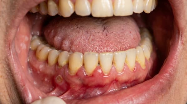
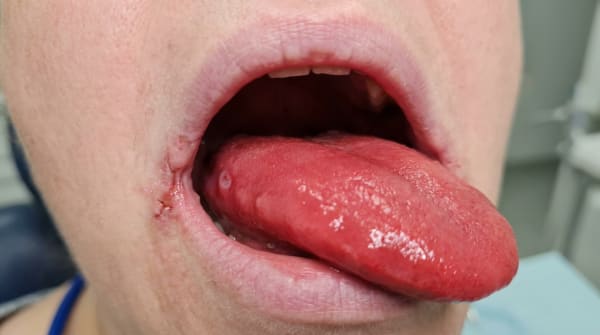
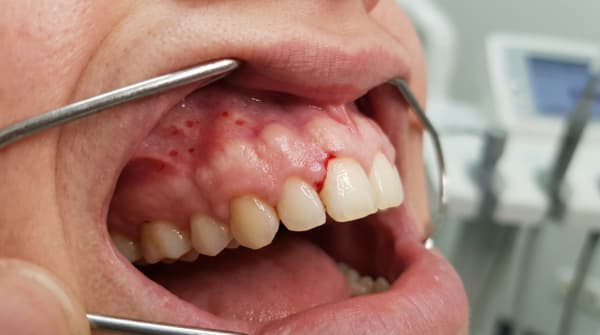
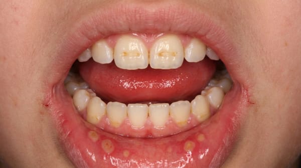
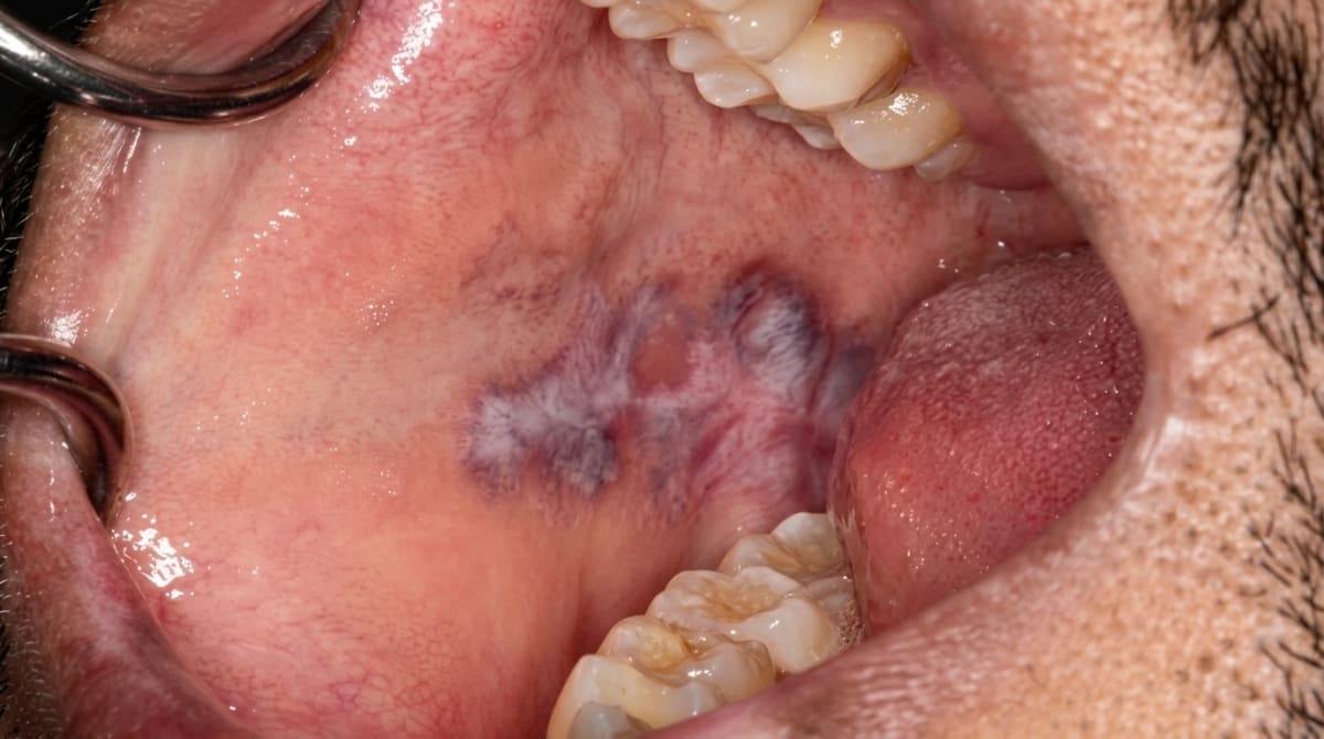
The classic oral lesion of lupus is a well-defined red (atrophic) central area surrounded by fine, delicate, radiating white lines or striae, a bit like the rays of a small sun. There may be small white dots or tiny dilated blood vessels (telangiectasia) within the red zone, and sometimes shallow ulcers or erosions.
The most commonly affected sites are:
The buccal mucosa (inner cheek), most common
The palate, particularly suggestive of lupus, because lichen planus tends to spare this site
The gingiva (gums)
The lower lip vermilion border, sometimes called lupus cheilitis, where a scaly, atrophic patch can form on the lip
The tongue
In systemic lupus, you may also see scattered painful erosions or ulcers, small areas of bleeding (petechiae), and a dry mouth. On the skin, the well-known butterfly rash spreads across the cheeks and bridge of the nose, sparing the folds beside the nostrils, and discoid lupus produces scaly, disc-shaped red patches on sun-exposed skin that may heal with scarring, loss of pigment, and (on the scalp) permanent hair loss.
What it feels like
Many discoid oral lesions are painless or only mildly uncomfortable, and a person may not notice them until a dentist points them out. When the surface breaks down into atrophic or ulcerated areas, the lesions can become sore, especially with acidic, salty, or spicy foods. Other oral symptoms reported in lupus include:
Burning or sore mouth (stomatodynia)
Dryness (xerostomia)
A change or distortion in taste (dysgeusia)
Recurrent oral candidiasis (thrush)
General symptoms of systemic lupus include tiredness, joint pain, fevers, weight loss, and a rash worsened by sunlight.
What an X-ray might show
Lupus does not usually produce specific dental X-ray changes, so imaging is not the main way it's identified. X-rays may be taken to help rule out other dental problems that could be confused with the mouth lesions.
What happens at the dentist?
Mouth lesions of lupus can look very similar to other conditions, particularly oral lichen planus, so making the diagnosis usually involves more than just a clinical exam. At ArtSmiles, assessment of unusual mouth changes is part of the standard examination across our general dental services.
A dentist may:
Take a careful history, including any sun-sensitive skin rashes, joint pain, fatigue, weight loss, kidney issues, or family history of autoimmune disease
Examine the mouth, lips, and visible skin, looking for the typical pattern of red atrophic patches with delicate radiating white striae, particularly on the buccal mucosa or palate, and asymmetric distribution
Recommend a biopsy, a small sample of the lesion may be sent for histopathology, often together with a special test called direct immunofluorescence, which can show characteristic deposits of antibodies and complement at the basement membrane (the so-called lupus band test)
Suggest blood tests through your GP, looking for antinuclear antibodies (ANA), anti-double-stranded DNA antibodies, and anti-Sm antibodies, which are very specific for systemic lupus
Refer for medical assessment, to a GP, dermatologist, or rheumatologist for full work-up, since oral lesions can sometimes be the first sign of systemic disease
If medications are suspected of triggering a lupus-like reaction, your medical team may review them.
Is this serious?
🟡 The seriousness depends on which form of lupus is present.
Discoid (chronic cutaneous) lupus is generally confined to the skin and oral mucosa and has a good outlook. About 50% of people see their disease eventually settle after several years. Around 5 to 15% may go on to develop systemic lupus over time, which is why ongoing review matters.
Systemic lupus is a serious multi-system disease. With modern treatment, about 95% of people are alive at 5 years and 78% at 20 years. Kidney involvement (lupus nephritis), lupus-related inflammation of the kidneys is the most important driver of long-term outcome.
Left unchecked, the oral lesions themselves are usually not dangerous, but they may continue to be painful, recur, and very rarely (in long-standing discoid lesions, especially on the lip) carry a small risk of malignant transformation. The bigger reason to seek assessment is that mouth changes can be an early flag for an underlying systemic condition that benefits from medical care.
If you've noticed any of these signs for more than two weeks, it's worth booking an assessment.
Could it be something else?
The oral signs of lupus overlap with several other conditions, and a dentist will usually consider the following alternatives. Differentials are drawn from Regezi, Neville, Cawson, and Laskaris.
Oral lichen planus, also produces white striae and red atrophic or eroded areas on the inner cheek. Lichen planus is typically symmetrical (both sides), with sharp white lacy lines (Wickham's striae), and tends to spare the palate. Lupus is often more asymmetric, the striae are more delicate and radiate from a central red zone, and the palate is more often involved.
Lichenoid drug reactions, medication-induced lesions that look very similar to lichen planus. Distinguished by a clear timing relationship with starting a new drug, and resolution when the drug is changed.
Pemphigus vulgaris, an autoimmune blistering disease that causes painful, ragged erosions in the mouth. Differentiated by Nikolsky's sign, fragile blisters, and characteristic biopsy and immunofluorescence findings.
Mucous membrane pemphigoid, produces sub-epithelial blisters and red, eroded gums (desquamative gingivitis). Distinguished by the deeper level of blister formation on biopsy and a different pattern on immunofluorescence.
Erythema multiforme, causes acute, painful lip crusting and oral ulcers, often after an infection or medication trigger. Differentiated by its sudden onset, target-shaped skin lesions, and self-limiting course.
Leukoplakia, a white patch that cannot be wiped off and has no obvious cause. Differentiated by its uniform white appearance without the red atrophic centre and radiating striae of lupus, and confirmed by biopsy.
Erythroplakia, a velvety red patch that can have a high risk of dysplasia. Differentiated by its uniformly red appearance without surrounding white striae, and biopsy is essential.
Oral candidiasis (thrush), particularly the erythematous form, which causes red patches especially on the palate. Distinguished by response to antifungal treatment and microbiological testing.
Recurrent aphthous stomatitis, common, painful round mouth ulcers. Differentiated by their well-defined yellow-grey base, red halo, and absence of radiating white striae or skin lesions.
Sjögren's syndrome, another autoimmune condition that causes dry mouth and dry eyes; it can coexist with lupus in around 30% of cases. Identified by salivary flow studies, lip biopsy, and antibodies to SS-A (Ro) and SS-B (La).
Geographic tongue (benign migratory glossitis), produces map-like red patches with white borders on the tongue. Differentiated by its location on the tongue dorsum, shifting pattern, and benign course.
Chronic ulcerative stomatitis, a rare condition with lesions resembling erosive lichen planus or lupus. Distinguished by characteristic speckled antinuclear antibody deposits on direct immunofluorescence.
Syphilis (mucous patches), secondary syphilis can produce greyish-white oral patches. Differentiated by serological testing and clinical history.
Oral leukoplakia, a persistent white patch that cannot be wiped off and does not fit another diagnosis. Distinguished by its uniformly white appearance and the absence of the radiating striae seen in lupus.
Oral thrush (pseudomembranous candidiasis), creamy white plaques that wipe off to leave a red base. Distinguished by the wipeable plaques and a positive fungal swab.
Erythroplakia, a velvety red patch that cannot be otherwise diagnosed. Distinguished by its uniformly red appearance and a high concern for early dysplasia, biopsy is typically recommended.
How is it treated?
Treatment depends on whether the disease is limited to the mouth and skin (discoid) or systemic, and is usually shared between your dentist and your medical team.
Self-care that may help:
Sun protection, broad-spectrum sunscreen, hats, and sun-protective clothing, since UV light can trigger flares
Smoking cessation, smoking can worsen disease activity and lip lesions
Gentle oral hygiene, soft-bristled toothbrush, mild fluoride toothpaste, and avoiding strong mouthwashes that may sting eroded areas
Avoiding trigger foods during flares, particularly acidic, salty, or spicy foods that aggravate sore patches
Regular dental check-ups, to monitor lesions and screen for any change
Professional care may include:
Topical corticosteroids applied to oral lesions, often in a paste or rinse, for discoid oral lupus and milder oral involvement of SLE
Topical calcineurin inhibitors (such as tacrolimus or pimecrolimus) for lesions resistant to topical steroids
Antimalarial drugs (such as hydroxychloroquine), an antimalarial tablet commonly used as a first-line lupus medication, prescribed by a GP, dermatologist, or rheumatologist, which are often very helpful for skin and oral lesions
Systemic corticosteroids for more active disease, particularly when other organs are involved
Other immunosuppressive or immunomodulating medications for severe or refractory disease
Treatment of associated dry mouth and thrush, saliva substitutes, sialagogues, and antifungal therapy where needed
Periodic monitoring for any malignant change in long-standing lip or oral lesions
Referral to a dermatologist or rheumatologist is appropriate when systemic features are present or when oral lesions don't settle with topical treatment.
What's the long-term outlook?
Outlook varies by form. For discoid lupus, the prognosis is generally good, most people respond to topical treatment, and around half see their disease resolve over several years. A small proportion (around 5 to 15%) develop systemic lupus over time, which is why ongoing review is sensible.
For systemic lupus, modern treatment has greatly improved survival, with around 95% of people alive at 5 years. Long-term outcome depends on which organs are involved (kidney involvement being the most important) and how often the disease flares. Oral lesions tend to come and go, often flaring with skin disease and settling with treatment.
Lupus is a long-term condition rather than something that is usually cured outright. With sun protection, well-chosen medications, and regular review by both your dental and medical teams, most people manage it effectively and keep oral symptoms to a minimum.
A note on this article
This article is for educational purposes only and does not constitute a clinical diagnosis. Please consult a registered dental practitioner for assessment and treatment advice.
The cover image above is an AI-generated illustration based on the most common visible features of this condition described in clinical pathology references. It is not a photograph of a real case and should not be used to diagnose or rule out the condition in your own situation. If you are concerned about something you have noticed, please book an assessment with a registered dental practitioner.
References
Regezi, J. A., Sciubba, J. J., & Jordan, R. C. K. (2017). Oral pathology: Clinical pathologic correlations (7th ed.). Elsevier. Chapter 3, White Lesions: Lupus Erythematosus, pp. 102 to 104.
Neville, B. W., Damm, D. D., Allen, C. M., & Chi, A. C. (2023). Oral and maxillofacial pathology (5th ed.). Elsevier. Chapter 16, Dermatologic Diseases: Lupus Erythematosus, pp. 798 to 801.
Cawson, R. A., & Odell, E. W. (2017). Cawson's essentials of oral pathology and oral medicine (8th ed.). Elsevier. Chapter 13, Diseases of the Oral Mucosa: Non-infective Stomatitis (Lupus Erythematosus), pp. 230 to 231; Chapter 25, The Medically Compromised Patient (Systemic Lupus Erythematosus), pp. 365 to 366.
Laskaris, G. (2003). Color atlas of oral diseases (3rd ed.). Thieme. Chapter 21, Autoimmune Diseases: Discoid and Systemic Lupus Erythematosus, pp. 188 to 191.
Frequently asked questions
What oral signs can lupus cause?
Lupus can cause oral lesions in around 25-40% of patients. Typical features are central red atrophic patches with white striae on the palate, cheeks or lips (resembling oral lichen planus), painful ulcers (often on the palate), desquamative gingivitis, lip discoid lesions and butterfly-rash extension onto the lips. Secondary Sjögren's syndrome causes dry mouth in many lupus patients.
How is oral lupus told apart from lichen planus?
They look alike clinically. Lupus lesions are often centrally atrophic with radiating white striae and tend to involve the hard palate, whereas oral lichen planus more often involves the cheek. Direct immunofluorescence on a biopsy shows a different pattern (granular deposits along the basement membrane in lupus vs. fibrinogen in lichen planus). Blood tests (ANA, anti-dsDNA, anti-Sm) support a systemic lupus diagnosis.
Do lupus medications affect the mouth?
Yes. Hydroxychloroquine can rarely cause oral pigmentation. Corticosteroids and immunosuppressants increase susceptibility to oral candidiasis and slow healing. Mycophenolate, methotrexate and biologics can cause mouth ulcers. NSAIDs can affect bleeding. Always update your dentist on current medications.
What about dental treatment during lupus flares?
Elective treatment is best deferred until disease is quiescent. Photo-sensitive flares can occur after prolonged dental light exposure in some patients. Lupus anticoagulant can prolong bleeding despite normal INR. Antibiotic prophylaxis is occasionally needed if there is associated heart valve involvement (Libman-Sacks endocarditis) — your rheumatologist and cardiologist will advise.