Compiled from clinical pathology references. Medically reviewed by Dr Cristian Dunker , Principal Dentist, ArtSmiles Cosmetic Dentistry.
Quick summary
Also called | OLP, lichen planus of the mouth, reticular/sore and raw (erosive)/atrophic/plaque/papular/bullous lichen planus |
How urgent? | 🟡 Worth a check-up, most cases are mild, but persistent ulcers or red patches deserve a proper assessment. |
Common or rare? | Relatively common, affects roughly 0.1% to 2% of adults. |
Who it affects | Mostly middle-aged adults, usually 30 to 60 years old. Women are affected more often than men (around 3 to 2). Children are rarely affected. |
Who treats it | General dentist for diagnosis, monitoring and most treatment. Specialist referral (oral medicine) for stubborn or atypical cases. |
Based on | Regezi, Neville, Cawson |
What is it?
Oral lichen planus is a long-running inflammatory condition of the lining of the mouth. It typically shows up as fine white lacy lines, red patches, or sore areas inside the cheeks, on the tongue, or on the gums. The condition tends to come and go in waves over many years.
It is not an infection and it is not contagious, even though the name sounds botanical, it has nothing to do with fungi or plants.
Who tends to get it?
Oral lichen planus is mostly a condition of middle age. The textbooks place most patients between 30 and 60 years old, with women making up roughly 65 per cent of cases, about a 3 to 2 female-to-male ratio. Children are very rarely affected.
Around 0.1 to 2 per cent of the adult population may have the oral form, which makes it one of the more common chronic mouth conditions a dentist sees. The skin form of lichen planus exists too, but most people who have oral lesions never develop skin lesions, and vice versa.
What causes it?
The exact cause is still unknown. What is well established is that oral lichen planus is an immune-driven condition. The body's own T-lymphocytes (a type of white blood cell) target the cells lining the mouth, leading to inflammation and surface changes.
A few factors can trigger or aggravate it:
Stress and anxiety, these don't cause the condition on their own, but they often parallel flare-ups.
Certain medications, some blood pressure tablets, anti-inflammatories, antimalarials, beta-blockers, oral diabetes medications and gold-based drugs can produce a near-identical reaction. When a drug is the trigger, the condition is usually called a lichenoid drug reaction rather than true lichen planus.
Dental restorations, particularly older or corroded amalgam fillings can cause a localised lichen-planus-like reaction in the mouth lining (mucosa) that touches the filling.
Hepatitis C infection, reported as a possible association in some Mediterranean populations, though not consistently in the United States, the United Kingdom or Australia.
Hypothyroidism, a possible link has been suggested in more recent studies.
Genetics, certain tissue-type genes appear to slightly increase susceptibility in some populations.
There is no clear link to diet, oral hygiene products in most patients, or smoking as a cause, although smoking does matter for long-term risk (more on that below).
How does it develop?
Think of the mouth's lining as a brick wall. The cells at the very base of that wall (the basal cells) sit on a foundation called the basement membrane (the thin layer that anchors the surface lining to the connective tissue underneath). In oral lichen planus, the body's T-cells gather along that foundation and start attacking the basal cells, causing them to die off in a controlled, programmed way (apoptosis).
The lining responds by trying to thicken its surface keratin layer, that's what creates the visible white lacy lines, called Wickham's striae. When the inflammation is severe, the base of the wall thins out so much that the surface can break down, leaving red patches (atrophic form) or open sores (erosive form). Occasionally the surface lifts off entirely as a blister (bullous form).
Unlike a typical allergy, the trigger sticks around for years, which is why the condition tends to be chronic and recurring.
What might you notice?
What it looks like
Oral lichen planus shows up in several different patterns, and it is common to see more than one form in the same mouth at the same time:
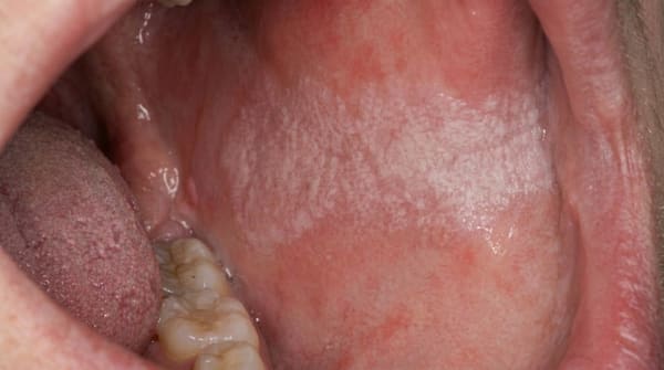
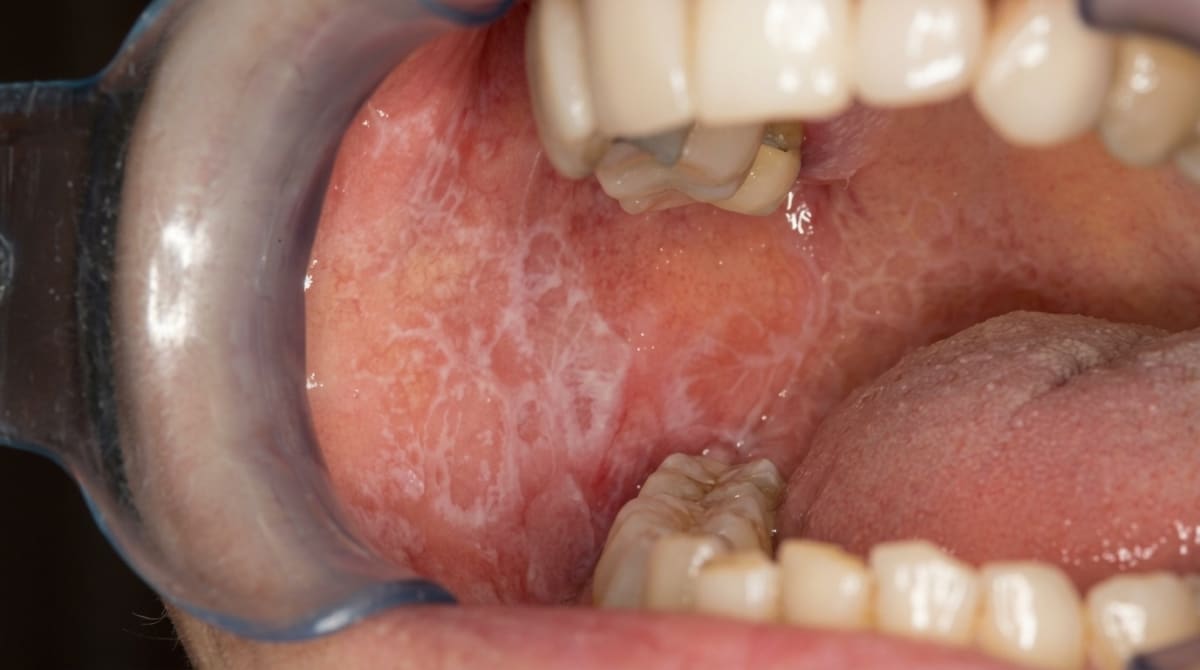
Reticular form, the most common and the most recognisable. Fine, interlacing white lines (Wickham's striae) form a lacy or net-like pattern, almost always on both inner cheeks symmetrically. Often there are no symptoms at all, and many people only learn about it when a dentist points it out.
Papular form, small white pinpoint dots, often mixed in with the lacy lines.
Plaque form, flatter, white patches that can look a bit like leukoplakia. Most common on the tongue and inner cheek.
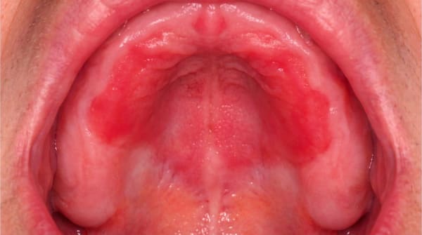
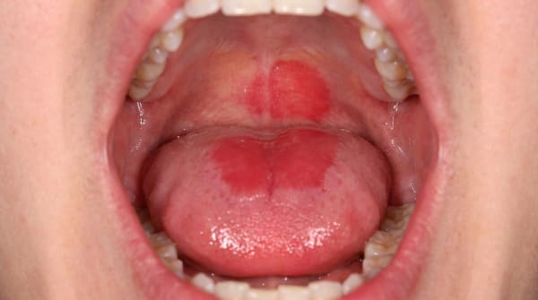
Atrophic (erythematous) form, red, thinned-looking patches with delicate white striae around the edges. Often involves the gums in all four quadrants. Can feel sensitive or burning.
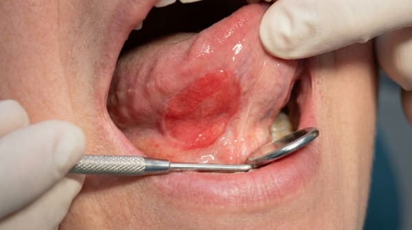
Erosive (ulcerative) form, red areas with central ulcers covered by a yellowish film. The edges usually still show the radiating white lines. This form is the most uncomfortable.
Bullous form, uncommon. Small to large blisters that quickly burst and leave painful sores, usually on the inner cheek near the back teeth.
The inner cheeks (buccal mucosa) are by far the most common location, and the lesions are almost always on both sides. The tongue, gums, lips and palate can also be involved. When the gums are affected, they often look red, shiny and tender, a pattern called desquamative gum (the gum surface peels and reddens, looking sore and raw).
What it feels like
The reticular and papular forms are usually completely painless. Many people are unaware they have anything until a dentist mentions it.
The atrophic form can feel sensitive, sore, or like a constant burning, especially when eating spicy, acidic or salty foods. The erosive and bullous forms are typically the most uncomfortable, patients describe rawness, soreness with eating and tooth brushing, and sometimes ongoing pain even at rest.
Gum involvement makes brushing tender, which can lead to plaque build-up, which in turn aggravates the lichen planus, a cycle that needs gentle but consistent oral hygiene to break.
What an X-ray might show
Oral lichen planus is a soft-tissue condition. It does not show up on X-rays, and imaging is not part of the diagnosis.
What happens at the dentist?
In many reticular cases, a dentist at ArtSmiles can make the diagnosis on appearance alone, symmetrical white lacy lines on both inner cheeks are highly suggestive. For atypical, plaque-type, erosive or one-sided lesions, more investigation is usually appropriate.
A typical assessment may include:
A careful clinical examination, looking at all parts of the mouth, including the tongue, gums and lips, and noting whether the pattern is symmetrical.
A medication review, a full list of current medications helps rule out a lichenoid drug reaction.
An assessment of nearby fillings, particularly older amalgams in contact with the affected area, since a localised lichenoid contact reaction can look identical.
A biopsy (a small tissue sample sent to the lab for testing), a small tissue sample may be recommended if the diagnosis is unclear, if the appearance is atypical, or if the lesions are confined to higher-risk sites such as the side or underside of the tongue or the floor of the mouth. Microscopic features include thickening of the surface keratin, breakdown of the basal cell layer, and a dense band of lymphocytes hugging the lining.
Direct immunofluorescence (DIF) (a special lab stain that highlights immune-system markers in tissue), a special stain done on biopsy tissue. In lichen planus, it typically shows a shaggy band of fibrinogen along the basement membrane in 90 to 100 per cent of cases, with no specific immunoglobulin pattern. This helps separate it from conditions like mucous membrane pemphigoid or pemphigus vulgaris, which show very different DIF patterns.
Specialist referral, appropriate when symptoms are severe, when the lesions don't respond to first-line treatment, or when the appearance is unusual.
Is this serious?
🟡 In most cases, oral lichen planus is a chronic but manageable nuisance rather than a dangerous condition. The reticular form, in particular, often produces no symptoms and only needs monitoring.
The two reasons a dentist takes it seriously are:
The erosive and atrophic forms can be genuinely painful, sometimes enough to interfere with eating, drinking and oral hygiene over long periods.
A small risk of malignant change exists. The textbooks place this risk at roughly 1% over five years (Regezi), with longer-term figures cited in older literature ranging from 1% to 4% over ten years, though the higher end is contested, because some of those cases may have been dysplastic lesions misdiagnosed as lichen planus rather than true OLP. Modern reviews settle around a 1% lifetime risk for most patients. The risk appears slightly higher in the erosive and atrophic forms, particularly in people who smoke. The reticular form carries the lowest risk.
If you've noticed any of these signs for more than two weeks, it's worth booking an assessment.
Because the condition can persist for a decade or more and can change in character over time, periodic dental review, typically every 6 to 12 months for stable cases, or more often for erosive cases, is the standard approach.
Could it be something else?
Quite a few conditions can look similar to oral lichen planus, which is why a careful examination (and sometimes a biopsy) matters. The textbooks consistently mention the following:
Lichenoid drug reaction, looks almost identical clinically and microscopically. The clue is timing, often a connection with starting a new medication, sometimes a one-sided or unusual distribution. Withdrawing or substituting the drug usually resolves the lesion.
Lichenoid contact reaction (often to dental amalgam), a localised lichen-planus-like patch confined to mucosa in direct contact with a filling, usually a corroded amalgam. Unlike true lichen planus, it does not migrate and resolves once the offending restoration is replaced.
Lupus erythematosus (oral lesions), can produce white striae and red patches, but the striae tend to radiate from a central area rather than form a lacy network, and there are usually skin or systemic findings. Direct immunofluorescence shows a different pattern.
Graft-versus-host disease (GVHD) (an immune reaction after a bone marrow transplant), appears in patients who have had a bone marrow transplant. The white striae can mimic lichen planus closely. The medical history is the giveaway.
Mucous membrane (cicatricial) pemphigoid, can also cause desquamative gingivitis and erosions, but typically lacks the classic lacy striae. DIF shows immunoglobulins and complement at the basement membrane rather than fibrinogen.
Pemphigus vulgaris, causes painful erosions and blisters. DIF shows a fishnet pattern of antibodies between the cells of the lining, which is quite different from lichen planus.
Chronic ulcerative stomatitis, can look almost identical to erosive lichen planus, including desquamative gingivitis. DIF shows antibodies against epithelial nuclei in the lower epithelium, and the condition responds better to antimalarial drugs than to steroids.
Leukoplakia (a persistent white patch) (especially proliferative verrucous leukoplakia), can resemble the plaque form of lichen planus. Biopsy is usually needed to tell them apart, and any dysplastic change tips the diagnosis away from lichen planus.
Discoid lupus erythematosus, causes plaques with central healing and white striae radiating outward, often with skin involvement on the face or scalp.
Cheek-chewing (morsicatio buccarum), produces a shaggy white surface on the inner cheek but does not have the symmetrical lacy striae, and the surface can usually be wiped or peeled in places.
Candidiasis, can co-exist with lichen planus and complicate the picture. Wiping or rubbing the white patches reveals red mucosa underneath in candida; lichen planus striae cannot be wiped off.
Hairy leukoplakia, vertical white corrugations on the side of the tongue in immunocompromised patients. Distribution and patient history separate it.
White sponge nevus, a hereditary condition with widespread white spongy mucosa, present from a young age and usually with a family history.
How is it treated?
There is currently no cure for oral lichen planus, but in most cases the condition can be controlled comfortably. The textbooks recommend a stepped approach.
At home (helpful for everyone):
Maintain meticulous but gentle oral hygiene with a soft toothbrush.
Reduce intake of spicy, acidic and salty foods during flare-ups.
Avoid smoking, which appears to increase the small risk of malignant change.
Note and report any new medications that coincide with a flare.
Manage stress where possible, since flares often follow periods of high stress.
Professional treatment may include:
Monitoring only, for asymptomatic reticular lesions. No active treatment is required, just regular review.
Topical corticosteroids, the mainstay of treatment for symptomatic forms. Potent topical preparations (such as clobetasol, betamethasone or fluocinonide) applied as a thin film several times a day usually settle a flare within one to two weeks.
Antifungal medication, often paired with corticosteroids, because steroids can encourage thrush (candida overgrowth) and because candida often complicates lichen planus.
Topical calcineurin inhibitors (another class of immune-modulating creams) (tacrolimus, pimecrolimus), another type of immune-modulating cream, useful when steroids don't help or aren't suitable.
Systemic corticosteroids, reserved for severe flare-ups under medical supervision.
Replacing a suspect amalgam, if a localised lichenoid contact reaction is suspected, replacing the filling with a different material (such as glass ionomer, composite or porcelain) often resolves the lesion.
Reviewing medications with the prescribing doctor, when a lichenoid drug reaction is likely, a substitute medication may be considered.
Specialist (oral medicine) referral, for complex, recalcitrant or atypical cases.
Gingival lichen planus is the trickiest form to manage because steroids are harder to keep in place on the gums. Adhesive pastes, custom trays for steroid delivery, and rigorous plaque control can all help.
What's the long-term outlook?
Oral lichen planus is a long-term condition. Untreated, it can persist for ten years or more, with flares and quieter periods. Most patients can expect their symptoms to be controlled, not necessarily eliminated, with appropriate care.
Key points about the longer view:
The reticular form often stays stable for years, sometimes indefinitely, with no symptoms and no treatment beyond occasional review.
The atrophic and erosive forms typically wax and wane. Topical treatments usually work, but the lesions tend to return, which is why long-term monitoring is sensible.
Skin lesions of lichen planus, when they occur alongside oral lesions, tend to resolve within one to two years on their own. Oral lesions usually persist longer.
Malignant transformation is uncommon (around 1% over five years in well-documented studies, with most modern reviews settling on roughly 1% lifetime risk) but real, particularly in the erosive form and in smokers. Periodic dental review allows any change in appearance to be picked up early.
With a clear diagnosis, sensible management of triggers, and ongoing monitoring, most people with oral lichen planus continue to eat, speak and live comfortably without major disruption.
A note on this article
This article is for educational purposes only and does not constitute a clinical diagnosis. Please consult a registered dental practitioner for assessment and treatment advice.
The cover image above is an AI-generated illustration based on the most common visible features of this condition described in clinical pathology references. It is not a photograph of a real case and should not be used to diagnose or rule out the condition in your own situation. If you are concerned about something you have noticed, please book an assessment with a registered dental practitioner.
References
Regezi, J. A., Sciubba, J. J., & Jordan, R. C. K. (2017). Oral pathology: Clinical pathologic correlations (7th ed.). Elsevier. Chapter 3, White Lesions, Lichen Planus, pp. 97 to 102.
Neville, B. W., Damm, D. D., Allen, C. M., & Chi, A. C. (2023). Oral and maxillofacial pathology (5th ed.). Elsevier. Chapter 16, Dermatologic Diseases, Lichen Planus, pp. 787 to 792.
Cawson, R. A., & Odell, E. W. (2017). Cawson's essentials of oral pathology and oral medicine (8th ed.). Elsevier. Chapter 13, Diseases of the Oral Mucosa: Non-infective Stomatitis, Lichen Planus, pp. 225 to 229.