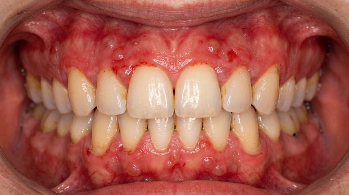
Compiled from clinical pathology references. Medically reviewed by Dr Cristian Dunker , Principal Dentist, ArtSmiles Cosmetic Dentistry.
Quick summary
Also called | MMP, cicatricial pemphigoid, benign mucous membrane pemphigoid, ocular pemphigoid, mouth lining (mucosa)l pemphigoid |
How urgent? | 🔴 See a dentist promptly, chronic blistering and erosion of the gums or mouth lining warrants assessment, and the eyes may also be at risk |
Common or rare? | Uncommon, but at least twice as common as pemphigus vulgaris |
Who it affects | Older adults, typically 50 to 80 years; women more often than men (about 2 to 1) |
Who treats it | General dentist for diagnosis and topical therapy, with referral to oral medicine, dermatology and ophthalmology where indicated |
Based on | Regezi, Neville, Cawson, Laskaris |
What is it?
Mucous membrane pemphigoid is a long-standing autoimmune blistering disease that mainly affects the lining of the mouth and the surface of the eyes. The body's own antibodies attack the layer of tissue that anchors the surface skin (surface lining), epithelium to the deeper tissue beneath, so the surface lifts away and forms blisters or raw, eroded patches.
In the mouth it most often shows up as red, peeling gums, a pattern called desquamative gum (the gum surface peels and reddens, looking sore and raw), but it can also affect the palate, cheeks, tongue and lips. Because untreated eye involvement can lead to scarring and even blindness, an early and accurate diagnosis really matters.
Who tends to get it?
Mucous membrane pemphigoid usually starts in middle-aged and older adults, with the average age of onset between 50 and 60 years and many patients in their 60s and 70s. Women are affected roughly twice as often as men.
The oral mucosa is involved in almost every patient, and in around 95% of cases the mouth is the first site to show changes. Childhood cases have been reported but are rare. The condition does not appear to favour any particular ethnic background, and there is no clear hereditary pattern.
What causes it?
Mucous membrane pemphigoid is an autoimmune disease, which means the immune system mistakenly produces antibodies against the body's own tissues. In this case, the antibodies target proteins in the basement membrane zone (the thin layer that glues the surface lining to the connective tissue underneath).
The main targets identified so far include:
BP180, a protein in the basement membrane that helps anchor the surface lining
Laminin 332 (also known as laminin 5), another anchoring protein
Alpha-6 / beta-4 integrin, building blocks of the tiny rivets (hemidesmosomes) that hold the surface cells in place
The trigger that sets off this misdirected immune response is unknown. There is no proven dietary cause, no infectious trigger and no clear lifestyle risk factor. One particular subtype, anti-laminin 332 mucous membrane pemphigoid, has been associated with an underlying solid tumour in around 30% of patients (which is why doctors sometimes screen for an internal cancer in this subtype), so investigation for a possible internal malignancy is sometimes recommended in this group.
How does it develop?
Think of the lining of your mouth as wallpaper bonded to a wall. The basement membrane is the glue holding the wallpaper on. In mucous membrane pemphigoid, antibodies attach to that glue and recruit inflammatory cells, which weaken the bond.
When the bond breaks, fluid collects underneath and lifts the surface away, creating a blister. Because the split happens beneath the entire thickness of the surface skin (a beneath the surface lining (subepithelial) split), the roof of the blister is relatively thick, so blisters can sometimes be seen intact in the mouth before they pop. This is different from pemphigus vulgaris, where the split is within the surface layer and the blisters are so fragile they almost always rupture before a dentist sees them.
Once the blister bursts, it leaves a raw, ulcerated patch that takes weeks or months to heal. On the eye and some other sites, healing happens with scarring (a cicatrix), which is where the older name cicatricial pemphigoid came from.
What might you notice?
What it looks like
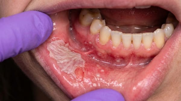
In the mouth, the most common pattern is desquamative gingivitis: gums that look bright red, shiny, friable and peeling, often around several teeth at once. The whole width of the attached gum may be affected, and tags of separating tissue can sometimes be seen at the edges.
Less often, you may notice:
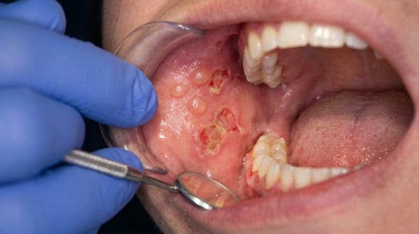
Intact blisters (vesicles or large blisters), bullae on the palate, cheeks or gums, these can be clear or blood-stained
Large, irregular, shallow ulcers after blisters rupture, with well-defined margins
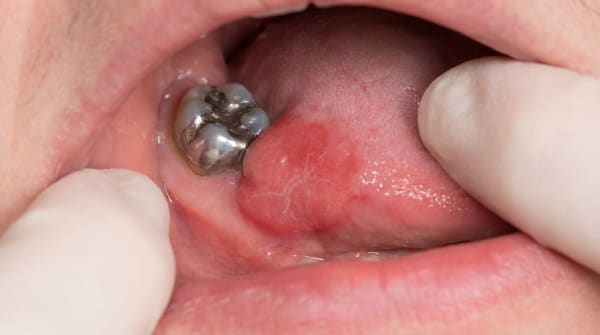
Erosions on the buccal mucosa (cheeks), palate, tongue or lips
Skin lesions in around 10,20% of cases, usually tense blisters on the head, neck or limbs
Eye redness, irritation, gritty sensation or in-turning eyelashes
What it feels like
Fresh ulcers are typically painful, with discomfort that ranges from mild stinging to severe soreness during eating, brushing or speaking. Brushing the gums tends to make them bleed easily. With time, the pain often becomes less intense, but the lesions persist.
Some people notice that pressing the gum or accidentally touching it with a fingernail causes the surface to peel away, this is called a positive Nikolsky sign (a sign where light pressure peels the surface). Eye involvement may begin with simple irritation or dryness; voice changes or breathing difficulty can occasionally signal involvement of the larynx.
What an X-ray might show
Mucous membrane pemphigoid is a soft-tissue condition, so an X-ray is not used to diagnose it. However, because the gums are sore and oral hygiene is often compromised, plaque can build up and contribute to gum and bone changes that may be visible on routine dental imaging.
What happens at the dentist?
If your dentist at ArtSmiles suspects mucous membrane pemphigoid, they will usually:
Take a careful history, how long the gums or mouth have been sore, whether blisters appear, whether the eyes, nose, throat, genitals or skin are also affected, and what medications you take.
Examine the whole mouth, looking at the gums, palate, cheeks, tongue and lips for redness, blisters, ulcers and peeling tissue, and gently testing whether the surface separates with light pressure.
Recommend a biopsy (a small tissue sample sent to the lab for testing) of perilesional tissue, meaning a small sample taken from clinically normal-looking mucosa right next to a lesion, rather than from the ulcer itself. This is important because the loosened epithelium often strips off during a biopsy of the lesion, and the diagnostic interface is then lost.
Send the sample for direct immunofluorescence (a special lab stain that highlights immune-system markers in tissue) (DIF), a special laboratory test that detects a continuous linear band of IgG and complement (C3) (immune-system markers) along the basement membrane zone. This pattern is positive in around 90% of patients and is the most reliable way to confirm the diagnosis.
Refer to an ophthalmologist, once the diagnosis is confirmed, a baseline eye examination is recommended for every patient, even without eye symptoms, because early conjunctival changes are difficult to detect without a slit-lamp (the special microscope eye doctors use).
Refer to oral medicine, dermatology or other specialists as needed if the throat, nose, oesophagus, genitals or skin are involved.
A biopsy and immunofluorescence test are essentially mandatory, clinical appearance alone is not enough, because several other conditions look very similar.
Is this serious?
🔴 See a dentist promptly. Mucous membrane pemphigoid is generally a slow, indolent disease that is rarely life-threatening, and the oral lesions usually heal without scarring. However, the condition is chronic, the course tends to be protracted and progressive rather than coming and going in clear remissions, and several complications matter:
Eyes, up to about 25% of people with oral lesions eventually develop ocular involvement, with scarring, in-turned eyelashes, dry eyes and (without treatment) blindness in severe cases
Larynx, rare laryngeal blisters can cause voice changes or, occasionally, airway problems
Genitals and oesophagus, pain with intercourse and difficulty swallowing can both occur
Mouth, chronic pain, difficulty eating and oral hygiene problems can all reduce quality of life
If you've noticed any of these signs for more than two weeks, it's worth booking an assessment.
Could it be something else?
Several other conditions can cause peeling gums, mouth ulcers or oral blisters, so a definitive diagnosis nearly always requires a biopsy with immunofluorescence. Differentials your dentist may consider include:
Pemphigus vulgaris, also causes painful oral erosions and a positive Nikolsky sign, but the split is within the epithelium rather than beneath it, blisters are very fragile, and IgG deposits sit between epithelial cells rather than along the basement membrane.
Erosive (atrophic) lichen planus, can produce desquamative gingivitis that looks almost identical, but usually has accompanying white lacy striae (Wickham striae) and shows a different microscopic pattern with characteristic immune-cell infiltration at the epithelial-connective tissue junction.
Bullous pemphigoid, the closely related skin counterpart of MMP. Mainly affects the skin in older adults, with the mouth involved less often and usually with periods of clear remission.
Linear IgA bullous dermatosis (a related blistering disease distinguished by which antibody class deposits along the basement membrane), shows a similar linear pattern on immunofluorescence, but only IgA (not IgG) is deposited along the basement membrane zone.
Epidermolysis bullosa acquisita, another subepithelial blistering disease, distinguished by autoantibodies against type VII collagen and a specific salt-split skin test result.
Erythema multiforme, typically affects younger people, with sudden onset, swollen and crusted lips and target-shaped skin lesions; usually self-limiting.
Paraneoplastic pemphigus, rare and associated with an underlying tumour; combines features of pemphigus and pemphigoid with severe, treatment-resistant lip and eye lesions.
Discoid lupus erythematosus (a localised form of lupus affecting skin and mucosa), can affect the gums and palate but usually has central atrophy with surrounding white striae and a different immunofluorescence pattern.
Dermatitis herpetiformis (an itchy blistering skin condition linked to coeliac disease); oral lesions are rare and immunofluorescence shows granular IgA at dermal papillae, not a linear band.
Angina bullosa haemorrhagica, produces sudden blood-filled blisters (often on the soft palate) that heal without scarring and have no immunological abnormalities.
Contact allergy / contact stomatitis, gum redness from a specific irritant or allergen, which improves once the trigger is removed.
Plasma cell gingivitis, causes red, shiny gums, but biopsy and immunofluorescence patterns are different.
Plaque-related gingivitis, looks completely different on close examination, follows the gum margin and resolves with professional cleaning and good home care.
How is it treated?
There is no single treatment that suits every patient. Your dental team will usually tailor therapy to how active the disease is, where it is located, and which other organs are affected. Treatment may include:
At home
Gentle but thorough oral hygiene with a soft toothbrush, plaque makes gum lesions worse
A chlorhexidine mouthrinse, which can reduce plaque when brushing is uncomfortable
Frequent professional cleans to control plaque while the gums are sore
Avoiding spicy, acidic, crunchy or very hot foods that aggravate the ulcers
Topical therapy (often first line for mild oral disease)
High-potency topical corticosteroid gels or creams (such as clobetasol, betamethasone, fluocinonide or desoximetasone) applied to the affected gums or mucosa
A custom-made flexible mouthguard / occlusal splint as a tray to hold the steroid against the gums for longer contact time
Antifungal therapy when needed, because long-term topical steroids can encourage Candida overgrowth
Systemic therapy (when topical treatment is not enough, or for eye, throat or skin involvement)
Dapsone, often considered before systemic steroids for mild-to-moderate oral disease
Tetracycline or minocycline combined with niacinamide (nicotinamide)
Systemic corticosteroids such as prednisone for moderate to severe disease
Steroid-sparing immunosuppressants (drugs that dampen the immune system), azathioprine, methotrexate, mycophenolate mofetil, cyclophosphamide or cyclosporine
Rituximab (an anti-CD20 monoclonal antibody) for recalcitrant or relapsed cases
Intravenous immunoglobulin (IVIG) in selected severe cases, particularly with active eye disease
Specialist input
Ophthalmology review for every patient at baseline, with ongoing monitoring if eye changes appear
Surgical correction of any eyelid scarring (symblepharon, where the eyelid scars onto the surface of the eye) only when the disease is quiet, as operating during a flare tends to provoke further inflammation
ENT, gynaecology or gastroenterology input where appropriate
Treatment is often long-term, and the goal is to control symptoms and prevent scarring rather than to cure the underlying autoimmune process.
What's the long-term outlook?
For most people, mucous membrane pemphigoid is a chronic but manageable condition. Oral lesions usually heal without scarring, and the overall outlook is good when the diagnosis is made early and treatment is consistent. Some patients experience slow spontaneous improvement; others have a long, fluctuating course with periods of relative quiet alternating with flares.
The key to a good long-term result is:
Early diagnosis through biopsy and immunofluorescence
Routine eye review to catch and treat ocular disease before scarring sets in
Ongoing topical or systemic therapy to keep flares under control
Excellent oral hygiene to limit plaque-driven aggravation of the gums
With this kind of coordinated care, most patients can expect to keep their teeth, their eyesight and a comfortable mouth, even though the underlying autoimmune tendency tends to persist for life.
A note on this article
This article is for educational purposes only and does not constitute a clinical diagnosis. Please consult a registered dental practitioner for assessment and treatment advice.
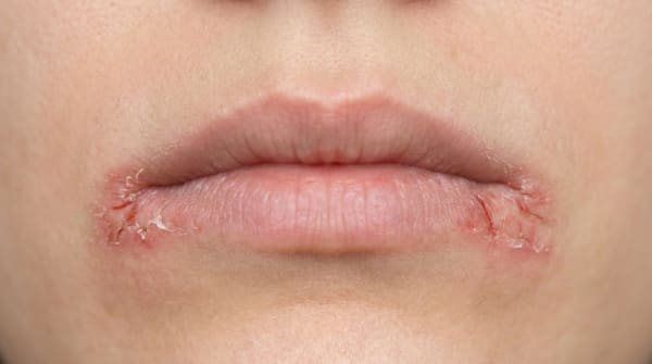
The cover image above is an AI-generated illustration based on the most common visible features of this condition described in clinical pathology references. It is not a photograph of a real case and should not be used to diagnose or rule out the condition in your own situation. If you are concerned about something you have noticed, please book an assessment with a registered dental practitioner.
References
Regezi, J. A., Sciubba, J. J., & Jordan, R. C. K. (2017). Oral pathology: Clinical pathologic correlations (7th ed.). Elsevier. Chapter 1, Vesiculobullous Diseases (Mucous Membrane Pemphigoid), pp. 15,18.
Neville, B. W., Damm, D. D., Allen, C. M., & Chi, A. C. (2023). Oral and maxillofacial pathology (5th ed.). Elsevier. Chapter 16, Dermatologic Diseases (Mucous Membrane Pemphigoid), pp. 775,779.
Cawson, R. A., & Odell, E. W. (2017). Cawson's essentials of oral pathology and oral medicine (8th ed.). Elsevier. Chapter 13, Diseases of the Oral Mucosa: Non-Infective Stomatitis (Mucous Membrane Pemphigoid), pp. 233,235.
Laskaris, G. Color atlas of oral diseases. Thieme. Chapter 22, Skin Diseases (Cicatricial Pemphigoid), pp. 207,210.
Frequently asked questions
What is mucous membrane pemphigoid?
Mucous membrane pemphigoid (MMP), also called cicatricial pemphigoid, is a chronic autoimmune blistering disease primarily affecting the mucous membranes — mouth, eyes, nose, throat, oesophagus, larynx, anus and genitals. In the mouth it usually presents as desquamative gingivitis with patchy or diffuse erosions and occasionally tense, fluid-filled blisters.
What does the mouth look like?
The most characteristic finding is desquamative gingivitis — bright red, glazed, sore gums that bleed easily on brushing. Erosions and tense blisters can also appear on the cheeks, palate or tongue. The lesions heal with scarring, which distinguishes MMP from pemphigus. Severe gum involvement can lead to chronic inflammation and tooth loss.
Why does eye involvement matter?
Eye (ocular) MMP can cause progressive scarring of the conjunctiva, leading to dry eyes, fused eyelids and ultimately blindness if untreated. Patients with oral MMP should be screened by an ophthalmologist. Other mucous membrane sites (throat, larynx, genital) should also be assessed.
How is MMP treated?
Treatment is shared between oral medicine, dermatology and ophthalmology. Mild oral disease may respond to topical corticosteroids (gels, sprays, custom mouthguards). Moderate-to-severe disease needs systemic immunosuppression — corticosteroids, dapsone, mycophenolate, azathioprine, methotrexate or rituximab. Aggressive treatment of eye involvement is essential to prevent blindness.