Compiled from clinical pathology references. Medically reviewed by Dr Cristian Dunker , Principal Dentist, ArtSmiles Cosmetic Dentistry.
Quick summary
Also called | PV, pemphigus, autoimmune pemphigus |
How urgent? | 🔴 See a dentist or doctor promptly, pemphigus vulgaris is a serious autoimmune disease and early diagnosis genuinely changes outcomes. |
Common or rare? | Uncommon, roughly one to five new cases per million people each year. |
Who it affects | Adults, most often between 40 and 60 years of age. More common in people of Mediterranean, Jewish, South Asian or Middle Eastern heritage. |
Who treats it | Both, usually a dentist or oral medicine specialist working alongside a dermatologist or physician experienced in immunosuppressive (drugs that dampen down the immune system) therapy. |
Based on | Regezi, Neville, Cawson, Laskaris |
What is it?
Pemphigus vulgaris is an autoimmune blistering disease, which means the body's own immune system mistakenly attacks the cells that hold the lining of the mouth and skin together. The result is fragile blisters that quickly burst to leave painful, ragged ulcers.
The mouth is usually the first place the disease shows up, often weeks or months before any skin involvement. Pemphigus vulgaris is uncommon, but it is a serious condition that needs prompt medical attention, without treatment, it can be life-threatening.
Who tends to get it?
Pemphigus vulgaris is the most common form of pemphigus, but the disease itself is not seen often. The textbooks estimate around one to five new cases per million people each year.
It usually appears in middle age, with most people diagnosed between their fourth and sixth decade, roughly 40 to 60 years old. The average age at diagnosis is around 50. Some reports describe equal numbers of men and women affected; others describe a slight female predominance, and Cawson notes that women aged 40 to 60 are predominantly affected.
Genetic and ethnic factors play a clear role. The disease is more common in people of Ashkenazi Jewish, Mediterranean (Greek, Italian), South Asian, and Middle Eastern descent, and is linked to certain inherited tissue-type (HLA) markers. Pemphigus vulgaris in children is rare but does occur.
What causes it?
Pemphigus vulgaris is caused by the immune system producing antibodies (specifically IgG antibodies (a type of immune-system protein)) that target a protein called desmoglein 3. Desmoglein 3 is part of the molecular "glue" that holds neighbouring cells of the mouth lining together. When the antibodies attach to this glue, the cells lose their grip on each other and the lining splits apart.
In people who go on to develop skin involvement as well, antibodies against a related protein, desmoglein 1, also appear.
Why the immune system starts producing these antibodies in the first place is not fully understood. Several factors are recognised as contributing or triggering:
Genetics, particular HLA tissue-type variants (genetic markers used to match tissue compatibility) run more commonly in affected families and ethnic groups.
Medications, drugs containing thiol groups (such as penicillamine and captopril), as well as some penicillins, cephalosporins, ACE inhibitors, rifampin and non-steroidal anti-inflammatory drugs, can occasionally trigger a pemphigus-like reaction.
Other autoimmune conditions, pemphigus vulgaris sometimes occurs alongside myasthenia gravis, lupus, rheumatoid arthritis, Hashimoto's thyroiditis, thymoma or Sjögren's syndrome.
Underlying malignancy, a separate form called paraneoplastic pemphigus is triggered by a hidden lymphoma or other cancer, although this is a distinct condition.
Nothing about lifestyle, diet, oral hygiene or dental work causes pemphigus vulgaris. It is not contagious and is not the patient's fault.
How does it develop?
A helpful way to picture it is to think of the lining of the mouth as a brick wall. Each "brick" is a cell, and the cells are held to each other by tiny rivets called desmosomes. Desmoglein 3 is the key component of those rivets in the deeper layer of the wall.
In pemphigus vulgaris, the immune system makes antibodies that lock onto desmoglein 3 and disable it. With the rivets out of action, the bricks lose contact with one another and a horizontal split develops just above the bottom row. The bottom row of cells stays anchored to the floor below, pathologists describe this remaining row as resembling a "row of tombstones", while everything above it lifts off as a fragile blister.
Because the split is so superficial inside the lining, the roof of the blister is thin. In the mouth it almost always tears within hours, leaving a raw, painful erosion behind. The same process can happen on the skin, where intact blisters are seen more often before they rupture.
What might you notice?
What it looks like
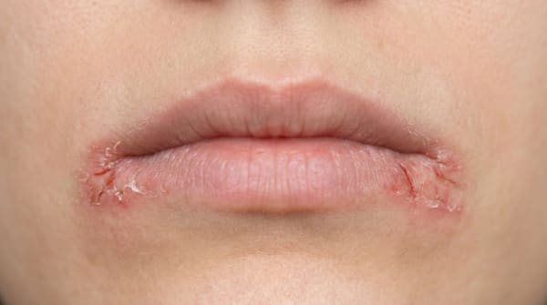
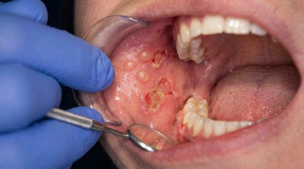
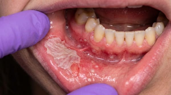
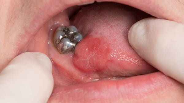
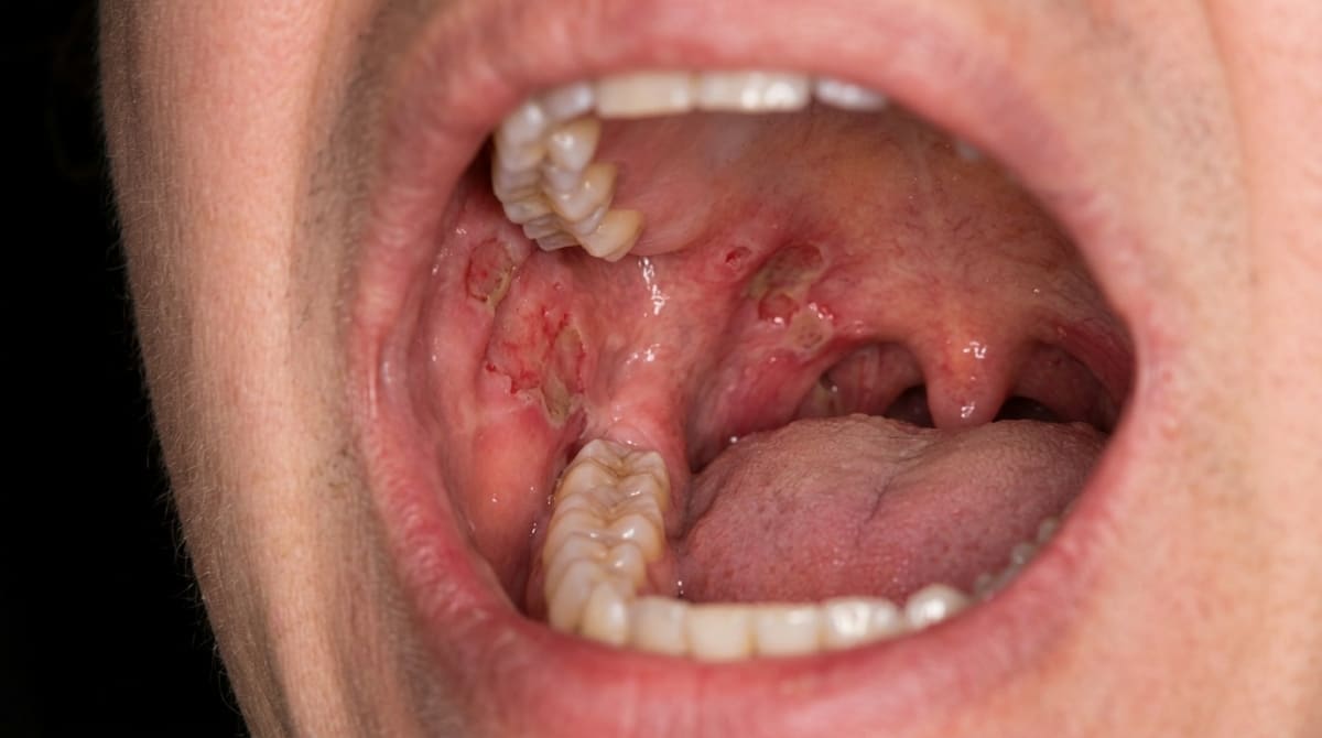
In around half to two-thirds of people, the very first sign of pemphigus vulgaris is in the mouth. Intact blisters are rarely caught in time, what most people actually see is one or more ragged, irregular, red erosions or ulcers. The edges often look frayed, with small tags of loose epithelium hanging at the margins.
Lesions can appear almost anywhere inside the mouth, but the soft palate, cheek lining, gums, lip mouth lining (mucosa) and the underside of the tongue are common sites. Skin blisters, when they appear, are flaccid (soft and floppy) and burst easily, leaving denuded (stripped of surface) red patches that crust over.
A telling clinical sign is Nikolsky's sign: gentle sideways pressure or rubbing on apparently healthy mucosa or skin can cause the surface layer to slide off, revealing a raw area underneath.
What it feels like
The ulcers are typically painful and tender. Eating, drinking, brushing and even speaking can become uncomfortable. When the soft palate, throat or many small areas of the cheek are involved, the discomfort can be considerable, and some people develop difficulty swallowing.
Untreated, the lesions tend to persist and gradually involve more surface area rather than healing on their own.
What an X-ray might show
Pemphigus vulgaris is a soft-tissue disease and does not show up on dental X-rays. Imaging is not used to diagnose it.
What happens at the dentist?
Because pemphigus vulgaris so often starts in the mouth, dentists at ArtSmiles and elsewhere are frequently the first health professionals to see it. Picking it up early genuinely matters, Neville notes that disease control is generally easier when the diagnosis is made early in the course.
A careful clinical examination is the first step. Your dentist may look at the distribution of the ulcers, the appearance of the edges, whether any blisters are intact, and whether Nikolsky's sign can be elicited gently on apparently healthy mucosa.
The next step is usually a biopsy (a small tissue sample sent to the lab for testing). Two samples may be taken from tissue immediately next to a lesion (this is called perilesional tissue):
One is sent for routine microscopic examination, where the pathologist looks for the characteristic split just above the basal cell layer (the bottom row of the surface lining) and the rounded, separated "Tzanck" cells floating within it.
The second is sent for direct immunofluorescence (a special lab stain that highlights immune-system markers in tissue) (DIF). This specialised test highlights the IgG antibodies (and often complement protein C3 (an immune-system marker)) sitting in the spaces between the surface cells, producing a striking pattern that is often described as "chicken wire" or "fishnet". This pattern is highly characteristic of pemphigus vulgaris.
A blood test (indirect immunofluorescence (a blood test that detects circulating autoantibodies) or ELISA, a blood test for specific antibodies) may also be used to detect circulating antibodies and to monitor disease activity over time.
Because pemphigus vulgaris is a systemic autoimmune disease, your dentist will refer you to a specialist team, most commonly an oral medicine specialist, dermatologist or physician with experience in immunosuppressive therapy, to coordinate treatment.
Is this serious?
🔴 Yes. Pemphigus vulgaris is one of the more serious conditions covered in oral pathology textbooks, and all four reference books are direct about that.
Before corticosteroid treatment was developed, the disease was fatal in 60% to 90% of cases, with patients dying from infection, fluid loss, electrolyte imbalance and malnutrition due to widespread ulcer formation. Modern immunosuppressive treatment has transformed the picture: current mortality rates reported in the textbooks are in the order of 5% to 10%, and continuing to fall as newer biologic therapies become more widely used.
The key message is that pemphigus vulgaris is treatable but not something to wait out. Mouth ulcers that look ragged, persist for more than a couple of weeks, or come and go in different places deserve a proper dental assessment.
If you've noticed any of these signs for more than two weeks, it's worth booking an assessment.
Could it be something else?
Many mouth conditions can cause persistent ulcers or erosions. The textbooks list a number of conditions that can look similar to pemphigus vulgaris and need to be distinguished from it, usually with the help of a biopsy and immunofluorescence.
Mucous membrane pemphigoid, also causes chronic erosions and a positive Nikolsky sign, and frequently affects the gums. The split, however, is deeper (below the lining rather than within it), the antibodies target the basement membrane rather than the cell-to-cell glue, and the disease is generally less aggressive.
Erosive lichen planus, can produce painful red and ulcerated areas of the cheek, tongue or gums. White lacy striae at the edges of the lesions and a different microscopic and immunofluorescence pattern help distinguish it.
Erythema multiforme, tends to come on suddenly in younger adults, often with crusted, bleeding lips and target-shaped lesions on the skin, rather than the slowly progressive ragged oral erosions of pemphigus.
Paraneoplastic pemphigus, a rare and severe variant linked to underlying lymphoma or other cancer. The picture is more polymorphic (varied in appearance), often involving the lips with hemorrhagic crusting, the eyes with scarring, and sometimes the lungs. Specific antibodies against plakin proteins and biopsy findings of mixed clefting help identify it.
Pemphigus vegetans, a rare variant of pemphigus vulgaris itself, where ulcers heal with thickened, warty (vegetating) growths, particularly at the corners of the mouth.
Pemphigus foliaceus and pemphigus erythematosus, superficial forms of pemphigus that affect the skin (and rarely the mouth), with crusting, scaly lesions rather than deep oral erosions.
Bullous pemphigoid, primarily a skin disease of older adults; the mouth is rarely involved, and when it is, the picture is usually milder.
Recurrent aphthous ulcers, common, round, well-defined, recurrent ulcers that heal within one to two weeks. Pemphigus erosions are typically larger, more ragged, more persistent, and often multiple.
Herpetic gingivostomatitis, caused by the herpes simplex virus, typically with crops of small vesicles, fever and tender lymph nodes; it resolves within one to two weeks rather than persisting and progressing.
Dermatitis herpetiformis and linear IgA disease, other autoimmune blistering diseases, distinguished primarily by their immunofluorescence patterns.
Chronic ulcerative stomatitis, a rare condition that can resemble erosive lichen planus but has a distinct pattern of speckled IgG antibodies on direct immunofluorescence.
Epidermolysis bullosa acquisita, another subepithelial (under the surface lining) blistering disease, distinguished by antibodies against type VII collagen.
Discoid lupus erythematosus, can produce chronic oral ulcers, but typically with a characteristic pattern of central erosion, surrounding white striae and radiating telangiectasia (small dilated blood vessels).
Toxic epidermal necrolysis and Stevens,Johnson spectrum, usually drug-related and acute, with widespread skin sloughing and severe systemic illness, very different in tempo from pemphigus.
Hailey,Hailey disease (chronic benign familial pemphigus), a rare inherited condition with skin erosions; oral involvement is uncommon and the pattern of inheritance and immunofluorescence findings differ.
How is it treated?
Pemphigus vulgaris is a systemic disease, so treatment has to be systemic, topical mouth treatments alone are not enough. The textbooks emphasise that care should be coordinated by a clinician experienced in immunosuppressive therapy, with the dentist or oral medicine specialist supporting management of the mouth lesions.
Typical components of treatment may include:
Systemic corticosteroids (most often prednisone or prednisolone) at moderate to high doses to bring the disease under control. A common starting regimen described in the textbooks is around 80 to 100 mg of prednisolone per day, often combined with another agent.
Steroid-sparing immunosuppressants such as azathioprine, mycophenolate mofetil, cyclophosphamide or dapsone. These are added to allow the corticosteroid dose to be reduced and to limit long-term side effects.
Rituximab, a monoclonal antibody that targets the B-lymphocytes responsible for producing the harmful antibodies. The textbooks describe rituximab as increasingly used as a first-line approach, often combined with a lower dose of corticosteroid, with faster and more durable remissions reported.
Other adjuncts, intravenous immunoglobulin (IVIg), plasmapheresis or immunoadsorption may be considered in severe or unresponsive cases.
Supportive oral care, gentle oral hygiene, antiseptic mouth rinses (such as chlorhexidine), and antifungal therapy if topical or systemic steroids are causing thrush. Soft, bland diets help while erosions are active.
Long-term monitoring, antibody levels can often be tracked in the blood and tend to mirror disease activity.
What home care can do is limited but worthwhile: keeping the mouth clean and reducing trauma (gentle brushing with a soft toothbrush, avoiding sharp or spicy foods during flare-ups) helps lesions settle and reduces secondary infection. Patients on long-term immunosuppression also need regular dental check-ups, because both the medications and the disease can affect oral health.
What's the long-term outlook?
The outlook for pemphigus vulgaris has changed dramatically in recent decades. Before corticosteroids, the disease was usually fatal. Today, with modern immunosuppressive and biologic therapy, most people achieve good control, and the textbooks describe long-term remission in a meaningful proportion of patients, Neville cites one study suggesting that up to 75% of patients may achieve disease resolution after about ten years of treatment, although most centres historically report remission in around 30%.
The condition is described as guarded rather than easily curable: relapses and flare-ups are common, and most people require ongoing low-dose maintenance therapy and lifelong specialist follow-up. Much of the morbidity now comes from the side effects of long-term corticosteroid use rather than the disease itself, which is why newer agents like rituximab have become so important.
The mouth lesions often respond more slowly than the skin lesions, they have been described as "the first to show, and the last to go." Mouth comfort, careful dental care and good communication between your dental team and your treating physician can make a real difference to day-to-day quality of life.
The most important point is one the textbooks return to repeatedly: early diagnosis matters. Pemphigus vulgaris that is recognised and treated early is much easier to control than disease that has been allowed to progress.
A note on this article
This article is for educational purposes only and does not constitute a clinical diagnosis. Please consult a registered dental practitioner for assessment and treatment advice.
The cover image above is an AI-generated illustration based on the most common visible features of this condition described in clinical pathology references. It is not a photograph of a real case and should not be used to diagnose or rule out the condition in your own situation. If you are concerned about something you have noticed, please book an assessment with a registered dental practitioner.
References
Regezi, J. A., Sciubba, J. J., & Jordan, R. C. K. (2017). Oral pathology: Clinical pathologic correlations (7th ed.). Elsevier. Chapter 1, Vesiculobullous Diseases (Pemphigus Vulgaris), pp. 11,15.
Neville, B. W., Damm, D. D., Allen, C. M., & Chi, A. C. (2023). Oral and maxillofacial pathology (5th ed.). Elsevier. Chapter 16, Dermatologic Diseases (Pemphigus), pp. 769,774.
Cawson, R. A., & Odell, E. W. (2017). Cawson's essentials of oral pathology and oral medicine (8th ed.). Elsevier. Chapter 13, Diseases of the Oral Mucosa: Non-infective Stomatitis (Pemphigus Vulgaris), pp. 232,233.
Laskaris, G. Pocket atlas of oral diseases. Thieme. Chapter on Skin Diseases (Pemphigus Vulgaris and variants), pp. 202,205.