Compiled from clinical pathology references. Medically reviewed by Dr Cristian Dunker, Principal Dentist, ArtSmiles Cosmetic Dentistry.
Quick summary
Also called | Drug-induced lichenoid reaction, oral lichenoid lesion, contact lichenoid reaction (amalgam-associated) |
How urgent? | 🟡 Not urgent but needs proper review, symptoms can be controlled, but follow-up is important |
Common or rare? | Uncommon but under-recognised; rising with multi-medication use in older adults |
Who it affects | Adults middle-aged and older, more often women, often on long-term cardiovascular, anti-inflammatory or diabetic medication |
Who treats it | General dentist for diagnosis; oral medicine specialist for biopsy and topical steroids; GP coordinates the trigger medicine change |
Based on | Neville, Cawson and Regezi |
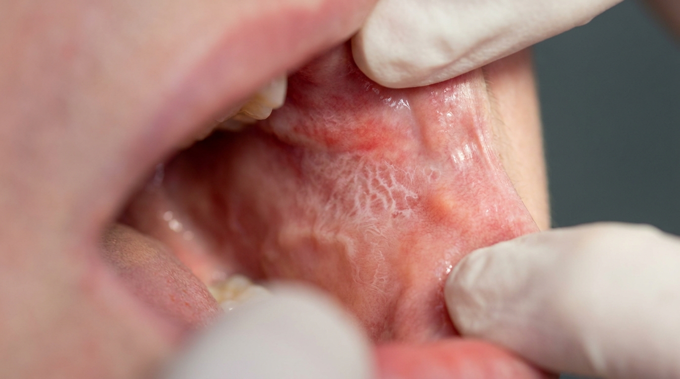
Some medicines that are prescribed to treat blood pressure, arthritis, diabetes or other conditions can quietly produce changes in the lining of the mouth that look like oral lichen planus, lacy white streaks, red atrophic (thinned and red) patches and, sometimes, painful ulcers. When these changes are caused by a medicine rather than by lichen planus itself, the condition is called a lichenoid drug reaction.
This article from the team at ArtSmiles, reviewed by Dr Cristian Dunker, explains what lichenoid drug reactions are, which medicines are most often involved, and how the condition is recognised and managed.
What is it?
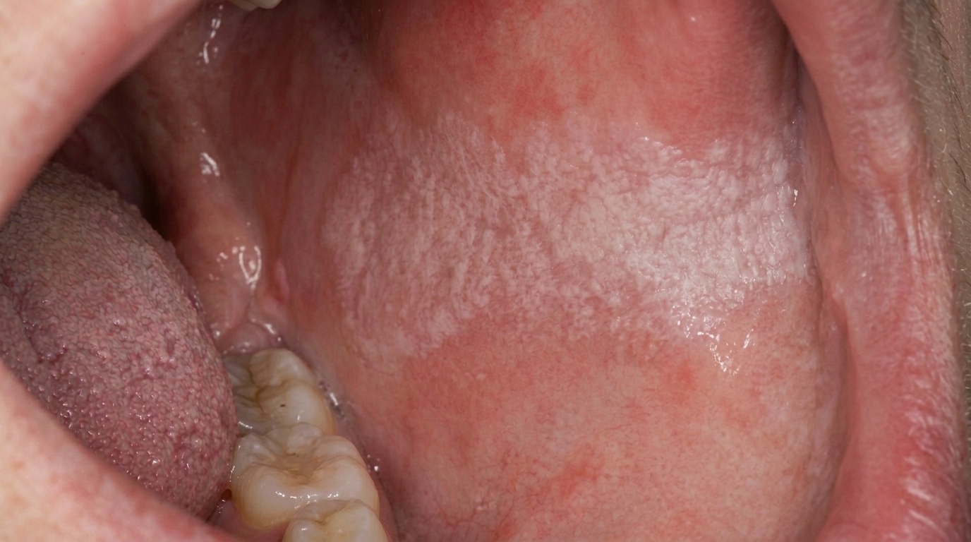
A lichenoid drug reaction is an inflammatory change in the lining of the mouth that clinically and microscopically resembles oral lichen planus but is triggered by a specific medication. The features include:
Lacy white streaks (Wickham striae (fine white lace-like lines)) on the cheek lining or tongue.
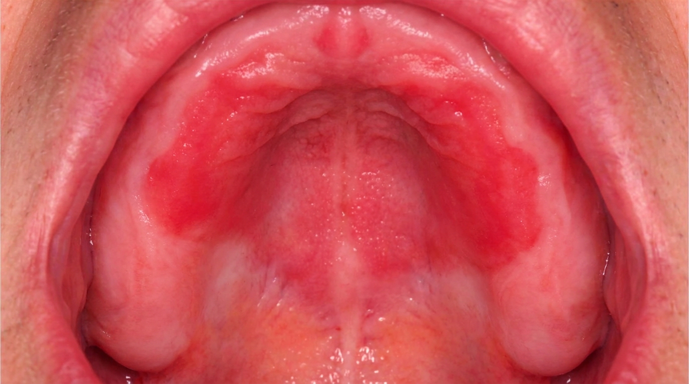
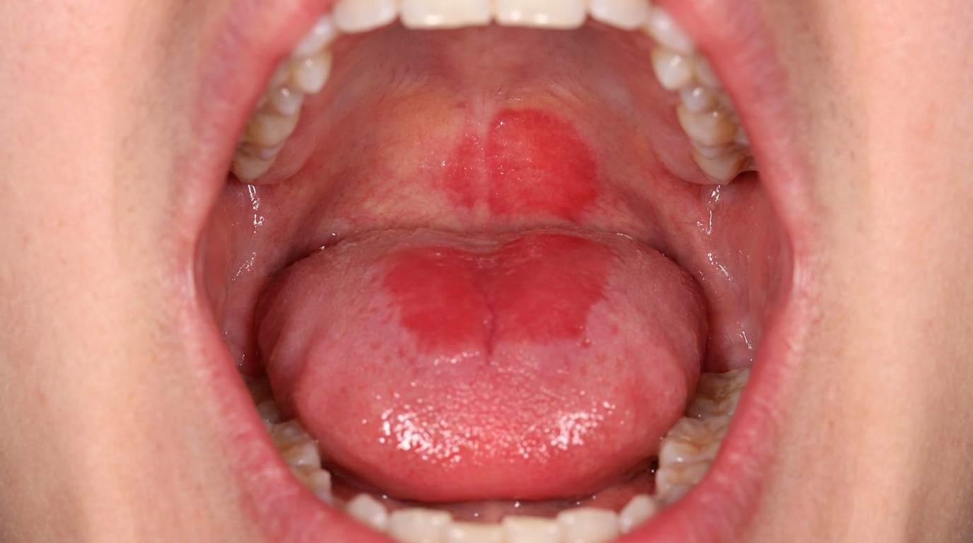
Red, atrophic areas alongside the white streaks.
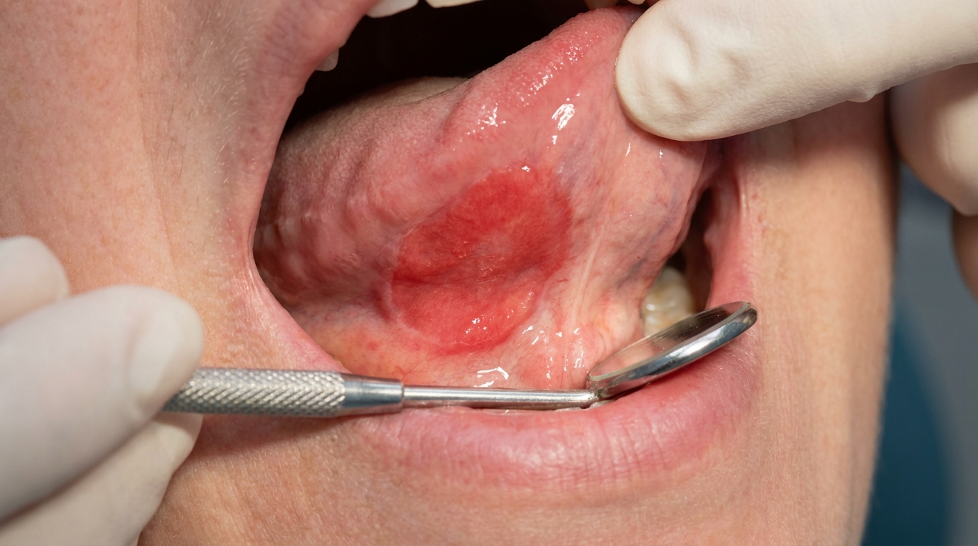
Painful ulcers or erosions in more severe cases.
A pattern that is often unilateral (on one side only) (one side of the mouth) or limited to areas in contact with a particular dental restoration.
Symptoms that began or worsened after a new medicine was started.
The link with medication is what separates this condition from idiopathic (no known cause) oral lichen planus.
Who tends to get it?
Lichenoid drug reactions are seen most often in:
Adults of middle age and older, who are more likely to be on long-term medication.
Women, who are slightly more affected than men.
Patients on multiple medicines, where teasing out the trigger can take time.
Patients with chronic conditions such as high blood pressure, arthritis, heart disease and diabetes.
Patients with old amalgam fillings, in whom the picture can also overlap with a contact lichenoid reaction (see below).
What causes it?
The dominant cause is a medication that triggers an immune response in the lining of the mouth. Drugs commonly implicated include:
Non-steroidal anti-inflammatory drugs (NSAIDs), ibuprofen, diclofenac, naproxen.
ACE inhibitors for blood pressure, captopril, enalapril, lisinopril.
Beta-blockers, propranolol, metoprolol, atenolol.
Antimalarials, hydroxychloroquine, quinine.
Diuretics, hydrochlorothiazide, furosemide.
Oral diabetes medicines, sulphonylureas.
Gold preparations for rheumatoid arthritis (used less now).
Allopurinol for gout.
Statins for cholesterol (less commonly).
A separate but related condition is the contact lichenoid reaction, where an amalgam (silver) filling causes a localised lichenoid change on the cheek lining or tongue immediately next to the filling. The pattern is more localised and the trigger is local rather than systemic.
How does it develop?
The course typically follows the medication:
A new medicine is started for a medical condition.
Over weeks to months, white streaks appear on the cheek lining, tongue or palate.
In susceptible patients, red atrophic patches and painful erosions follow.
The patient notices the change and brings it to a dental visit.
After a careful history, the suspected medicine is identified and (with the prescribing doctor's agreement) substituted with an alternative.
The mouth lining gradually returns to normal over weeks to several months, sometimes longer.
Confirmation of the link can take time because medicines that trigger lichenoid reactions often have to be replaced with a similar drug from a different class, and the change may take many weeks to show in the mouth.
What might you notice?
Common things people notice include:
White streaks or net-like patterns on the inside of the cheek or on the tongue.
Red, raw-looking patches alongside the white streaks.
Painful ulcers that come and go.
Burning sensation when eating spicy or acidic food.
A metallic taste, particularly when amalgam is involved.
Symptoms on one side of the mouth only, or strictly next to a particular filling.
A clear time link with starting a new medicine, although this is not always remembered.
What an X-ray might show
Lichenoid drug reactions involve only the soft tissue lining of the mouth and do not show on X-rays.
What happens at the dentist?
When a lichenoid drug reaction is suspected at ArtSmiles, the visit usually involves:
A detailed history including all current medicines, doses and start dates.
A thorough examination of the cheek lining, tongue, gums, palate and floor of mouth.
A note about restorations in the area of any localised changes.
Photography for the file.
Referral to an oral medicine specialist or oral and maxillofacial surgeon for biopsy. The microscopic features of lichen planus and lichenoid drug reactions are similar but careful examination by an oral pathologist can sometimes give clues.
Communication with your prescribing doctor. With your permission, we discuss the possibility that a particular medicine is contributing, and explore alternatives.
Topical corticosteroid therapy, prescribed by an oral medicine specialist, to relieve symptoms while the diagnosis is confirmed.
Replacement of suspected amalgam fillings in carefully selected cases of contact lichenoid reaction.
Long-term review to make sure the lining returns to normal and to monitor for any persistent changes.
Is this serious?
Lichenoid drug reactions are not life-threatening, but they deserve careful attention because:
Symptoms can significantly affect eating, speaking and quality of life.
The medicine causing the reaction may also be causing reactions on the skin or elsewhere.
Long-standing erosive lesions carry a small risk of malignant transformation (rare progression to cancer), similar to oral lichen planus, so follow-up is recommended.
Misdiagnosis as ordinary lichen planus can lead to ongoing exposure to the trigger drug.
Could it be something else?
Conditions that look or feel similar include:
Idiopathic oral lichen planus, the same appearance, but no medication trigger.
Lupus erythematosus, can produce mucosal patches with central erythema and surrounding white striae.
Graft-versus-host disease, in patients after stem-cell transplant, mucosal changes that resemble lichen planus.
Chronic hyperplastic candidiasis, white patches that do not rub off, particularly at the corners of the cheek.
Frictional keratosis, a localised white area at a site of repeated rubbing (cheek bite, sharp tooth).
Leukoplakia, a persistent white patch with potential for malignant change.
Squamous cell carcinoma, particularly important to exclude if a single area is firm, ulcerated and persistent.
Biopsy and a careful clinical history sort these out.
How is it treated?
Treatment is built around three steps:
Identify and remove the trigger. With the prescribing doctor's agreement, the suspected medicine is changed to an alternative. Replacing amalgam fillings can resolve a contact lichenoid reaction.
Symptomatic relief. Topical corticosteroids (gels, mouth rinses or custom trays) reduce inflammation and pain. A non-alcohol mouthwash, soft-bristled brush and gentle hygiene reduce irritation.
Long-term review by both the dental team and your medical doctor. Most patients see significant improvement within weeks to months of changing the trigger.
Patients on multiple medicines that cannot easily be changed will be supported with topical therapy and regular follow-up.
What's the long-term outlook?
The outlook is generally good. When the trigger can be identified and changed, the lining usually returns to normal over weeks to months. When the trigger cannot be removed (because the medicine is essential), symptoms can almost always be controlled with topical therapy and regular review.
Long-term follow-up is recommended to monitor for any persistent changes and to detect any rare malignant transformation early. If you have noticed white streaks or red patches in your mouth and you take regular medicines, please book a visit so we can investigate together.
A note on this article
This article is for educational purposes only and does not constitute a clinical diagnosis. Please consult a registered dental practitioner for assessment and treatment advice.
The cover image above is an AI-generated illustration based on the most common visible features of this condition described in clinical pathology references. It is not a photograph of a real case and should not be used to diagnose or rule out the condition in your own situation. If you are concerned about something you have noticed, please book an assessment with a registered dental practitioner.
References
Neville, B. W., Damm, D. D., Allen, C. M., & Chi, A. C. (2016). Oral and maxillofacial pathology (4th ed., Ch. 16: Dermatologic Diseases, Lichenoid Drug Reactions). Elsevier.
Cawson, R. A., & Odell, E. W. (2017). Cawson's essentials of oral pathology and oral medicine (8th ed., Ch. 21: Mucocutaneous Diseases). Elsevier.
Regezi, J. A., Sciubba, J. J., & Jordan, R. C. K. (2017). Oral pathology: clinical pathologic correlations (7th ed., Ch. 3: Red and White Lesions). Elsevier.