Compiled from clinical pathology references. Medically reviewed by Dr Cristian Dunker , Principal Dentist, ArtSmiles Cosmetic Dentistry.
Quick summary
Also called | Erythroplasia, erythroplasia of Queyrat (genital form), oral erythroplakia |
How urgent? | 🔴 Important, almost all true erythroplakias show severe dysplasia (microscopic changes that can signal an increased cancer risk), carcinoma in situ (early cancer confined to the surface layer), or invasive cancer on biopsy. Worth assessing without delay. |
Common or rare? | Uncommon, point prevalence around 1 in 2,500 adults, much less common than leukoplakia, but with a higher risk per lesion |
Who it affects | Most often middle-aged and older adults; no strong sex predilection. Smokers, heavy alcohol users, and betel quid (a chewed mixture of areca nut, tobacco and slaked lime, common in parts of Asia) users are particularly at risk. |
Who treats it | General dentist for diagnosis and biopsy, with referral to an oral medicine specialist or oral and maxillofacial surgeon for definitive treatment |
Based on | Cawson, Neville, with cross-references in Regezi |
What is it?
Erythroplakia is the formal name for a persistent velvety red patch in the mouth that cannot be explained by any other specific disease. Like its better-known white cousin, oral leukoplakia, it is a clinical diagnosis of exclusion, used only when other recognisable causes of a red patch (such as candidiasis, lichen planus or trauma) have been ruled out. The term originally comes from Queyrat's description of a similar precancerous red lesion on the penis. The reason erythroplakia matters is the very high rate of dysplasia or cancer found on biopsy: almost every true erythroplakia shows severe dysplasia, carcinoma in situ or invasive squamous cell carcinoma. This makes it one of the most clinically significant lesions in the mouth, despite being relatively uncommon.
Who tends to get it?
The textbooks describe a fairly consistent profile:
Middle-aged to older adults, with the peak prevalence in the United States reported at 65-74 years and in India at 45-54 years.
No significant sex predilection, both men and women are affected.
Smokers and former smokers, tobacco use is a major risk factor.
Heavy alcohol users, particularly when combined with smoking.
Betel quid and areca nut users, which is why the prevalence is higher in South Asia.
People with previous oral cancer, who are at higher risk of developing erythroplakia and other red and white patches.
Multiple lesions are possible, and erythroplakia frequently occurs alongside areas of leukoplakia (called erythroleukoplakia or speckled leukoplakia).
The estimated point prevalence among general adult populations is around 1 case per 2,500 adults, with reported rates in epidemiological studies ranging from 0.01% to 0.83% depending on the population.
What causes it?
The textbooks describe the same risk factors that drive oral squamous cell carcinoma:
Tobacco use in any form, cigarettes, cigars, pipes, smokeless tobacco, and betel quid.
Alcohol, particularly when combined with tobacco.
Areca nut and betel quid use, directly associated with both erythroplakia and oral submucous fibrosis.
Genetic susceptibility, accumulating molecular changes in the lining cells underlie the high cancer risk.
Previous oral cancer, a strong predictor of further high-risk lesions.
Idiopathic in some cases, erythroplakia can occur in people with no identifiable risk factor, in which case the underlying mechanism is presumed to be similar but the trigger is unknown.
How does it develop?
The red colour of erythroplakia comes from two combined changes in the lining of the mouth:
Atrophy, the surface epithelium becomes thinner than normal and no longer produces its usual layer of keratin.
Dysplasia, carcinoma in situ or invasive carcinoma, the deeper cells show abnormal architecture and may have already crossed the threshold into early cancer.
Because the surface layer is thinned and unkeratinised (without the usual protective surface layer), the underlying blood vessels in the connective tissue show through and produce the velvety red appearance. The textbooks describe this combination of atrophy (thinning of the surface layer) plus dysplasia as the microscopic hallmark of erythroplakia. The clinical lesion can stay relatively stable for months or years, but the tendency over time is for the dysplasia to deepen and for invasive carcinoma to develop, sometimes at the original site and sometimes at adjacent areas.
What might you notice?
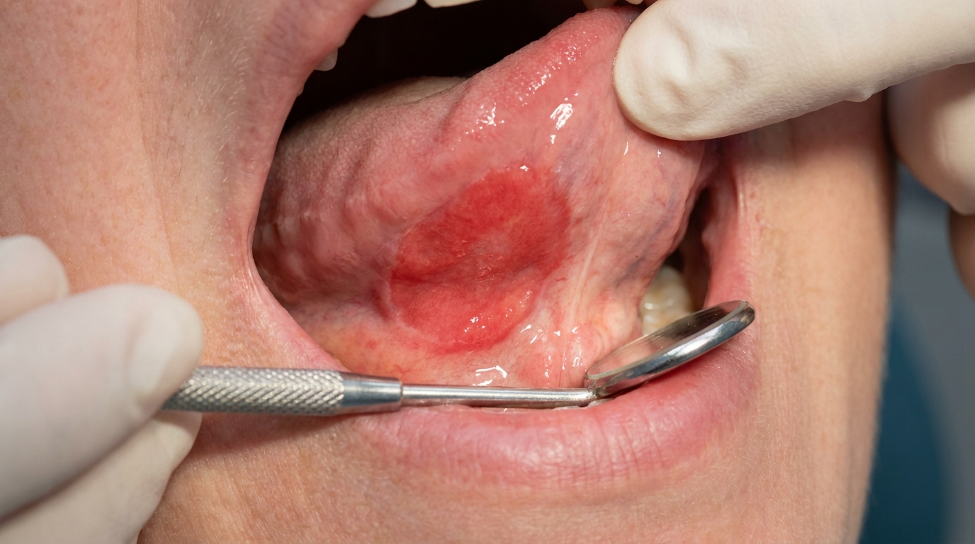
What it looks like
The classic appearance is well described:
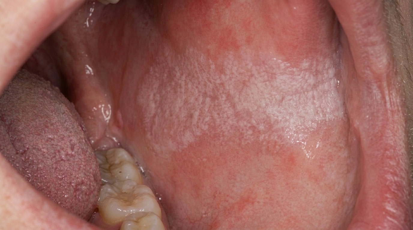
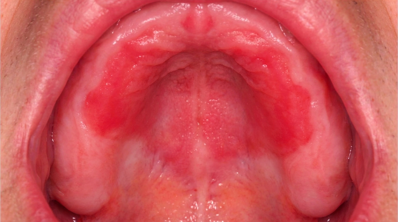
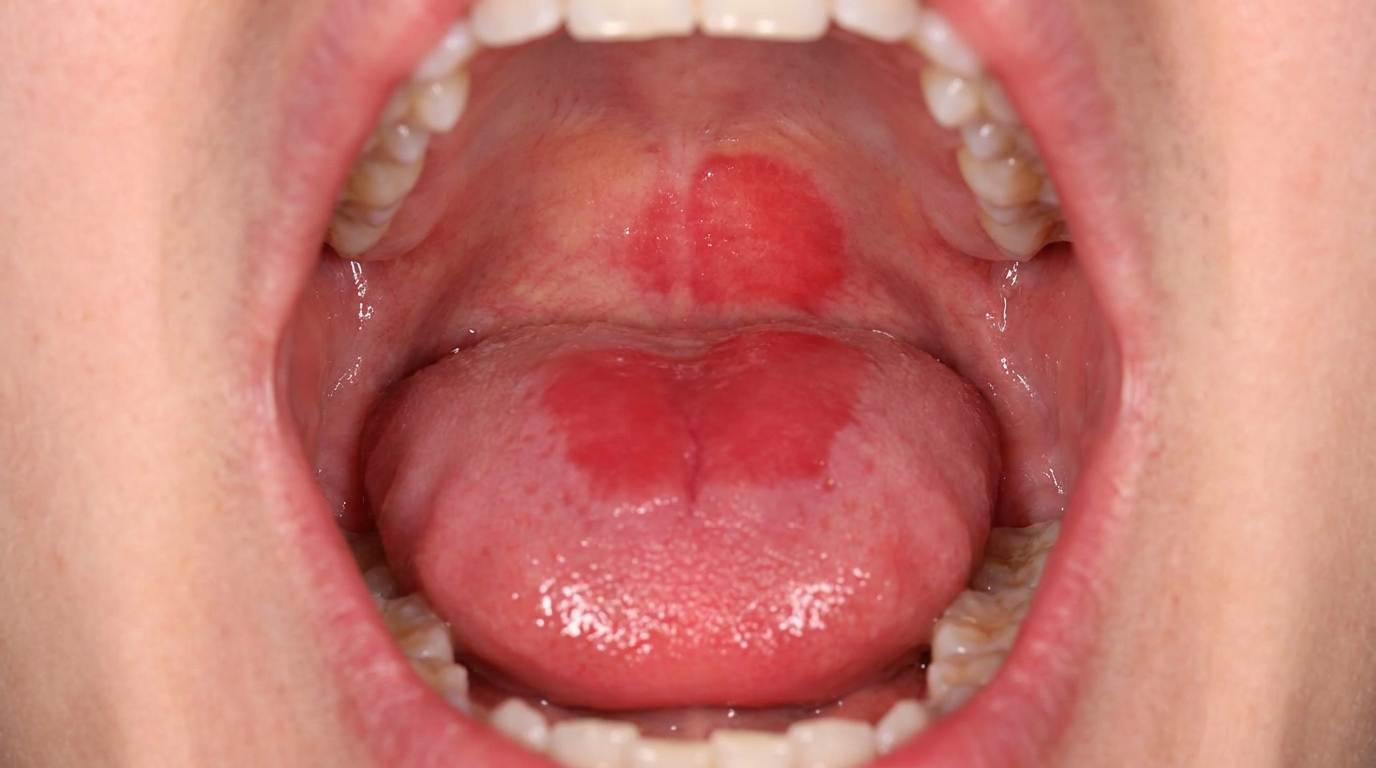
A well-demarcated red patch or plaque in the mouth.
A velvety, smooth or finely granular surface texture.
The patch may be slightly depressed compared to the surrounding mucosa, particularly on the floor of the mouth and the underside of the tongue.
The most common sites are the floor of the mouth, the side and underside of the tongue, the soft palate and the inside of the cheeks.
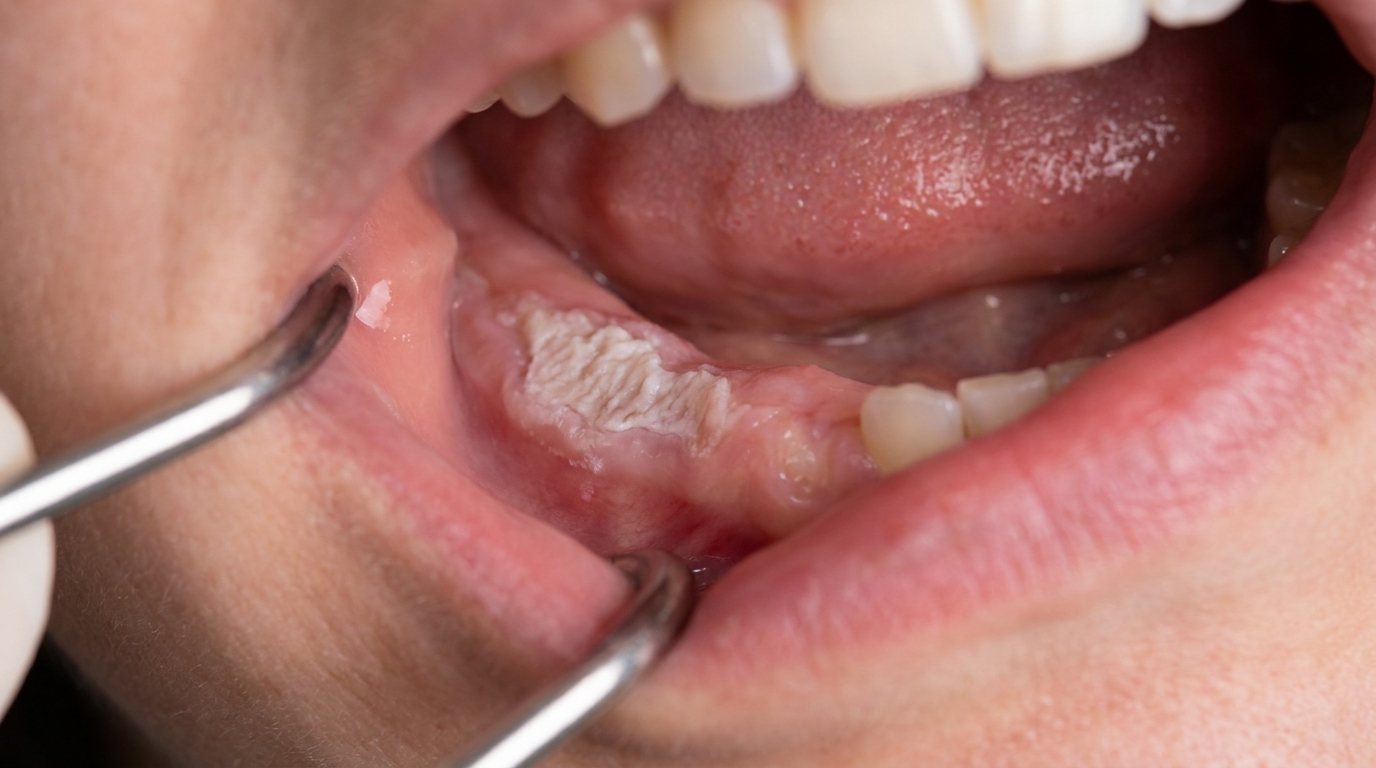
Mixed red-and-white areas, known as erythroleukoplakia or speckled leukoplakia, are particularly suspicious for high-grade dysplasia or carcinoma.
Multiple lesions can occur in the same mouth.
What it feels like
Most erythroplakias are entirely painless. Symptoms when present may include:
Mild burning or soreness with hot or spicy foods, particularly in lesions with a thinner surface.
A rough sensation felt with the tongue.
Bleeding with brushing or eating, when the surface is particularly atrophic.
No symptoms at all in most patients, which is why these lesions are easily missed unless the dentist actively looks for them.
What an X-ray might show
X-rays are not used to diagnose erythroplakia. They may be relevant if there is concern about an underlying tumour invading the bone or adjacent structures.
What happens at the dentist?
A persistent red patch should always be carefully examined and almost always biopsied. A dentist at ArtSmiles, typically as part of a dental check-up and clean, will commonly:
Examine the patch carefully under good light, noting its size, location, surface texture and edges.
Take a careful history, including tobacco and alcohol use, betel quid habits, recent illnesses, medications, and any past oral cancer.
Look for an obvious local cause, a sharp tooth, an ill-fitting denture, or recent burn, that could explain the redness. Where such a cause is found, the lesion may be reviewed in 2 weeks to see whether it resolves.
Photograph and measure the lesion as a baseline.
Recommend biopsy in nearly all cases, since the only reliable way to determine the level of cellular change is on histopathology (microscopic examination of tissue). The textbooks specifically advise that "red lesions of the oral mucosa, especially those of the oral floor and ventrolateral tongue, should be viewed with suspicion, and a biopsy should be performed."
Refer to an oral medicine specialist or oral and maxillofacial surgeon for definitive management, particularly when biopsy shows moderate or worse dysplasia.
Coordinate cessation support for any tobacco, alcohol or betel quid use.
Is this serious?
🔴 Erythroplakia is one of the most clinically significant lesions in the mouth. The textbooks describe it as carrying a much greater risk for malignancy than leukoplakia, with almost all true erythroplakias showing severe dysplasia, carcinoma in situ or invasive squamous cell carcinoma at the time of biopsy. The good news is that, when caught early, the cancers that arise within or alongside erythroplakia are often at a stage where they can be effectively treated. The key practical message is that any persistent red patch in the mouth deserves prompt assessment.
If you have noticed a red patch in the mouth that has not gone away in two to three weeks, particularly on the floor of the mouth, the side or underside of the tongue, or with a velvety surface, it is worth booking an assessment so the right biopsy and follow-up can be arranged without delay.
Could it be something else?
Several conditions can produce red patches in the mouth and need to be distinguished from erythroplakia. The textbooks list these as the main differentials:
Erythematous (atrophic) candidiasis, produces redness, often on the palate beneath an upper denture, and responds rapidly to antifungal treatment.
Erosive or atrophic oral lichen planus, usually has lacy white edges adjacent to red atrophic areas, and tends to be symmetrical.
Lupus erythematosus, can produce red patches with white striae or ulceration; often associated with skin changes and systemic features.
Local trauma or chemical burn, a clear history of biting, denture rub or chemical exposure usually identifies these, and they resolve with removal of the cause.
Denture stomatitis, generalised red change of the mucosa beneath an upper denture.
Vascular lesions, haemangiomas, varices, telangiectasias and pyogenic granulomas can all produce red lesions, but they have distinct clinical and histological features.
Oral leukoplakia with a partly red component, when red and white areas mix, the lesion is called erythroleukoplakia or speckled leukoplakia and behaves more like erythroplakia in its risk.
How is it treated?
Treatment is guided by the histopathology, the size and site of the lesion, and the patient's overall health and risk factors.
At-home measures and habits:
Stop smoking and reduce alcohol, the most important single change a patient can make.
Stop using betel quid, paan or smokeless tobacco.
Maintain excellent oral hygiene to limit any inflammatory contribution.
Address any source of chronic friction, a sharp tooth, a poorly fitting denture, or a habit of cheek-biting.
Eat a balanced diet with plenty of fruit and vegetables to support general mucosal health.
Professional steps your dentist may consider:
Biopsy, almost universally indicated, since most lesions show severe dysplasia, carcinoma in situ or invasive carcinoma. Excisional biopsy is often preferred so that the entire lesion can be examined microscopically.
Surgical excision or laser ablation of dysplastic or carcinomatous lesions, with adequate margins.
Cryotherapy or photodynamic therapy in selected cases.
Treatment as oral squamous cell carcinoma, including specialist surgical and oncological care, when invasive carcinoma is identified.
Smoking cessation and harm-reduction support through your GP and quit programmes.
Long-term follow-up at 3-6 month intervals, since recurrence and new lesions are common. The textbooks specifically note that "recurrence and multifocal involvement are common; hence, long-term follow-up is suggested."
A patient-centred approach is particularly important here, where the diagnosis of "premalignant" or "early cancer" can be overwhelming. Honest, unhurried explanation of what the biopsy means, what treatment achieves, and how follow-up will work is itself part of effective care, values that sit at the heart of our clinical philosophy.
What's the long-term outlook?
The outlook depends primarily on three things, the histological grade of the lesion, whether the underlying causes can be removed, and how well the patient engages with long-term follow-up.
Severe dysplasia or carcinoma in situ caught and excised early has a very good outcome, although careful follow-up is essential because new lesions can develop.
Early invasive carcinoma identified within an erythroplakia is usually treated as oral cancer with conventional surgical and oncological care, with outcomes that depend on the size and stage at diagnosis.
Continued smoking, alcohol or betel quid use dramatically worsens the long-term outlook by driving fresh lesions in adjacent or new sites.
The single most important factor across all of these is regular dental review and a strong patient-clinician partnership over many years. With consistent follow-up and risk-factor change, the very real risks of erythroplakia can be effectively managed.
A note on this article
This article is for educational purposes only and does not constitute a clinical diagnosis. Please consult a registered dental practitioner for assessment and treatment advice.
The cover image above is an AI-generated illustration based on the most common visible features of this condition described in clinical pathology references. It is not a photograph of a real case and should not be used to diagnose or rule out the condition in your own situation. If you are concerned about something you have noticed, please book an assessment with a registered dental practitioner.
References
Neville, B. W., Damm, D. D., Allen, C. M., & Chi, A. C. (2023). Oral and maxillofacial pathology (5th ed.). Elsevier. Chapter 10, Epithelial Pathology: Erythroplakia (Erythroplasia; Erythroplasia of Queyrat), with detailed clinical and histopathologic features and treatment recommendations, pp. 390 to 391.
Cawson, R. A., & Odell, E. W. (2017). Cawson's essentials of oral pathology and oral medicine (8th ed.). Elsevier. Chapter 16, Oral Premalignancy: Erythroplasia ('erythroplakia') and Speckled Leukoplakia, pp. 262 to 263.
Regezi, J. A., Sciubba, J. J., & Jordan, R. C. K. (2017). Oral pathology: Clinical pathologic correlations (7th ed.). Elsevier. Chapter 6, Premalignant and Malignant Soft Tissue Lesions: Erythroplakia coverage.