Compiled from clinical pathology references. Medically reviewed by Dr Cristian Dunker , Principal Dentist, ArtSmiles Cosmetic Dentistry.
A sore tongue, cracked corners of the mouth, or an unusually pale lining inside your cheeks can sometimes be the first visible sign of an underlying blood problem. Iron deficiency, vitamin B12 (a vitamin needed for healthy nerves and blood cells) deficiency, and folate deficiency all show up in the mouth before many people realise something is wrong with their blood, and a dentist is often the first clinician to spot the change.
Quick summary
Also called | anaemic glossitis, atrophic glossitis (smooth, thinned, sore tongue), Hunter's glossitis, Moeller's glossitis; Plummer-Vinson (Paterson-Kelly) syndrome when combined with dysphagia |
How urgent? | 🟡 Worth a check-up, these signs usually mean a blood test is needed; they are rarely emergencies but should not be ignored |
Common or rare? | Common, iron deficiency (low body iron stores) anaemia is the most common anaemia worldwide; B12 and folate deficiencies are also frequent |
Who it affects | Adults of all ages; women of child-bearing age, pregnant women, older adults, vegetarians and vegans, and people with gastrointestinal disease are most at risk |
Who treats it | Both, your dentist may notice the oral changes first, then a GP confirms the cause with blood tests and manages the underlying anaemia |
Based on | Regezi, Neville, Cawson, Laskaris |
What is it?
Anaemia is a general term for a drop in the number of red blood cells, or in the amount of haemoglobin those cells carry. Because red blood cells are responsible for delivering oxygen to every tissue in the body, the lining of the mouth, which renews itself rapidly and needs a steady oxygen supply, is one of the first places the shortage tends to show.
When the body runs short of iron, vitamin B12, or folate, the tongue and mouth can change in distinctive ways. The tongue may turn smooth and red, the corners of the mouth may crack, and the cheeks and gums may look unusually pale. These changes are not a disease in themselves; they are signals that something is happening in the blood that needs investigating.
Who tends to get it?
The pattern depends on which type of anaemia is involved.
Iron deficiency anaemia is the most common form worldwide and the most common cause of anaemia in Australia. It usually affects women of child-bearing age, often as a result of menstrual blood loss or pregnancy. Men can also be affected, typically through gastrointestinal blood loss from peptic ulcers, haemorrhoids, or other digestive disease. Babies, growing children, and older adults whose diets are limited can also become iron deficient.
Pernicious anaemia (B12 deficiency) chiefly affects adults from the age of about 30 onward, and is more common in older people of Northern European heritage, although it has been reported across all populations. Strict vegans can also develop B12 deficiency because the vitamin comes mainly from animal foods. People who have had stomach or bowel surgery, or who have coeliac or Crohn's disease, are at higher risk.
Folate deficiency tends to occur in younger adults, particularly during pregnancy, and in people with malabsorption, alcohol-related illness, or certain medications such as phenytoin and methotrexate.
Sickle cell anaemia mainly affects people of African, Afro-Caribbean, Indian, Mediterranean, or Middle Eastern background. Thalassaemia is more common in those whose families come from the Mediterranean, parts of Africa, the Middle East, India, and Southeast Asia.
What causes it?
The oral signs of anaemia all share a common root: there are not enough healthy red blood cells doing their job. The reason for that shortage varies.
Low iron intake or absorption. Iron-poor diets, fussy eating in childhood, and diets in older adults that lack red meat, leafy vegetables, or legumes can all reduce iron levels. Coeliac disease and inflammatory bowel disease (especially Crohn's) can stop the gut absorbing iron properly.
Chronic blood loss. Heavy menstrual periods, gastrointestinal bleeding from ulcers or haemorrhoids, or long-term use of medications such as aspirin can slowly drain iron stores.
Increased demand. Children going through growth spurts and women during pregnancy need more iron than usual.
Loss of intrinsic factor. Pernicious anaemia is an autoimmune condition where the body attacks the cells in the stomach lining that make intrinsic factor, the protein needed to absorb vitamin B12.
Diet without animal products. B12 is found mainly in meat, fish, eggs, and dairy. Strict vegans who do not supplement can develop deficiency over time.
Folate-poor diet, pregnancy, or certain medications. Alcohol, methotrexate, and phenytoin can interfere with folate metabolism.
Inherited haemoglobin disorders. Sickle cell anaemia and thalassaemia are genetic conditions in which the haemoglobin molecule itself is abnormal or made in reduced amounts.
How does it develop?
Think of red blood cells as delivery vans carrying oxygen around the body. To build a working van, the bone marrow needs raw materials: iron for the haemoglobin (the oxygen container), and vitamins B12 and folate for the DNA that lets the marrow churn out new cells.
If any of those raw materials run short, the production line slows down or produces faulty vans. In iron deficiency, the cells that are made are smaller and paler than normal. In B12 or folate deficiency, the cells that are made are unusually large and don't survive as long. Either way, fewer working red cells reach the tissues, and oxygen delivery drops.
The lining of the tongue and mouth turns over very quickly, the cells on the surface are replaced every few days. Tissues that renew themselves this fast are the first to suffer when raw materials are short. The tiny papillae that give the tongue its rough, velvety texture begin to flatten and disappear, leaving the tongue smooth, red, and tender. The corners of the mouth, where skin and lip meet, can break down and let yeast move in, causing angular cheilitis (cracking and soreness at the corners of the lips). The mucosa loses its usual pink colour and starts to look pale.
What might you notice?
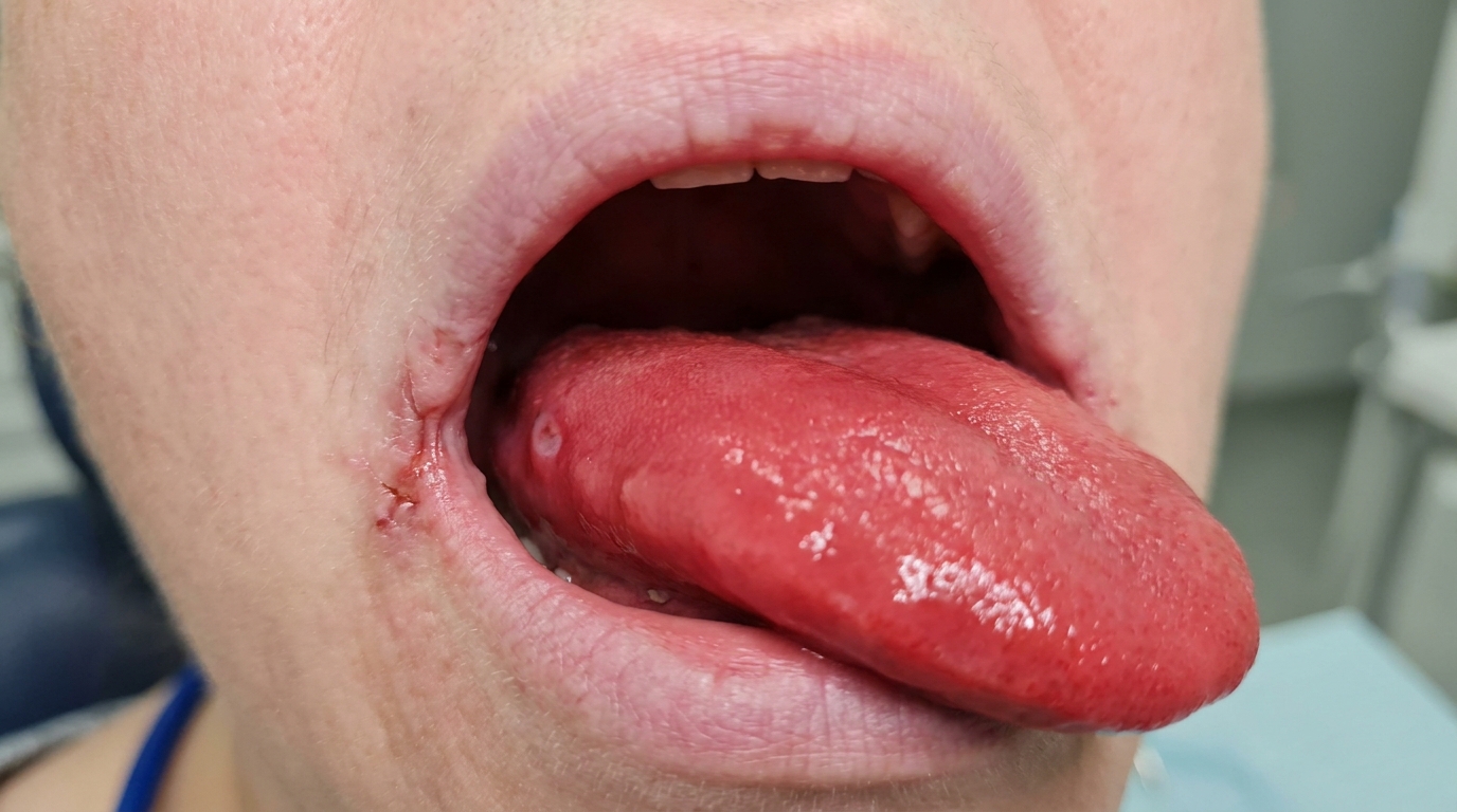
What it looks like
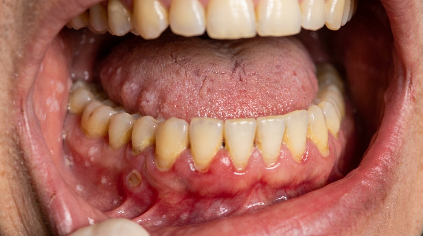
A smooth, red, shiny tongue. The small bumps (filiform and fungiform papillae) that normally cover the top of the tongue gradually flatten until the surface looks polished. This is sometimes called atrophic glossitis, Hunter's glossitis (in B12 deficiency), or Moeller's glossitis.
Cracked, sore corners of the mouth. Angular cheilitis appears as red, fissured patches at the corners, often with a yellowish crust. It is a classic sign of iron deficiency, and is usually colonised by Candida yeast.
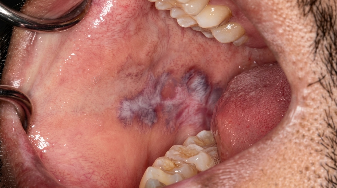
Pale lining inside the mouth. The cheeks, palate, and gums may look washed-out compared with normal. The undersides of the lower eyelids and the nail beds often look pale at the same time.
Spoon-shaped, brittle nails. This curved-spoon nail change (called koilonychia) is a long-standing sign of chronic iron deficiency.
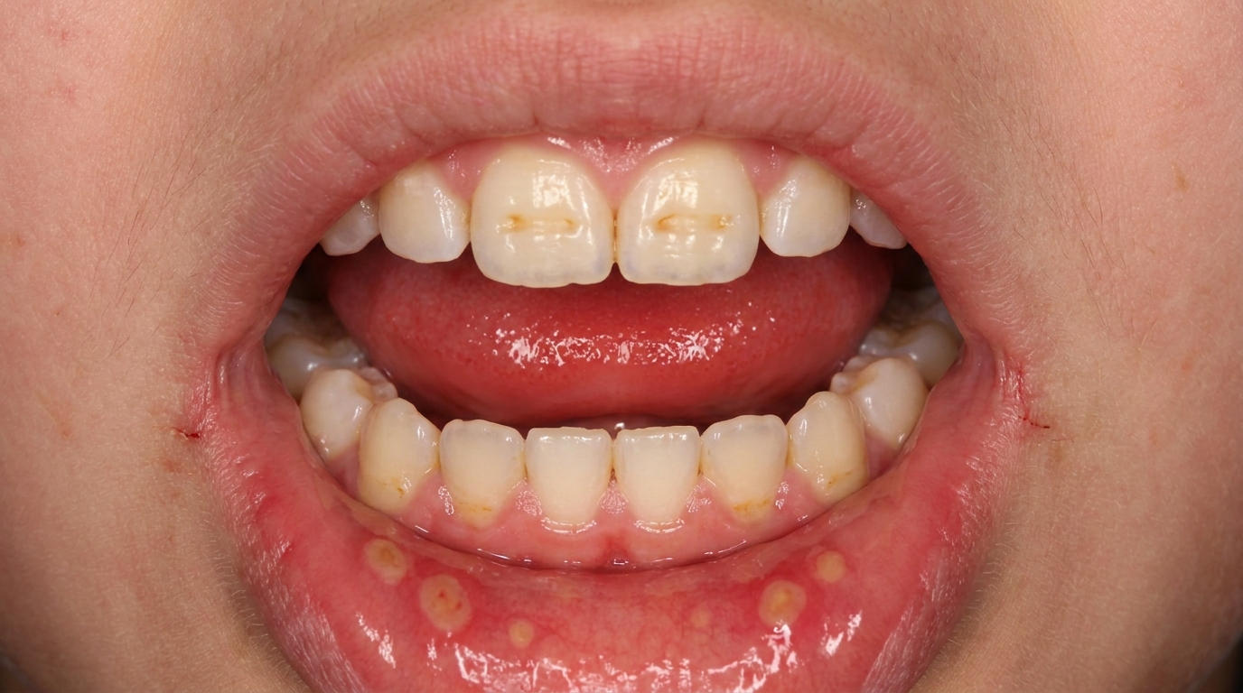
Recurrent mouth ulcers. Aphthous ulcers that start, or get worse, later in life can sometimes be linked to a hidden B12, folate, or iron deficiency.
What it feels like
Many people describe a burning, tingling, or sore tongue, particularly with hot or spicy foods. The pain may come and go, or be present all the time. In B12 deficiency, the soreness can begin before the haemoglobin level drops far enough to be picked up on routine blood tests.
Other common symptoms include a change in taste, a dry mouth feeling, and tenderness when wearing dentures. People with iron deficiency may notice difficulty swallowing, particularly with dry foods, which can be a feature of Plummer-Vinson (Paterson-Kelly) syndrome.
General signs of anaemia often appear alongside the oral changes: tiredness, breathlessness on exertion, palpitations, headache, and lightheadedness.
What an X-ray might show
For most types of anaemia, the mouth changes are confined to the soft tissues, so dental X-rays are usually unremarkable. Two exceptions are worth noting. In sickle cell disease, X-rays of the skull or jaw can show a thinned trabecular pattern, a "hair-on-end" appearance of the calvarium, and occasional bone infarcts (areas of dead bone from blocked blood supply) that can mimic osteomyelitis (bone infection). In thalassaemia major, expansion of the bone marrow can produce marked enlargement of the maxilla and mandible, a wispy trabecular pattern, and a more pronounced "hair-on-end" skull appearance, sometimes giving the face a "chipmunk" appearance.
What happens at the dentist?
A dentist at ArtSmiles may be the first health professional to suspect anaemia, because the mouth changes can appear before someone realises they feel run-down. At your appointment, your dentist may:
Examine the tongue carefully, looking for loss of papillae, redness, and atrophy.
Check the corners of the mouth for angular cheilitis, and look for signs of oral candidosis.
Inspect the lining of the cheeks, palate, and gums for unusual pallor.
Look at your nails and the inner surface of your lower eyelid, both of which can show pallor or koilonychia.
Ask about general symptoms, fatigue, breathlessness, heavy periods, gastrointestinal symptoms, dietary patterns, and any history of bowel surgery or autoimmune disease.
If an underlying anaemia is suspected, your dentist will usually recommend a referral to your GP for blood investigations. The textbooks emphasise that blood tests are essential before treating the oral symptoms, because the mouth findings will not resolve until the underlying deficiency is corrected. Typical investigations include a full blood count with red cell indices (mean corpuscular volume, the average size of red blood cells), serum iron and ferritin (a blood marker of iron stores), vitamin B12, and folate levels. Sometimes additional tests are needed to find the cause, for example, looking for coeliac disease, antibodies against intrinsic factor, or a source of gastrointestinal bleeding.
In cases where the tongue or mouth changes are unusual or persistent despite treatment, a biopsy may occasionally be recommended to rule out other conditions.
Is this serious?
🟡 Worth a check-up. Most cases of iron, B12, or folate deficiency are very treatable once the cause is found. The oral changes themselves are reversible, and the tongue, lips, and lining of the mouth usually return to normal within a few weeks of starting appropriate therapy.
The reason these signs deserve attention is what they may point to. An undiagnosed iron deficiency may signal a slow gastrointestinal bleed; a B12 deficiency, if left untreated, can cause permanent nerve damage and walking difficulties; and Plummer-Vinson syndrome carries a higher long-term risk of cancer of the mouth, throat or oesophagus (the food pipe). Sickle cell anaemia and thalassaemia are lifelong conditions that need specialist medical care.
If you've noticed any of these signs for more than two weeks, it's worth booking an assessment.
Could it be something else?
A red, smooth, or sore tongue, or cracked mouth corners, can have several other explanations. Your dentist will usually consider the following:
Geographic tongue (erythema migrans), patchy red areas on the tongue can look similar, but they characteristically migrate, change shape over days, and have a slightly raised pale border. Anaemia, by contrast, produces uniform, persistent atrophy.
Oral candidosis (thrush), can cause a red, sore tongue and angular cheilitis. The clinical picture overlaps significantly, and candidosis often coexists with iron deficiency. A swab and a blood test help separate them.
Lichen planus (atrophic form), can flatten the tongue papillae and produce a smooth surface, but is usually painless at this stage and often shows white striae elsewhere in the mouth.
Burning mouth syndrome, produces burning and soreness with a normal-looking mouth. Anaemia must be excluded by blood tests before this diagnosis is accepted, because deficiency states are listed among its causes.
Sjögren's syndrome, can cause a red, cobblestoned tongue along with dry mouth and dry eyes. Specific blood tests for autoantibodies (anti-Ro, anti-La) help distinguish it.
Atrophic glossitis from tertiary syphilis, historically described, now rare. Serological testing differentiates.
Pellagra (niacin deficiency), can cause glossitis along with skin rash, diarrhoea, and changes in mood. The pattern of accompanying symptoms is the giveaway.
Riboflavin or other B-vitamin deficiency, can cause angular cheilitis and glossitis. Dietary history and blood tests help separate it from iron or B12 deficiency.
Recurrent canker-like (aphthous) stomatitis, when ulcers start or worsen later in life, an underlying haematological deficiency should be excluded by blood tests.
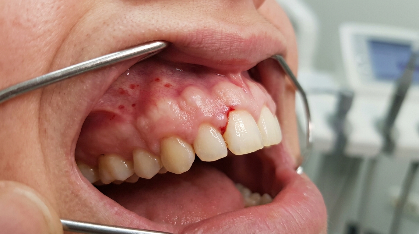
Leukaemia and aplastic anaemia, can cause mucosal pallor along with gum bleeding, swelling, ulceration, and bruising. A full blood count is the first step to rule these in or out.
Plummer-Vinson syndrome, should be considered when iron deficiency, glossitis, dysphagia, and koilonychia appear together, because of its association with cancer of the upper digestive tract.
How is it treated?
Treatment has two parts: correcting the underlying deficiency, and managing any oral problems that have developed.
Steps you can take at home
Eat a varied diet that includes good sources of iron (red meat, poultry, fish, legumes, leafy greens), B12 (meat, fish, eggs, dairy, or fortified foods), and folate (leafy vegetables, legumes, citrus, fortified cereals).
Keep the mouth clean and gentle. Avoid very spicy, salty, or acidic foods while the tongue is sore.
If you wear dentures, remove them at night and clean them carefully, a sore tongue plus dentures plus iron deficiency is a common combination for thrush.
Don't self-diagnose. Vitamin and iron supplements bought over the counter can mask a deficiency, change blood test results, and delay finding the real cause.
Professional treatment may include
Blood tests through your GP to confirm the type and cause of the anaemia. This is the essential first step.
Iron supplementation, usually oral ferrous sulfate, taken for several weeks to months. Parenteral iron (iron given by injection or drip) may occasionally be used in severe cases or where oral iron is not tolerated.
Vitamin B12 replacement, traditionally given as monthly intramuscular injections of cyanocobalamin (the standard injectable form of vitamin B12), although high-dose oral B12 has been shown to be equally effective in many cases. Oral lesions typically begin to improve within the first week or two of starting therapy, though it can take longer for the tongue to fully recover its normal surface.
Folate supplementation, given as oral folic acid. It is important to distinguish folate from B12 deficiency before treating, because giving folate alone in B12 deficiency can mask the anaemia while allowing nerve damage to progress.
Treatment of associated oral problems, antifungal therapy may be recommended for angular cheilitis or oral thrush; topical lubricants may help if dry mouth is a feature.
Investigation of the underlying cause, your GP may arrange further tests (such as endoscopy, tests for coeliac disease, or autoimmune screens) to find why the deficiency developed.
Specialist referral, to a haematologist for inherited anaemias such as sickle cell disease or thalassaemia, or to a gastroenterologist if absorption problems are suspected.
Periodic monitoring in Plummer-Vinson syndrome, because of the long-term risk of upper aerodigestive tract cancer.
What's the long-term outlook?
For the most common forms, iron deficiency, B12 deficiency, and folate deficiency, the outlook is genuinely encouraging. Once the cause is identified and replacement therapy is started, the oral changes usually resolve within a few weeks. Glossitis settles, the papillae regenerate, the mouth lining regains its normal pink colour, and angular cheilitis heals. Most patients also notice their general energy returning.
The key is finding and addressing the cause. If iron supplements are taken without identifying a slow gastrointestinal bleed, the anaemia will return. If B12 deficiency is left untreated, neurological problems such as numbness, tingling, and difficulty walking can become permanent, even after the blood picture is corrected. People with pernicious anaemia have a slightly higher long-term risk of gastric cancer, so periodic medical follow-up is sensible.
For inherited anaemias, sickle cell disease and thalassaemia, the long-term picture depends on the severity of the genetic condition, the quality of medical care, and how well crises and complications are prevented. These patients benefit from working closely with both their medical team and a dentist who is aware of their condition, because routine dental treatment needs special precautions.
In most patients, the mouth is an early warning system rather than the main problem. Listening to it, getting the blood tests, and treating the underlying deficiency is what makes the difference.
A note on this article
This article is for educational purposes only and does not constitute a clinical diagnosis. Please consult a registered dental practitioner for assessment and treatment advice.
The cover image above is an AI-generated illustration based on the most common visible features of this condition described in clinical pathology references. It is not a photograph of a real case and should not be used to diagnose or rule out the condition in your own situation. If you are concerned about something you have noticed, please book an assessment with a registered dental practitioner.
References
Regezi, J. A., Sciubba, J. J., & Jordan, R. C. K. (2017). Oral pathology: Clinical pathologic correlations (7th ed.). Elsevier. Chapter 4, Red-Blue Lesions: Vitamin B Deficiencies, Pernicious Anemia, Iron Deficiency Anemia, Burning Mouth Syndrome, pp. 124 to 127.
Neville, B. W., Damm, D. D., Allen, C. M., & Chi, A. C. (2023). Oral and maxillofacial pathology (5th ed.). Elsevier. Chapter 13, Hematologic Disorders, pp. 583 to 588; Chapter 17, Oral Manifestations of Systemic Diseases, pp. 830 to 833.
Cawson, R. A., & Odell, E. W. (2017). Cawson's essentials of oral pathology and oral medicine (8th ed.). Elsevier. Chapter 14, Tongue Disorders: Sore Tongue and Glossitis, pp. 246 to 248; Chapter 22, Anaemias, Leukaemias and Lymphomas, pp. 336 to 339.
Laskaris, G. (2003). Color atlas of oral diseases (3rd ed.). Thieme. Section 23, Hematologic Disorders, pp. 226 to 228.