Compiled from clinical pathology references. Medically reviewed by Dr Cristian Dunker , Principal Dentist, ArtSmiles Cosmetic Dentistry.
Quick summary
Also called | Idiopathic leukoplakia |
How urgent? | 🟡 Worth checking, most leukoplakias are harmless, but a small minority can transform into oral cancer over time, and the only way to tell is to investigate |
Common or rare? | Relatively common, overall prevalence in adults is around 1-3%, with higher rates among smokers and heavy alcohol users |
Who it affects | Most often middle-aged and older adults; smokers, heavy alcohol users, and users of tobacco-containing betel quid (paan) are particularly at risk |
Who treats it | General dentist for diagnosis and biopsy, often in coordination with an oral medicine specialist or oral and maxillofacial surgeon for management |
Based on | Cawson, Neville, Regezi |
What is it?
Oral leukoplakia is the formal name for a persistent white patch on the lining of the mouth that cannot be wiped off and cannot be explained by any other specific disease. The textbooks describe it as a diagnosis of exclusion: leukoplakia is the label given when frictional, infectious, immune-mediated and other recognisable causes of a white patch have been ruled out. The reason it matters is that, although most leukoplakias remain benign for many years, a small minority show abnormal cell changes (dysplasia (microscopic changes that can signal an increased cancer risk)) and a small proportion of these progress to oral squamous cell carcinoma. This means every persistent unexplained white patch deserves a careful look.
Who tends to get it?
The textbooks describe a fairly consistent profile:
Adults over the age of 40, with prevalence rising with age. Idiopathic leukoplakia is uncommon in young adults and rare in children.
Slightly more common in men in many studies, although the difference has narrowed as smoking patterns have changed.
Strongly associated with tobacco use in any form, cigarettes, cigars, pipes, smokeless tobacco, and tobacco-containing betel quid (paan).
Heavy alcohol use is a recognised additional risk factor, particularly when combined with smoking.
Areca nut and betel quid users in South Asia and the South Asian diaspora have particularly high rates.
People with chronic friction in a specific area, from a sharp tooth, an ill-fitting denture, or a habit of cheek-biting, can develop white patches. Most of these are not true leukoplakia, but persistent ones may behave similarly.
People with previous oral cancer are at higher risk of developing further leukoplakias and are followed closely.
What causes it?
The textbooks describe leukoplakia as idiopathic (no known cause) by definition, that is, by the time a patch is called leukoplakia, no specific cause has been found. Recognised contributors and associations include:
Tobacco, by far the strongest single contributor, in any form (smoked or smokeless).
Areca nut and betel quid use, directly associated with both leukoplakia and oral submucous fibrosis, and a major contributor to oral cancer in South Asia.
Heavy alcohol use, synergistic with tobacco.
Chronic friction, although patches caused by clear, ongoing trauma are often called frictional hyperkeratosis (thickened keratin on the surface) rather than true leukoplakia.
Candida albicans infection, chronic hyperplastic candidiasis can produce a similar-looking plaque, sometimes superimposed on a leukoplakia.
Genetic susceptibility, accumulating molecular changes in the surface cells appear to underlie the small risk of malignant transformation.
Idiopathic cases, many leukoplakias occur in people without any of the recognised risk factors.
It is important to note that leukoplakia is not caused by poor oral hygiene, vitamin deficiency or stress on its own, and it is not contagious.
How does it develop?
The white appearance of a leukoplakia comes from increased keratin (the same protein that makes up skin, hair and nails) on the surface of the oral mucosa. Microscopically, there is hyperkeratosis or parakeratosis (different forms of keratin thickening), often with thickening of the underlying epithelium (acanthosis). In some patches, the deeper cells show dysplasia, a microscopic disorder of cell shape, size and arrangement that signals a step toward cancer. The textbooks classify dysplasia as mild, moderate or severe depending on how much of the epithelial thickness is involved. Most leukoplakias have no dysplasia at all, but the lesions that do progress to cancer almost always show dysplasia first. Cawson notes that the malignant transformation rate of idiopathic leukoplakia is around 1-2% in 5 years, with higher rates for lesions that show dysplasia, are large, are speckled (white with red flecks mixed in) (red-and-white), occur on the floor of mouth or tongue, or are seen in non-smokers.
What might you notice?
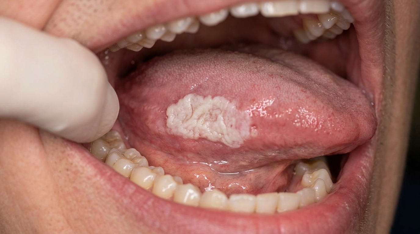
What it looks like
The textbooks describe several patterns of leukoplakia:
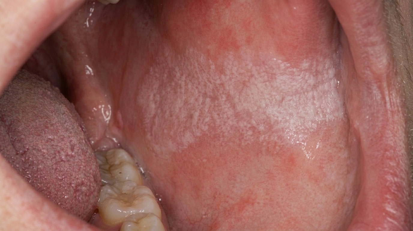
Homogeneous leukoplakia, a flat or slightly raised white patch with a uniform white surface. Lower risk of cancer change overall.
Non-homogeneous (uniformly flat and white) leukoplakia, patches with mixed colour or surface texture, including:
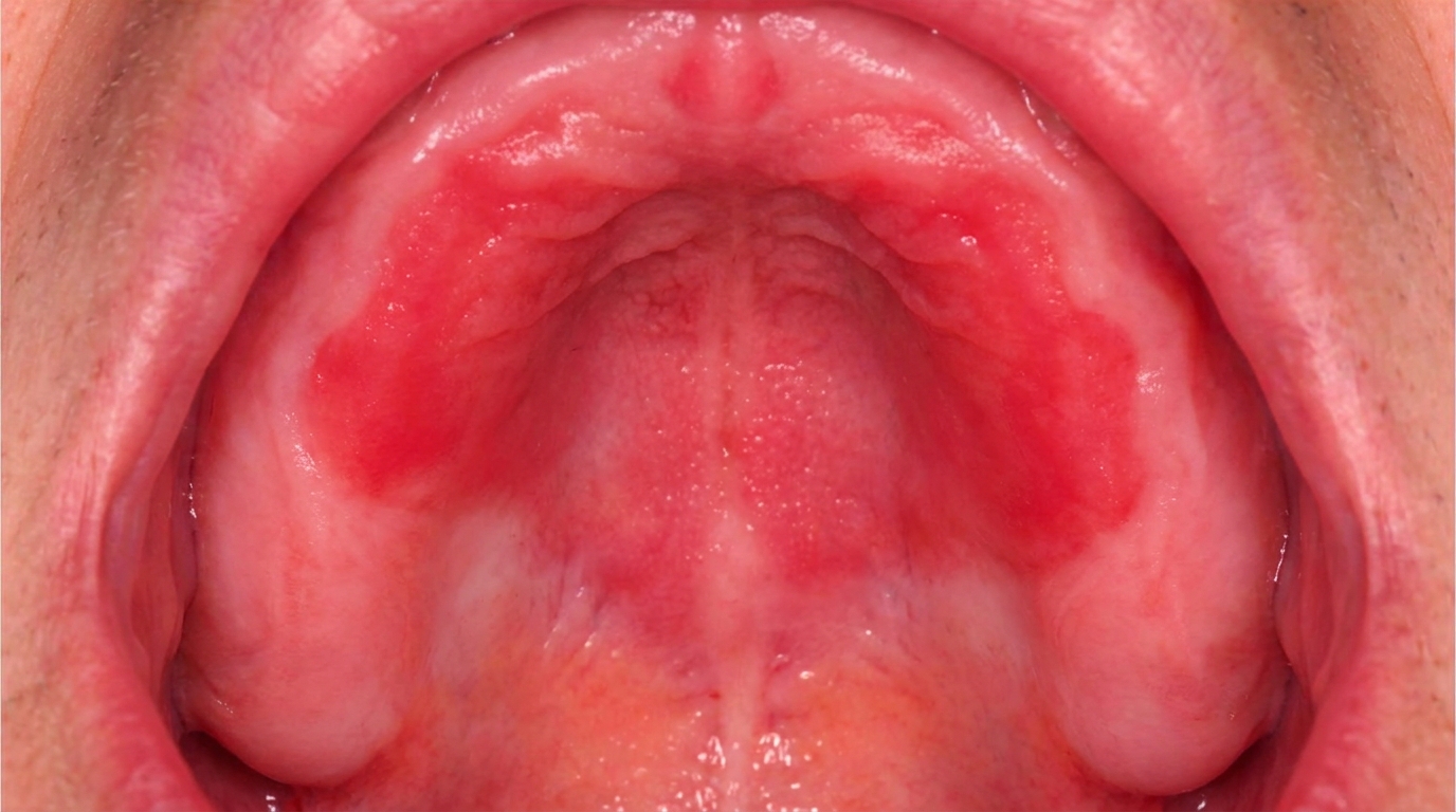
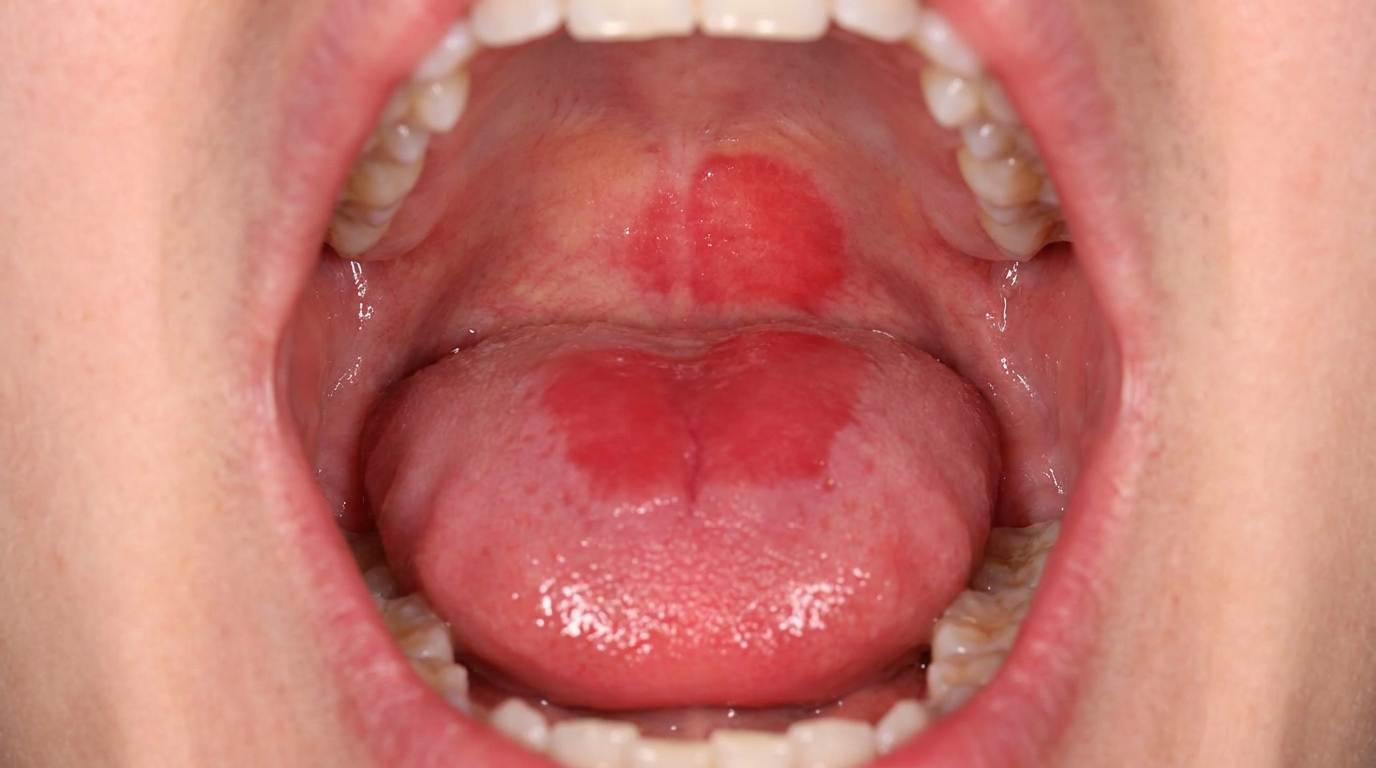
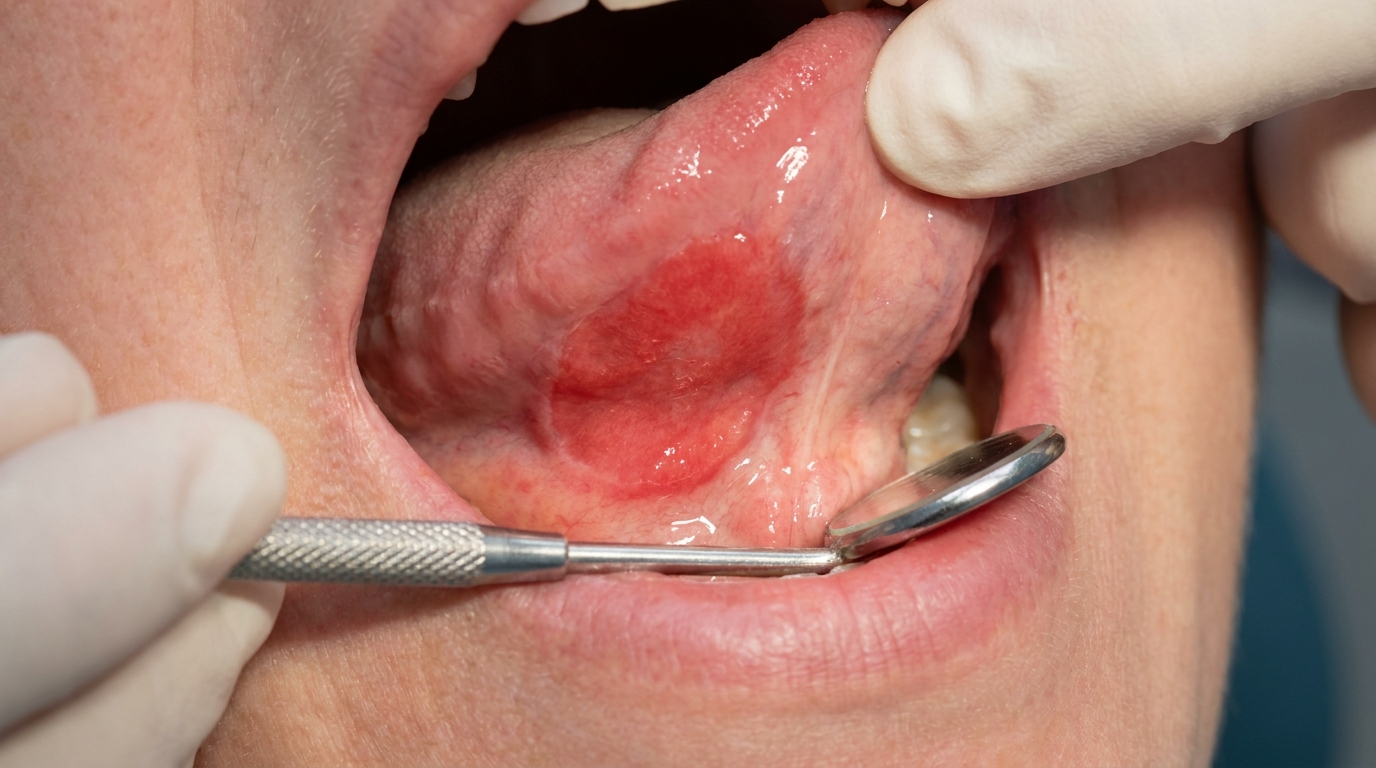
Speckled leukoplakia, alternating white and red areas. Higher risk of dysplasia and cancer change.
Verrucous leukoplakia, a thick, warty, cauliflower-like surface.
Nodular leukoplakia, small white lumps within a red background.
Common sites include the inside of the cheek (buccal mucosa), the area behind the wisdom teeth (retromolar region), the floor of the mouth, and the borders of the tongue.
The patch does not wipe off with gauze.
The patch is persistent, it has usually been present for several weeks at least.
The textbooks specifically caution that small, innocent-looking patches may still show dysplasia on biopsy, so size alone is not a reliable guide to risk.
What it feels like
Most leukoplakias are painless. When symptoms occur they may include:
Mild roughness felt with the tongue.
Tightness of the area.
Burning or soreness with hot or spicy foods, particularly in speckled lesions.
Loosening of an adjacent tooth or sudden discomfort, these can signal that the lesion has changed and warrant urgent reassessment.
What an X-ray might show
X-rays are not used to diagnose leukoplakia. They may be relevant if there is concern about an underlying tumour or related dental disease, but the diagnosis of leukoplakia itself is made on examination and biopsy.
What happens at the dentist?
A persistent white patch should always be carefully examined. A dentist at ArtSmiles, typically as part of a dental check-up and clean, will commonly:
Examine the patch carefully under good light, noting its size, location, surface texture, colour and edges.
Test whether the patch wipes off with gauze (it should not, in true leukoplakia).
Take a careful history, including any tobacco use, alcohol intake, betel quid or paan habit, recent illnesses, medications, and previous oral lesions.
Examine the rest of the mouth and the neck for other suspicious areas, enlarged lymph nodes or related findings.
Photograph and measure the lesion as a baseline.
Recommend a biopsy in nearly all cases, it is the only reliable way to determine whether dysplasia is present and to rule out an early carcinoma. A small piece of the lesion is removed under local anaesthetic and sent to an oral pathologist.
Address any associated factors, for example, a sharp tooth or ill-fitting denture causing chronic friction, or a candidal overgrowth that needs antifungal treatment.
Refer to an oral medicine specialist or oral and maxillofacial surgeon for higher-risk lesions or when more extensive treatment is being considered.
Is this serious?
🟡 Most oral leukoplakias are benign and never cause problems. However, leukoplakia carries the highest risk of oral cancer development of any common white patch in the mouth, with around 1-2% transforming to cancer over 5 years overall, and higher rates for speckled lesions, lesions with dysplasia, lesions on the floor of the mouth or tongue, and lesions in non-smokers. The textbooks specifically stress that even small, innocent-looking patches may harbour dysplasia, which is why biopsy is so important. The key message is not that every leukoplakia is dangerous, most are not, but that the only way to know is to investigate.
If you have noticed a white patch in the mouth that has not gone away in two to three weeks, particularly if it is on the floor of the mouth, the tongue, or has a mixed red-and-white appearance, it is worth booking an assessment so the right examination and biopsy can be arranged.
Could it be something else?
Several conditions can produce a white patch in the mouth that needs to be distinguished from leukoplakia. The textbooks list these as the main differentials:
Frictional hyperkeratosis, a white thickening from chronic biting, rubbing or denture pressure. It usually has a clear cause and resolves when the cause is removed.
Pseudomembranous oral candidiasis (thrush), a creamy-white plaque that wipes off, leaving red mucosa underneath.
Chronic hyperplastic candidiasis (candidal leukoplakia), a tough, adherent plaque, often at the corner of the mouth or on the cheek. Distinguished by biopsy and response to antifungals.
Hairy leukoplakia, a corrugated white patch on the side of the tongue caused by Epstein-Barr virus, usually in the context of immunosuppression.
Oral lichen planus, typically has fine white lacy lines (Wickham striae) and may affect both cheeks.
Lupus erythematosus, rare in the mouth but can produce white patches with red atrophic centres.
White sponge naevus, a hereditary, generalised white-spongy appearance present from childhood.
Leukoedema, a faint, milky-white change in the inside of the cheeks that disappears when the cheek is stretched.
Smoker's keratosis (palatal nicotine stomatitis), a thickened white area of the hard palate in heavy smokers.
Squamous cell carcinoma, invasive cancer can sometimes mimic leukoplakia, especially when an early carcinoma sits within a long-standing white patch. Biopsy distinguishes them.
Erythroplakia, the red counterpart of leukoplakia, with a much higher risk of harbouring dysplasia or carcinoma.
Proliferative verrucous (warty, with a rough, bumpy surface) leukoplakia, a particularly aggressive form with multiple white patches and a high transformation rate.
How is it treated?
Treatment depends on the histopathology, the size and site of the lesion, and the patient's risk factors. The textbooks describe a clear hierarchy:
At-home measures and habits:
Stop smoking and reduce alcohol intake. This is the most powerful single change a patient can make. Many non-dysplastic leukoplakias regress completely once tobacco use stops.
Stop using betel quid, paan or any smokeless tobacco product.
Maintain excellent oral hygiene to reduce overall mucosal inflammation.
Address any source of chronic friction, sharp teeth, ill-fitting dentures, or cheek-biting habits.
Eat a balanced diet with plenty of fruit and vegetables; while not a treatment, good general nutrition supports mucosal health.
Professional steps your dentist may consider:
Biopsy to assess the histology and determine whether dysplasia is present.
Antifungal therapy for several weeks if candidal infection is contributing to the appearance.
Removal of any obvious cause, for example, smoothing a sharp tooth or adjusting a denture, followed by reassessment of the lesion in 2-4 weeks.
Surgical excision or laser ablation (removal of the surface layer with a laser) of dysplastic lesions, particularly those with moderate or severe dysplasia, those on high-risk sites, or those with non-homogeneous (mixed, with bumpy, nodular or speckled areas) features.
Cryotherapy in selected cases.
Smoking cessation support, through your GP, specialist quit programmes, or pharmacotherapy.
Long-term follow-up with regular reviews at 3-12-month intervals depending on the pathology, even after successful excision, since recurrence and new lesions are well documented.
A patient-centred approach is particularly important here. A diagnosis of "premalignant" can be frightening, even when the actual numbers reassure. Calm, unhurried explanation of what dysplasia means, what the realistic risk is, and how monitoring works is itself part of effective care, values that sit at the heart of our clinical philosophy.
What's the long-term outlook?
The outlook depends primarily on three things, whether dysplasia is present on biopsy, whether the underlying cause (especially tobacco) can be removed, and whether the patient remains under regular review.
Non-dysplastic leukoplakia in a non-smoker has a low long-term cancer risk, particularly when followed up regularly.
Non-dysplastic leukoplakia in a smoker often regresses completely with smoking cessation.
Mild to moderate dysplasia can sometimes regress with risk-factor removal but warrants ongoing review and often surgical excision.
Severe dysplasia is treated as if early carcinoma is already present, usually with surgical excision and close follow-up.
Already transformed lesions are managed as oral cancer, with specialist care.
The single most important factor across all of these is a strong patient-clinician partnership over many years. With consistent follow-up, dental teams catch the very small minority of leukoplakias that progress at the earliest possible stage, when the chance of cure is highest.
A note on this article
This article is for educational purposes only and does not constitute a clinical diagnosis. Please consult a registered dental practitioner for assessment and treatment advice.
The cover image above is an AI-generated illustration based on the most common visible features of this condition described in clinical pathology references. It is not a photograph of a real case and should not be used to diagnose or rule out the condition in your own situation. If you are concerned about something you have noticed, please book an assessment with a registered dental practitioner.
References
Cawson, R. A., & Odell, E. W. (2017). Cawson's essentials of oral pathology and oral medicine (8th ed.). Elsevier. Chapter 16, Oral Premalignancy: Idiopathic Leukoplakia, Speckled Leukoplakia, Sublingual Keratosis, Proliferative Verrucous Leukoplakia and Smokeless Tobacco-Induced Keratoses, pp. 264 to 267.
Neville, B. W., Damm, D. D., Allen, C. M., & Chi, A. C. (2023). Oral and maxillofacial pathology (5th ed.). Elsevier. Chapter on Epithelial Pathology: Leukoplakia and oral epithelial dysplasia (microscopic changes in the surface layer that can signal an increased cancer risk).
Regezi, J. A., Sciubba, J. J., & Jordan, R. C. K. (2017). Oral pathology: Clinical pathologic correlations (7th ed.). Elsevier. Chapter 3, White Lesions: Idiopathic Leukoplakia, pp. 84 to 87.