Compiled from clinical pathology references. Medically reviewed by Dr Cristian Dunker, Principal Dentist, ArtSmiles Cosmetic Dentistry.
Heart disease and dental care are more closely linked than many people realise. The mouth can show signs of cardiac medications, bleeding tendencies and the broader effects of long-term illness. At the same time, dental visits influence the heart, through stress, the use of local anaesthetics, the small amount of bacteria that enters the bloodstream during cleaning, and the management of blood thinners.
This article from the team at ArtSmiles, reviewed by Dr Cristian Dunker, explains how heart conditions can show up in the mouth, what to watch for, and how a careful dental visit fits into your overall medical care.
Quick summary
At a glance | Detail |
|---|---|
Also called | Cardiac disease and the mouth; oral effects of heart medications |
How urgent? | 🟡 Worth discussing at every dental visit, the dental plan depends on your current heart status and medications |
Common or rare? | Common, almost any cardiac patient on long-term medication shows some oral findings |
Who it affects | Adults with hypertension, atrial fibrillation, ischaemic heart disease, valve disease, heart failure, or congenital heart disease |
Who treats it | Coordinated care, dentist working with your cardiologist and GP |
Based on | Neville, Cawson, Regezi |
What is it?
The oral effects of cardiac disease are not a single condition, they are a group of related findings, each linked to a specific aspect of heart health or its treatment:
Drug-related changes, gum overgrowth from calcium channel blockers, lichenoid reactions (white-streaked patches that can resemble lichen planus) from ACE inhibitors and beta-blockers, taste change from several heart medicines.
Bleeding tendencies, easier gum bleeding and bruising in patients on antiplatelet medication (drugs that stop platelets from sticking together, like aspirin or clopidogrel) or anticoagulant medication (drugs that slow the clotting cascade, like warfarin, apixaban or rivaroxaban).
Infection risk, infective endocarditis (an infection of the inner lining of the heart, especially the valves), a rare but serious condition in which oral bacteria play a role.
Signs of heart failure, pale, blue-tinged or cyanotic (low-oxygen, bluish) gums and lips when oxygen levels are low.
Chest pain or angina triggered by dental anxiety or pain.
Together, these are sometimes referred to as oral manifestations of cardiac disease, a useful umbrella term for everything dental teams need to think about in cardiac patients.
Who tends to get it?
Almost anyone with cardiac disease, especially those with several conditions or on multiple medicines, can have some oral findings. Common groups include:
Patients with high blood pressure on long-term medication.
Patients after a heart attack on antiplatelet therapy.
Patients with atrial fibrillation on long-term anticoagulants.
Patients with valve disease, particularly after valve replacement.
Patients with congenital heart disease, especially complex cyanotic conditions.
Patients with heart failure, who may also be on multiple medicines.
Older adults, in whom several conditions and medicines often coexist.
What causes the oral changes?
The oral manifestations come from a few overlapping mechanisms:
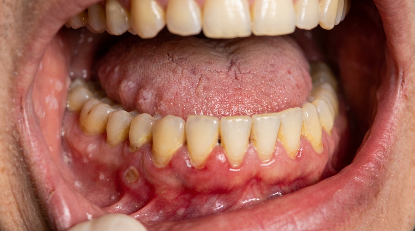
Medicine side-effects. Calcium channel blockers cause gingival overgrowth (the gum tissue grows thicker and more fibrous than normal) in some patients. ACE inhibitors and beta-blockers can produce lichenoid reactions (white-streaked, sometimes ulcerated areas of the lining of the mouth). Some medicines reduce saliva flow, leading to dry mouth and a higher rate of decay.
Bleeding effects. Antiplatelets and anticoagulants slow clotting; the gum tissue, with its constant low-grade inflammation, often shows the effect first.
Bacterial spread. Bacteria that live around the gums can enter the bloodstream during chewing, brushing and dental procedures. In a small number of high-risk patients, these bacteria can settle on a damaged heart valve and cause infective endocarditis.
Reduced perfusion. Perfusion (the flow of oxygenated blood through tissue) drops in heart failure or cyanotic congenital disease, giving the lips and gums a bluish tinge.
Stress and anxiety of dental visits, which can trigger angina or arrhythmia (an abnormal heart rhythm) in susceptible patients.
How does it develop?
Each oral effect develops in its own way. A typical pattern for a long-standing cardiac patient might be:
First medications are prescribed for blood pressure or rhythm.
Mild dry mouth or taste change is noticed within a few weeks.
Gum changes (overgrowth or bleeding) appear over months as plaque accumulates.
Lichenoid changes of the cheek lining or tongue may develop after many months on certain medicines.
Bleeding from the gum when brushing becomes more noticeable as antiplatelet or anticoagulant therapy is added.
Cyanosis appears in advanced heart failure or cyanotic congenital disease.
Each of these can be addressed individually, but the picture often makes more sense when the dental and medical teams talk to each other.
What might you notice?
Common things people notice include:
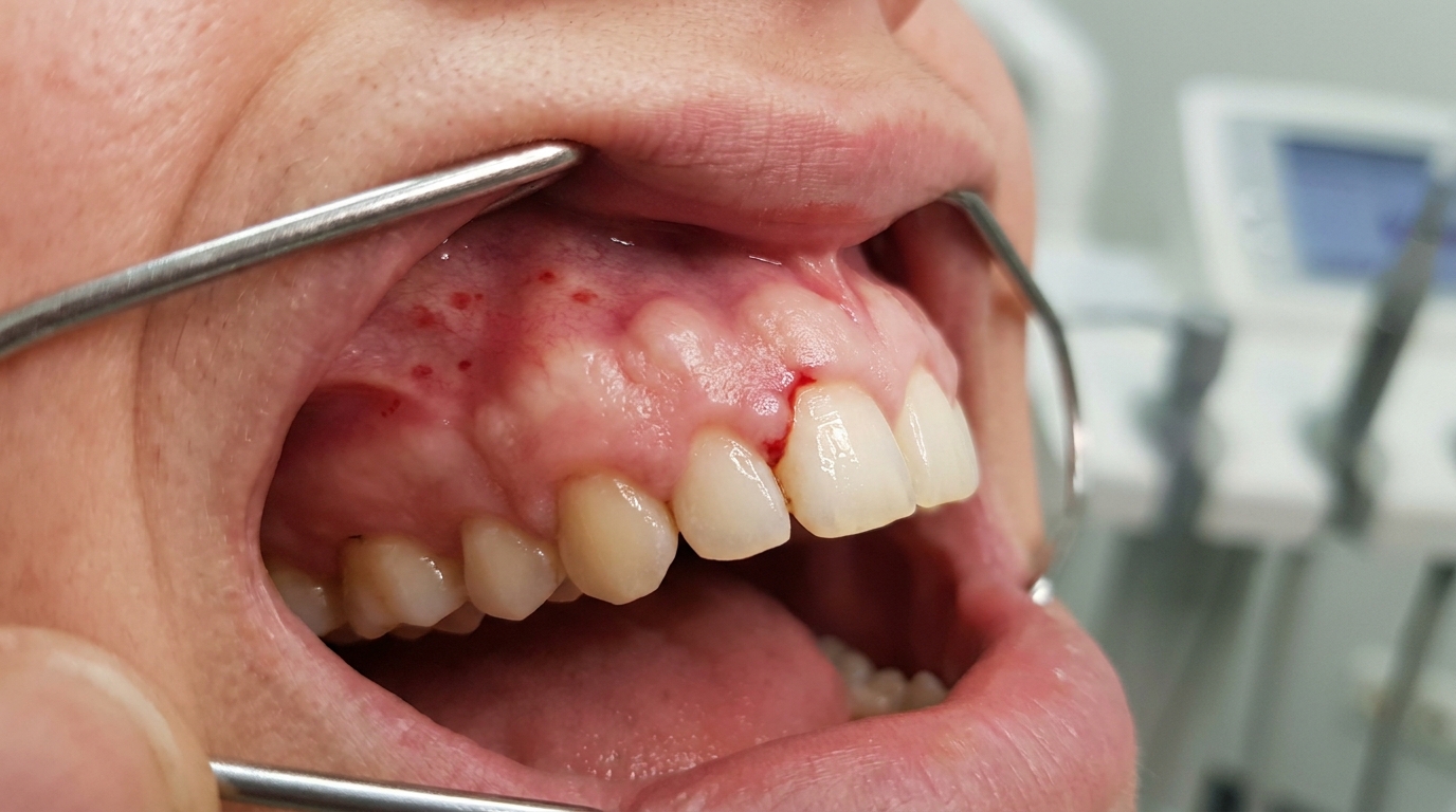
Bulky, fibrous gum overgrowth, especially between the front teeth.
Easy gum bleeding with brushing or flossing.
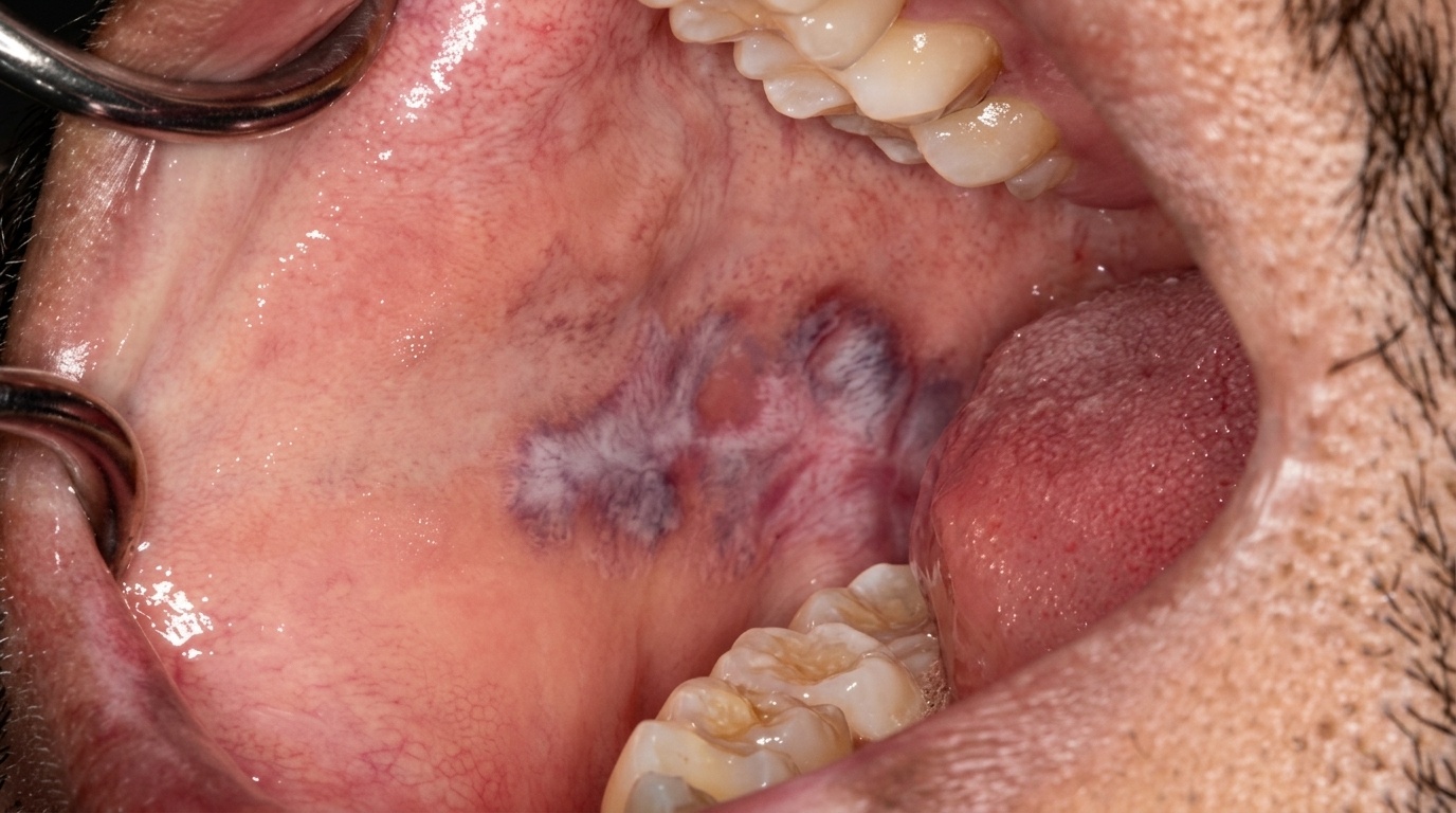
Bruising or red spots on the soft palate or inside the cheek.
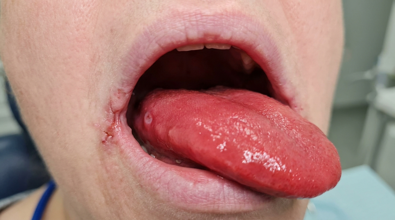
Lacy white streaks or ulceration on the cheek lining or tongue (lichenoid reaction).
Persistent dry mouth with difficulty swallowing dry foods.
Altered taste, sometimes a metallic or bitter feel.
Pale or bluish gums and lips, more noticeable in cold weather or after exertion.
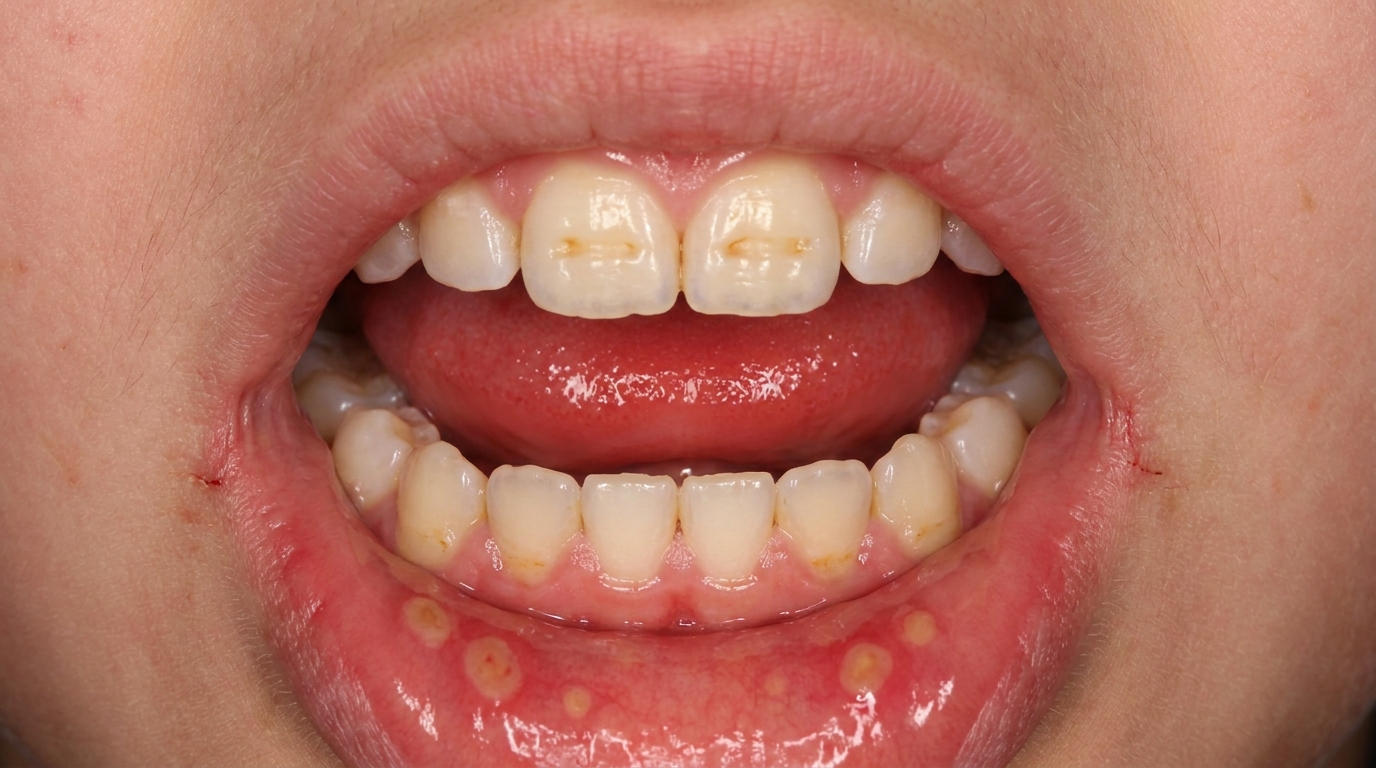
Increased dental decay, especially around the gum line.
What happens at the dentist?
A dental visit for a patient with cardiac disease is planned carefully. At ArtSmiles, a typical visit involves:
A detailed medical history. Heart conditions, medicines, recent hospital admissions, and any planned procedures.
Communication with your cardiologist or GP when needed, to confirm the safest plan and to clarify any antibiotic prophylaxis or anticoagulant decisions.
A careful oral examination, including the gums, palate, cheek lining and tongue, with attention to the patterns described above.
Blood pressure and pulse check, particularly before procedures.
A reduced stress experience, short, comfortable appointments, especially in the morning when many patients feel best, with breaks as needed.
Conservative use of adrenaline in local anaesthetic for patients with severe heart conditions; profound anaesthesia is still important, but the technique is adjusted.
Local measures for bleeding, careful suturing, oxidised cellulose, tranexamic acid (a mouth rinse that helps stabilise clots) instead of routinely stopping anticoagulants.
A clear plan for emergencies, with monitored equipment, oxygen and emergency medications on hand.
Is this serious?
🟡 The oral findings on their own are usually not life-threatening. The reasons coordinated care still matters are:
Infective endocarditis can be life-threatening, even though it is rare.
Bleeding and clotting decisions need to be made jointly with the prescribing doctor.
Cardiac events during dental visits, angina, arrhythmia, hypertensive crisis, are uncommon but possible.
Drug-induced changes can become a quality-of-life issue if not addressed.
Reduced oral function affects nutrition, which in turn affects general health.
In short, every cardiac patient benefits from a dental team that understands their full picture.
Could it be something else?
Many of the findings described above can have other causes:
Drug-induced gingival overgrowth can also come from anti-seizure medicines and ciclosporin, not only calcium channel blockers.
Lichenoid reactions can be caused by many medicines and by amalgam contact, not just ACE inhibitors.
Bleeding gums can be a sign of anaemia, vitamin C deficiency, leukaemia or liver disease, not just anticoagulant therapy.
Cyanosis can come from severe lung disease as well as cardiac disease.
Dry mouth has dozens of causes, only some of which are cardiac medicines. See Living with Dry Mouth for the patient-side response.
A careful history, combined with input from your medical team, sorts these out.
How is it treated?
Treatment is shaped to the specific finding:
Drug-induced overgrowth, improved hygiene, professional cleaning, drug substitution where possible, and gingivectomy (a minor surgical trimming of overgrown gum tissue) when needed.
Lichenoid reaction, biopsy if uncertain, topical corticosteroid where symptomatic, and discussion with the prescribing doctor about alternatives.
Dry mouth, saliva substitutes, frequent water sips, sugar-free chewing gum, fluoride support and short recall intervals.
Bleeding management, local measures rather than stopping blood thinners for most procedures.
Infective endocarditis prevention, outstanding daily oral hygiene, regular dental cleanings, and antibiotic prophylaxis only for the small high-risk group as defined by current guidelines.
The single most powerful preventive measure for cardiac patients is excellent daily oral hygiene, twice-daily brushing with fluoride toothpaste and daily interdental cleaning. Gum health reduces the bacterial load that can enter the bloodstream and lowers the chance of infective endocarditis.
What's the long-term outlook?
The long-term outlook for the oral findings is generally good when the dental and medical teams work together. Most drug-induced changes can be managed without changing essential medicines. Bleeding can almost always be controlled with local measures. Infective endocarditis remains rare, especially in patients who keep their mouth healthy and attend regularly.
If you have any heart condition, please tell us at every appointment, even if it has been mentioned before. Cardiac status changes over time, and the safest dental visit is one based on your most recent medical picture.
A note on this article
This article is for educational purposes only and does not constitute a clinical diagnosis. Please consult a registered dental practitioner for assessment and treatment advice.
The cover image above is an AI-generated illustration based on the most common visible features of this condition described in clinical pathology references. It is not a photograph of a real case and should not be used to diagnose or rule out the condition in your own situation. If you are concerned about something you have noticed, please book an assessment with a registered dental practitioner.
References
Neville, B. W., Damm, D. D., Allen, C. M., & Chi, A. C. (2023). Oral and maxillofacial pathology (5th ed.). Elsevier. Chapter 4, Periodontal Disease: Drug-Influenced Gingival Enlargement.
Cawson, R. A., & Odell, E. W. (2017). Cawson's essentials of oral pathology and oral medicine (8th ed.). Elsevier. Chapter 31, Medical Emergencies and Cardiovascular Disease.
Regezi, J. A., Sciubba, J. J., & Jordan, R. C. K. (2017). Oral pathology: Clinical pathologic correlations (7th ed.). Elsevier. Chapter 5, Connective Tissue Lesions; Chapter 7, Periodontal Disease.