Compiled from clinical pathology references. Medically reviewed by Dr Cristian Dunker, Principal Dentist, ArtSmiles Cosmetic Dentistry.
Quick summary
Also called | Atrophic gingivitis, erosive gingivitis, chronic desquamative gingivitis |
How urgent? | 🟡 Not urgent, but persistent peeling gums need investigation to find the underlying cause |
Common or rare? | Uncommon but characteristic; most often in adults over 40 |
Who it affects | Predominantly middle-aged and older women; often linked to an underlying autoimmune mucocutaneous (affecting both the mouth lining and the skin) disease |
Who treats it | General dentist for initial assessment; oral medicine specialist or oral maxillofacial surgeon for biopsy and diagnosis; GP or dermatologist for systemic management |
Based on | Neville, Cawson and Regezi |
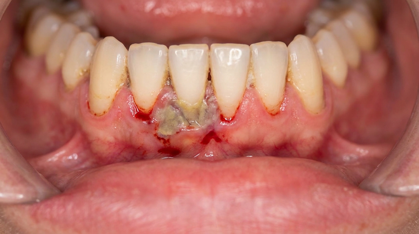
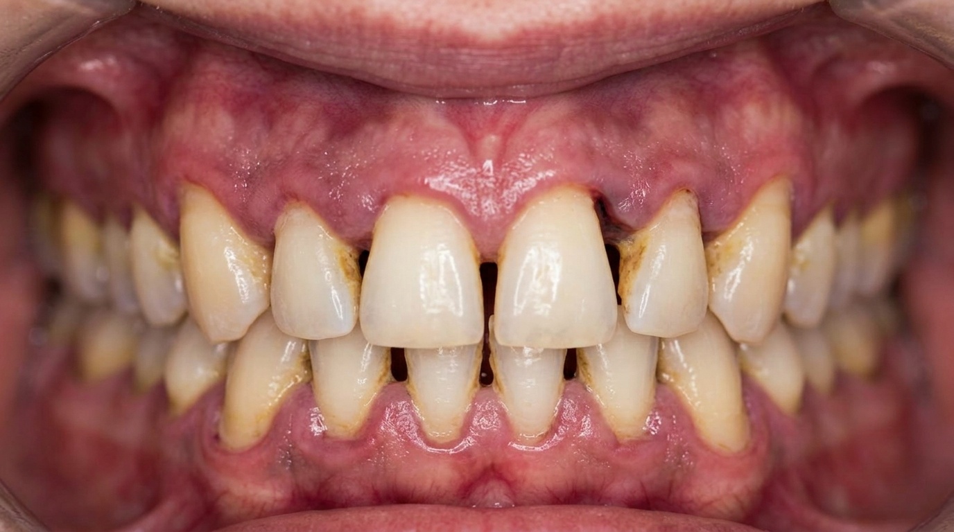
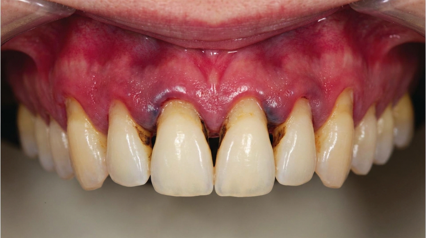
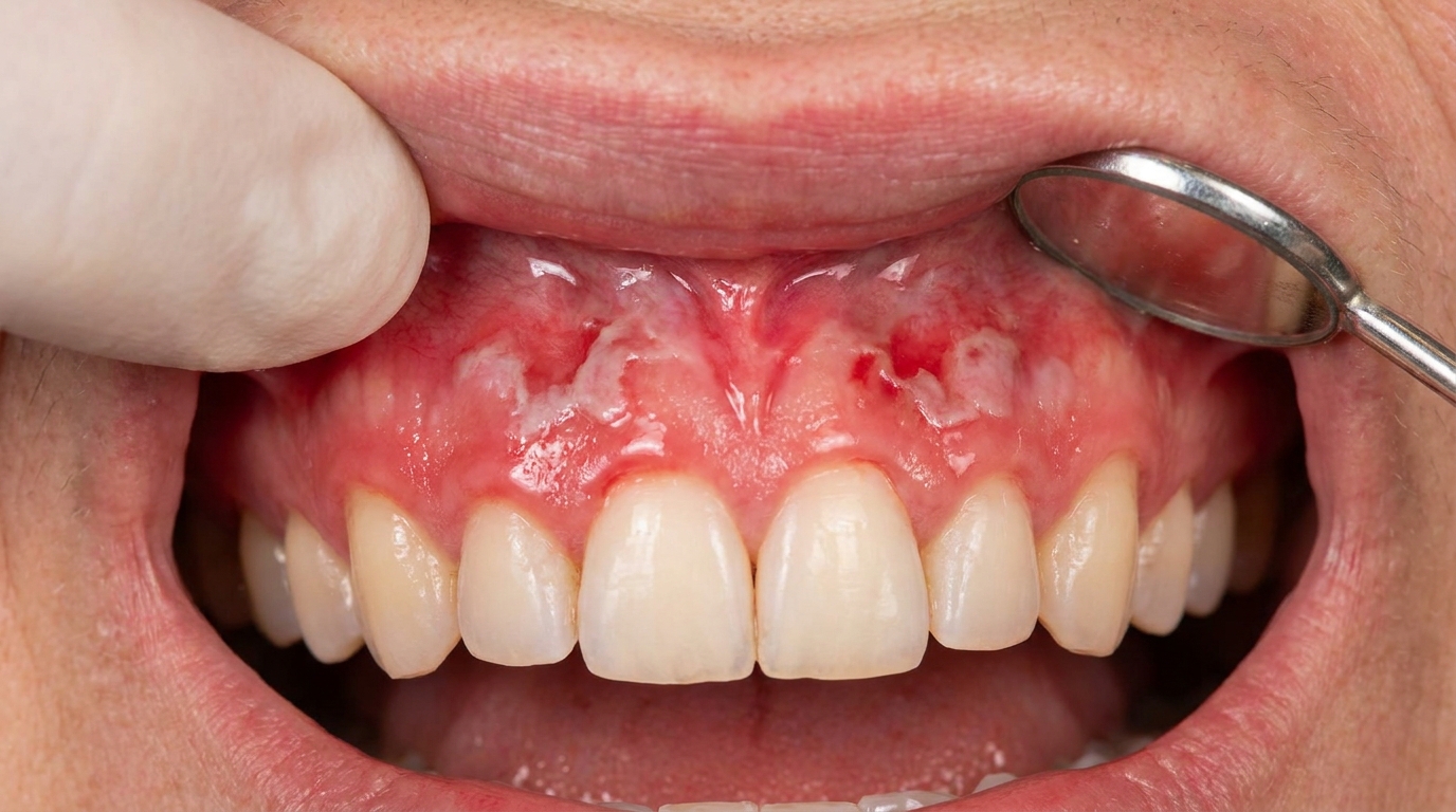
Bright red, glossy gums that look almost like they are peeling, and are often very sore even with the gentlest brushing, are the hallmark of desquamative gingivitis. The name simply means "gum tissue that is shedding its surface layer". It looks dramatic, and it can be uncomfortable, but the most important thing to understand is that it is a sign, not a disease in itself.
This article from the team at ArtSmiles, reviewed by Dr Cristian Dunker, explains what desquamative gingivitis is, what conditions can cause it, and how it is investigated and treated.
What is it?
Desquamative gingivitis is a clinical description of a particular gum appearance:
Generalised redness across the attached gum (rather than only at the gum margin).
Glossy and atrophic (thinned and shiny) surface, thin, shiny, and lacking the normal stippling.
Fragile epithelium that peels away with light pressure, leaving raw red areas.
Tender or burning with brushing, eating spicy food or drinking acidic liquids.
Often involves the upper and lower front gum more obviously than the back.
Plaque-related gingivitis, by contrast, is mostly limited to the gum line and improves quickly with cleaning. Desquamative gingivitis affects a wider area of the gum and does not respond fully to cleaning alone.
Who tends to get it?
Desquamative gingivitis is most often seen in:
Middle-aged and older women, who make up the majority of patients.
Patients with another mucocutaneous disease, even if it has not yet been diagnosed.
Patients on multiple medicines, including some that can themselves trigger lichenoid mucosal reactions.
Patients with a personal or family history of autoimmune disease.
It is uncommon in children and young adults.
What causes it?
Desquamative gingivitis is caused by an underlying mucocutaneous (mouth and skin) disease. The three commonest causes are:
Mucous membrane pemphigoid (MMP). An autoimmune blistering disease in which antibodies attack the structure that holds the surface lining to the underlying tissue. The lining lifts away easily, producing the typical desquamative picture.
Oral lichen planus (atrophic and erosive forms). A chronic immune-mediated inflammation that can produce reddened, eroded gums alongside the more typical lacy white streaks elsewhere in the mouth.
Pemphigus vulgaris. A more serious autoimmune blistering disease that often starts in the mouth before affecting the skin.
Less common causes include:
Lichenoid drug reactions.
Contact reactions to flavourings (cinnamon), toothpaste ingredients or dental materials.
Systemic lupus erythematosus.
Plasma cell gingivitis.
Linear IgA disease, dermatitis herpetiformis and other rare blistering conditions.
The exact diagnosis matters because the treatments differ, and pemphigus, in particular, is a serious condition that benefits from early specialist care.
How does it develop?
The course is gradual:
The underlying immune-mediated process begins to affect the gum lining.
The lining becomes thinner and more fragile.
Even gentle pressure (brushing, eating, suction at the dentist) lifts the surface away.
The patient notices red, sore, easily bleeding gums and avoids brushing, which leads to plaque accumulation, worsening the picture.
Over months, the appearance becomes the persistent picture described above, often prompting a dental visit.
What might you notice?
Common things people notice include:
Red, glossy gums that have changed in appearance over months.
Pain or burning when eating spicy, acidic or hot food.
Difficulty brushing, with bleeding even with a soft brush.
Peeling of the gum surface when wiped or rubbed.
White streaks or lace-like patterns elsewhere in the mouth (a clue to lichen planus).
Blisters or ulcers in other parts of the mouth or on the skin (a clue to a blistering disease).
Eye dryness or genital sores in some patients (a clue to mucous membrane pemphigoid affecting other lining surfaces).
What an X-ray might show
Desquamative gingivitis affects only the surface lining of the gum and does not show on X-rays. Imaging is reserved for ruling out bone changes if a separate periodontal problem is suspected.
What happens at the dentist?
When desquamative gingivitis is suspected at ArtSmiles, the visit usually involves:
A detailed history including duration, triggers, other affected areas (skin, eyes, genitals), medical conditions and medicines.
A careful examination of all the gum surfaces, the rest of the mouth, the lips and the visible skin.
A Nikolsky test in some cases, gentle pressure to see whether the surface lining lifts away, which can hint at a blistering disease.
Photography for the file.
Referral to an oral medicine specialist or oral and maxillofacial surgeon for biopsy. Two samples are usually taken, one for routine pathology, one fresh for direct immunofluorescence (a stain that lights up autoantibodies in tissue), which detects autoantibodies in the lining.
Coordination with your GP and dermatologist if other body areas are involved.
A gentle hygiene plan with soft brush, low-abrasive paste and chlorhexidine mouthwash for symptomatic relief.
Is this serious?
Desquamative gingivitis itself is not life-threatening, but the underlying condition can be serious in some cases:
Pemphigus vulgaris can affect skin and lining surfaces throughout the body and may be life-threatening if untreated.
Mucous membrane pemphigoid can cause scarring of the eye lining (with risk to vision) and other lining surfaces.
Severe lichen planus has a small but recognised risk of malignant transformation, requiring long-term follow-up.
The reason desquamative gingivitis deserves prompt attention is to identify and treat the underlying disease early.
Could it be something else?
Several conditions look or feel similar:
Plaque-related gingivitis. Marginal redness limited to the gum line, settling with cleaning.
Acute necrotising ulcerative gingivitis. Painful, ulcerated gum margins with grey slough, more sudden in onset.
Burning mouth syndrome. Burning sensation without visible lining change.
Allergic contact reaction. Confined to the area touching the trigger (a flavoured toothpaste, mouthwash, or dental material).
Erythroplakia. A persistent red patch with malignant potential, usually localised rather than generalised.
Anaemia. Pale rather than red gums with smooth tongue.
Vitamin C or other deficiencies. Bleeding swollen gums and other systemic signs.
This is why a specialist biopsy is so important.
How is it treated?
Treatment is directed at the underlying cause once a diagnosis is made:
Topical corticosteroids in custom trays, gels or rinses are the mainstay for lichen planus and many cases of mucous membrane pemphigoid. They reduce immune activity locally without major systemic effects.
Systemic immunosuppressive therapy under specialist care for pemphigus vulgaris and severe pemphigoid (corticosteroids, immunosuppressants, biologic therapies).
Identification and removal of triggers for lichenoid drug or contact reactions.
Excellent gentle oral hygiene, ultra-soft brush, low-abrasive paste, careful flossing.
Saliva substitutes for dryness, especially when systemic medicines reduce saliva flow.
Frequent dental cleanings under careful technique to keep plaque levels low.
Long-term follow-up with both dental and medical teams.
Patients with pemphigus and severe pemphigoid will be cared for primarily by a specialist (oral medicine, dermatology or rheumatology), with the dentist providing complementary support.
What's the long-term outlook?
The outlook depends on the underlying condition:
Mucous membrane pemphigoid is usually controllable with topical or low-dose systemic therapy.
Oral lichen planus is a chronic condition that fluctuates over years; long-term follow-up is recommended.
Pemphigus vulgaris has a much better outlook today than in the past, with modern immunotherapy producing lasting remission in many patients.
Throughout, regular dental care plays an important role: keeping plaque to a minimum, maintaining the gum tissue, and watching for any changes that may need further investigation. If you have noticed red, peeling, sore gums that do not respond to cleaning, please book a visit so we can investigate the cause and connect you with the right specialist.
A note on this article
This article is for educational purposes only and does not constitute a clinical diagnosis. Please consult a registered dental practitioner for assessment and treatment advice.
The cover image above is an AI-generated illustration based on the most common visible features of this condition described in clinical pathology references. It is not a photograph of a real case and should not be used to diagnose or rule out the condition in your own situation. If you are concerned about something you have noticed, please book an assessment with a registered dental practitioner.
References
Neville, B. W., Damm, D. D., Allen, C. M., & Chi, A. C. (2016). Oral and maxillofacial pathology (4th ed., Ch. 16: Dermatologic Diseases, Lichen Planus, Mucous Membrane Pemphigoid, Pemphigus Vulgaris). Elsevier.
Cawson, R. A., & Odell, E. W. (2017). Cawson's essentials of oral pathology and oral medicine (8th ed., Ch. 21: Mucocutaneous Diseases). Elsevier.
Regezi, J. A., Sciubba, J. J., & Jordan, R. C. K. (2017). Oral pathology: clinical pathologic correlations (7th ed., Ch. 1: Vesiculobullous Diseases). Elsevier.
Frequently asked questions
What is desquamative gingivitis?
Desquamative gingivitis is a descriptive term for red, sore, peeling gums that 'shed' their surface layer. It is a sign rather than a diagnosis: most cases are caused by an underlying mucocutaneous disease such as oral lichen planus, mucous membrane pemphigoid or pemphigus vulgaris.
Why is desquamative gingivitis painful?
The outer layer of the gum has separated from the deeper tissue, leaving raw, exposed connective tissue. Brushing, acidic foods, spicy meals and even toothpaste foaming agents can sting or burn. Plaque control is harder, which can worsen ordinary gingivitis on top of the underlying condition.
How is desquamative gingivitis diagnosed?
Diagnosis usually requires a small biopsy of involved tissue, often with direct immunofluorescence testing, to identify the underlying autoimmune disease. Blood tests and a careful review of skin and other mucosal surfaces (eyes, genitals, oesophagus) help build the full picture.
How is it managed?
Treatment is two-pronged. The first prong is meticulous gentle oral hygiene with soft brushing and mild toothpaste to control plaque. The second is medical treatment of the underlying disease, often topical or systemic corticosteroids or immunomodulators prescribed in shared care with a dermatologist or oral medicine specialist.