Compiled from clinical pathology references. Medically reviewed by Dr Cristian Dunker , Principal Dentist, ArtSmiles Cosmetic Dentistry.
Quick summary
Also called | Oral signs of coeliac disease, oral features of gluten-sensitive enteropathy, oral manifestations of non-tropical sprue |
How urgent? | 🟡 Worth a check-up, the mouth changes are usually painless, but they can be the first clue to an undiagnosed gut condition that needs medical follow-up |
Common or rare? | Coeliac disease is one of the most common genetic disorders; oral signs are common in those affected |
Who it affects | Children and adults of any age; first-degree relatives of people with coeliac disease are at higher risk |
Who treats it | General dentist often spots the signs; diagnosis and management are led by a GP and gastroenterologist, with dental support |
Based on | Cawson, Neville, Regezi, Laskaris |
What is it?
Coeliac disease is a long-term reaction to gluten, a protein found in wheat, barley and rye. In people with the condition, eating gluten triggers the immune system to damage the lining of the small bowel, which can stop the body absorbing nutrients properly.
While coeliac disease is mainly a gut condition, it often shows up in the mouth as well. The dentist may, in fact, be the first health professional to spot the signs, sometimes long before any tummy symptoms appear.
Who tends to get it?
Coeliac disease is one of the most common genetic disorders. It can begin in childhood but is frequently silent for years, only being recognised in adult life when complications develop.
It runs in families. First-degree relatives (parents, siblings, children) of someone with coeliac disease have a notably higher risk and are themselves more likely to show enamel defects (uneven or pitted enamel) on their teeth, even when their bowel symptoms are mild or absent.
Both men and women are affected. The oral changes can appear at any age, although the symmetrical enamel defects develop only while the teeth are forming, so they are most often noticed in children, teenagers and young adults.
What causes it?
The underlying cause is an inherited sensitivity to gluten. Almost all people with coeliac disease carry one of two specific tissue-type genetic markers (HLA-DQ2 or HLA-DQ8, the same kind of markers used in tissue matching for transplants), which is why the condition clusters in families.
When gluten is eaten, the immune system mistakenly treats it as a threat and attacks the lining of the small intestine. Over time this flattens the tiny finger-like projections (villi) that absorb food, leading to malabsorption (poor uptake of nutrients from the gut).
The oral changes are usually not caused by gluten directly. Instead, they reflect the knock-on effects of poor absorption, particularly shortages of iron, folate (folic acid) and vitamin B12, and, in growing children, disruption of the cells that build tooth enamel.
How does it develop?
Think of the lining of the small bowel as a shaggy carpet of tiny absorbent fibres. In coeliac disease, gluten triggers the immune system to mow that carpet flat, leaving a smooth surface that cannot soak up nutrients properly.
When iron, folate or vitamin B12 run low, the cells that line the mouth, which are among the fastest-renewing cells in the body, are some of the first to suffer. The tongue can lose its normal velvety surface, the corners of the mouth can crack, and recurrent ulcers may break out on the cheeks and lips.
In children, there is a second mechanism. While the permanent teeth are still forming under the gum, the cells that lay down enamel (called ameloblasts) are sensitive to upsets in calcium balance and to the immune disturbance of coeliac disease itself. If the disease is active during a particular window of tooth development, that window can leave a permanent fingerprint on the enamel, symmetrical, on matching teeth on both sides of the mouth, at the level the teeth had reached at the time.
What might you notice?
What it looks like
Several different changes can occur, often together:
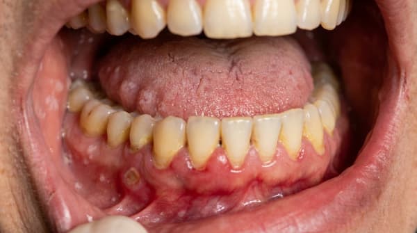
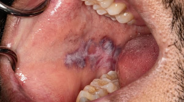
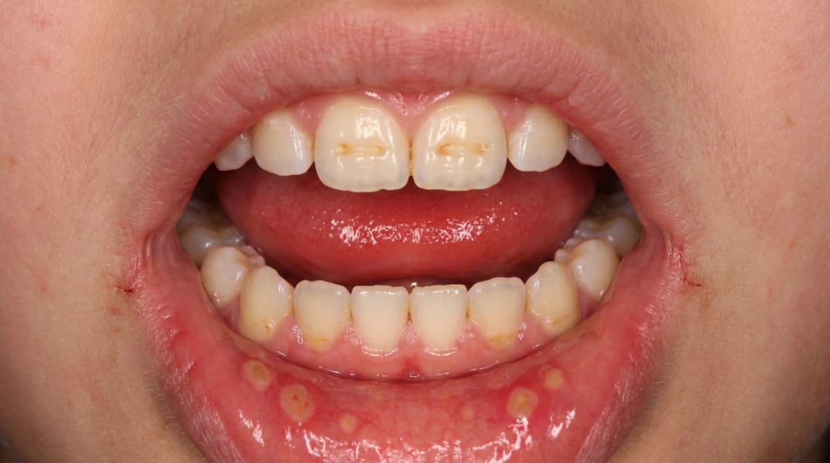
Recurrent mouth ulcers (aphthae). These are the most common oral feature. Round or oval ulcers, 5,10 mm across, with a yellowish floor and a red rim, appear on the inner cheeks, lips, under-surface of the tongue, soft palate or floor of the mouth. They tend to come back every few weeks and heal in 7,14 days.
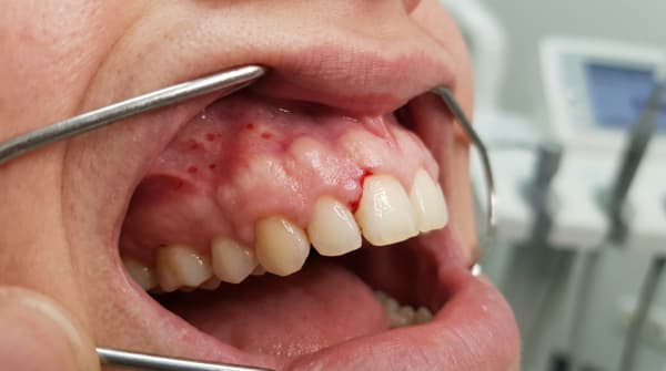
Enamel defects on the permanent teeth. Bands or patches of chalky white, cream, yellow or brown enamel that run horizontally across matching teeth on both sides of the mouth. Sometimes there are pits, grooves or thinner areas. Front teeth and first molars are most often affected.
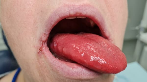
A smooth, red, sore tongue (atrophic glossitis), smooth, thinned, sore tongue. The dorsal surface of the tongue can lose its normal texture as the small papillae wear away, giving a glassy or beefy-red appearance.
Cracked, sore corners of the mouth (angular cheilitis). Red, fissured or crusted areas at one or both corners of the lips.
Delayed eruption of the baby or adult teeth in some children.
More dental decay than expected, particularly where pitted enamel is hard to keep clean.
What it feels like
Mouth ulcers can be sharply painful, particularly when eating acidic, spicy or salty foods. A sore tongue may sting or burn, and the mouth can feel generally tender. The enamel defects themselves are usually painless, but exposed or pitted enamel may feel sensitive to cold or sweet foods. Many people have no oral pain at all and only notice cosmetic changes, discoloured patches on the front teeth, or a smoother-looking tongue.
What an X-ray might show
Dental X-rays may show enamel that is thinner or less dense than normal in a symmetrical pattern across matching teeth, and there may be more decay than expected for the patient's age and oral hygiene. In children, X-rays sometimes show that several adult teeth have been slow to erupt without an obvious local cause.
What happens at the dentist?
A dentist at ArtSmiles who suspects coeliac disease will usually start by listening, asking about recurrent mouth ulcers, the family history, any tummy symptoms, growth in childhood, and whether anyone else in the family has been diagnosed.
They will then examine the lips, cheeks, tongue and palate, and look closely at the teeth for the symmetrical, chronological enamel pattern (often called Aine-type defects, after the researcher who first described them in coeliac disease). They may take photographs and X-rays to map which teeth are affected.
If the picture is suggestive, the dentist will typically recommend a referral to a GP for blood tests. The standard screening tests include anti-tissue transglutaminase (anti-tTG) antibodies, anti-endomysial antibodies (EMA) and a total IgA level, these are the markers that point to gluten sensitivity. A confirmed diagnosis usually requires a small-bowel biopsy carried out by a gastroenterologist.
It is important not to start a gluten-free diet before testing, because doing so can hide the disease and lead to a false-negative result.
Is this serious?
🟡 Worth a check-up, but not an emergency. The oral changes themselves are not dangerous. What matters is what they may be pointing to. Untreated coeliac disease can lead to long-term anaemia, weakened bones, fertility problems, and a small but real increase in the risk of certain bowel cancers. In children, it can affect growth and development.
The good news is that, once recognised, coeliac disease is highly manageable. A strict, lifelong gluten-free diet usually settles the bowel inflammation, restores normal absorption, and, in most cases, calms the recurrent mouth ulcers, glossitis and angular cheilitis as well. The enamel defects on already-formed teeth are permanent, but no new defects appear once the diagnosis is made and the diet is followed.
If you've noticed any of these signs for more than two weeks, it's worth booking an assessment.
Could it be something else?
Many conditions can cause similar oral signs. Some of the more important ones:
Recurrent canker-like (aphthous) stomatitis, recurrent ulcers identical to those of coeliac disease, but most cases occur in otherwise healthy people. The dentist may suggest blood tests if attacks start in adult life or worsen, to look for underlying causes including coeliac disease, iron, folate or vitamin B12 deficiency.
Crohn's disease, another gut condition associated with mouth ulcers, but typically also produces cobblestone swelling of the cheeks, tag-like folds in the sulcus and lip swelling, which are not features of coeliac disease.
Iron-deficiency anaemia, also produces a smooth, sore tongue and angular cheilitis, but without the symmetrical enamel pattern. Blood tests separate the two; coeliac disease is one of the causes of the anaemia.
Pernicious anaemia (vitamin B12 deficiency), gives a beefy-red, atrophic tongue much like that of coeliac disease, but is caused by a different absorption problem in the stomach. Specific blood tests distinguish them.
Plummer-Vinson syndrome, iron deficiency with glossitis, angular cheilitis and difficulty swallowing due to oesophageal webs (thin tissue folds in the food pipe). Distinguished by the swallowing symptoms and characteristic spoon-shaped fingernails.
Behçet's syndrome, recurrent mouth ulcers along with genital ulcers, eye inflammation and joint or skin involvement, none of which are features of coeliac disease.
Erythema migrans (geographic tongue), has been linked statistically with coeliac disease in some studies, but typically presents as moving map-like patches rather than a uniformly smooth red tongue.
Dermatitis herpetiformis, a very itchy blistering skin rash on the elbows, knees and buttocks. It is now considered the skin manifestation of coeliac disease, and most people with it have the same gut sensitivity to gluten. Oral lesions can occur and may resemble aphthae.
Environmental enamel hypoplasia from other causes, symmetrical enamel defects can also follow childhood fevers, severe infections, certain medications, premature birth or other systemic illnesses. The pattern alone cannot prove coeliac disease; blood tests are needed.
Amelogenesis imperfecta, a hereditary enamel disorder that affects all the teeth fairly uniformly, rather than a chronological band, and runs in families with a clear inheritance pattern.
Dental fluorosis, diffuse white flecks or patches across the enamel, linked to fluoride intake during tooth formation; usually lacks the sharp horizontal banding and is not associated with bowel symptoms.
How is it treated?
The core treatment for coeliac disease is medical, not dental: a strict, lifelong gluten-free diet, supervised by a GP, gastroenterologist and dietitian. Once gluten is removed, the bowel lining heals over weeks to months, nutrient absorption normalises, and most of the oral signs improve.
At home, what helps:
Following the gluten-free diet consistently, even small amounts of gluten can keep symptoms going.
Brushing twice a day with a fluoride toothpaste and flossing between teeth, paying particular attention to pitted or grooved areas where plaque collects.
Using a soft-bristled toothbrush during a flare of mouth ulcers or sore tongue.
Avoiding sodium lauryl sulfate-containing toothpastes if mouth ulcers seem triggered by them.
Keeping well hydrated and eating a varied, nutrient-rich diet within the gluten-free framework.
At the dentist, treatment may include:
A thorough check-up and clean to map enamel defects, identify decay, and reinforce the enamel where possible.
Application of fluoride varnish or sealants to vulnerable surfaces.
A simple filling for any cavities that have formed in pitted enamel.
For visible discolouration on front teeth, cosmetic options such as resin infiltration, microabrasion, composite bonding or veneers may be discussed once the patient is old enough and the rest of the dentition has stabilised.
Topical treatments for recurrent ulcers, for example, a chlorhexidine mouthwash, or a topical corticosteroid prescribed for short-term use during a flare.
Management of angular cheilitis with antifungal or antibacterial creams if a secondary infection is present.
Referral to a GP for coeliac blood tests, and to a paediatric or restorative specialist where the dental impact is extensive.
Where a child with confirmed coeliac disease is still developing, regular dental review every six months helps catch new decay early and supports the parents in keeping the gluten-free diet on track from a dental perspective.
What's the long-term outlook?
The outlook is good when coeliac disease is recognised and managed. A strict gluten-free diet usually settles the recurrent ulcers, restores the tongue's normal surface, clears the cracked corners of the mouth, and prevents any new enamel defects forming on teeth still developing under the gum.
The enamel defects that have already formed are permanent, enamel cannot regrow once it is laid down, but they are very treatable cosmetically and functionally.
For adults diagnosed later in life, ulcers and glossitis often improve substantially within a few months of going gluten-free, especially as iron, folate and vitamin B12 levels recover. People who continue to eat gluten, knowingly or not, tend to have ongoing mouth symptoms as well as ongoing gut problems, which is why follow-up with the GP and dietitian is so important.
From the dental side, the most important long-term step is regular check-ups. A dentist familiar with the condition can monitor the enamel, manage any decay early, support good home care, and stay alert for signs that gluten exposure may be slipping back in.
A note on this article
This article is for educational purposes only and does not constitute a clinical diagnosis. Please consult a registered dental practitioner for assessment and treatment advice.
The cover image above is an AI-generated illustration based on the most common visible features of this condition described in clinical pathology references. It is not a photograph of a real case and should not be used to diagnose or rule out the condition in your own situation. If you are concerned about something you have noticed, please book an assessment with a registered dental practitioner.
References
Cawson, R. A., & Odell, E. W. (2017). Cawson's essentials of oral pathology and oral medicine (8th ed.). Elsevier. Chapter 29, Gastrointestinal and liver disease (Coeliac disease, p. 393); Chapter 13, Soft tissue disease (Recurrent aphthae, pp. 222 to 224); Chapter 2, Disorders of development of the teeth and related tissues (p. 39).
Neville, B. W., Damm, D. D., Allen, C. M., & Chi, A. C. (2023). Oral and maxillofacial pathology (5th ed.). Elsevier. Chapter 9, Allergies and Immunologic Diseases (Recurrent Aphthous Stomatitis, pp. 322 to 324); Chapter 2, Abnormalities of Teeth (environmental enamel hypoplasia and delayed eruption, pp. 52 to 72); Chapter 17, Oral Manifestations of Systemic Diseases (Iron-Deficiency Anemia and celiac sprue, pp. 830 to 832).
Regezi, J. A., Sciubba, J. J., & Jordan, R. C. K. (2017). Oral pathology: Clinical pathologic correlations (7th ed.). Elsevier. Chapter 2, Ulcerative Conditions (Aphthous ulcers and gluten-sensitive enteropathy, pp. 38 to 40); Chapter 1, Vesiculobullous Diseases (Dermatitis herpetiformis and coeliac disease, pp. 18 to 19).
Laskaris, G. (2003). Color atlas of oral diseases (3rd ed.). Thieme. Chapter 22, Skin Diseases (Dermatitis herpetiformis and gluten-sensitive enteropathy, pp. 212 to 214; Aine-type dental enamel defects, p. 353).
Frequently asked questions
What oral signs can coeliac disease cause?
Coeliac disease can cause symmetrical enamel hypoplasia of the permanent teeth (especially incisors and first molars), recurrent aphthous ulcers, glossitis (a sore, atrophic tongue), angular cheilitis and delayed tooth eruption in children. Dentists sometimes notice these signs before the diagnosis is made elsewhere.
Why does coeliac affect the teeth?
Coeliac disease damages the small intestine and impairs absorption of calcium, vitamin D, iron, vitamin B12 and folate. If the damage is present during tooth formation in childhood, the developing enamel is laid down poorly, producing the characteristic pitted, banded or discoloured enamel of the permanent teeth.
How is coeliac disease diagnosed?
Diagnosis is by blood tests (anti-tissue transglutaminase IgA, anti-endomysial antibodies, total IgA) while the patient is still eating gluten, followed by a duodenal biopsy in most adults. Genetic testing (HLA-DQ2/DQ8) is sometimes used to rule out the diagnosis.
What does treatment involve?
Treatment is a strict, lifelong gluten-free diet. Most oral symptoms (ulcers, glossitis, angular cheilitis) improve once the diet is established. Enamel defects already present in the teeth are permanent and may need cosmetic restoration. Recurrent unexplained mouth ulcers, especially in children, should prompt coeliac screening.