Compiled from clinical pathology references. Medically reviewed by Dr Cristian Dunker , Principal Dentist, ArtSmiles Cosmetic Dentistry.
Quick summary
Also called | Focal hyperkeratosis, frictional hyperkeratosis, alveolar ridge keratosis, contact keratosis |
How urgent? | 🟡 Worth a check-up, usually harmless, but a dentist needs to confirm the cause and rule out look-alikes |
Common or rare? | Very common |
Who it affects | Adults of any age, particularly people with sharp teeth, ill-fitting dentures, or chewing habits; also seen on edentulous (without teeth) (toothless) ridges |
Who treats it | General dentist |
Based on | Regezi, Neville, Cawson, Laskaris |
What is it?
Frictional keratosis is a white patch inside the mouth that develops when an area of soft tissue is rubbed or chafed over and over. The mouth lining responds to this constant friction the same way the skin on your hand responds to gripping a tool, it builds up an extra layer of keratin (the tough protein that makes up calluses, fingernails and the outer skin layer) to protect itself, and that thickened layer looks white when wet with saliva.
In other words, it is essentially a callus inside the mouth. There is no cancer risk attached to a true frictional keratosis, and once the source of rubbing is removed, the patch usually fades on its own.
Who tends to get it?
Frictional keratosis is one of the most common white patches seen during routine dental examinations at ArtSmiles. It can happen at any age but tends to appear in adults whose mouths have something repeatedly rubbing the lining, a sharp or broken tooth, a denture clasp, an edge of restoration, or a long-standing habit such as cheek chewing or tongue thrusting.
People who wear dentures are particularly prone to it, especially along edentulous (toothless) ridges where the gums are squeezed against food during chewing. The patches often turn up on the cheeks along the bite line, the lateral edges of the tongue, the lips, and the gum ridges where teeth used to sit. If a denture is the source of the rubbing, addressing the poor fit or pressure points is usually the first step.
What causes it?
The single underlying cause is repeated low-grade trauma to one spot of mouth lining. The trauma can come from many places:
A sharp or broken tooth, or a rough filling edge that scrapes the cheek or tongue.
A denture that does not fit well and rubs the gum or vestibule.
Chronic cheek, lip, or tongue chewing, sometimes a stress habit, sometimes done unconsciously during sleep.
A tongue-thrusting habit, where the tongue is constantly pushed against the inside of the front teeth.
Chewing on the gum ridge in areas where teeth are missing, particularly with hard or crusty foods.
An ill-fitting orthodontic appliance or partial denture clasp pressing on one spot.
It is not caused by smoking, alcohol, infection, or any inherited condition, although those things can produce other white patches that look similar, which is why a careful examination matters.
How does it develop?
The lining of the mouth is a layered tissue, with a surface layer that can thicken in response to wear. When the same patch of lining is rubbed day after day, the surface cells respond by laying down extra keratin, the tough protein that makes up calluses on the feet and the outer skin of the heel.
As that keratin layer grows, it traps saliva and reflects light differently from the surrounding pink mucosa (the soft tissue lining of the mouth), so the area starts to look opaque and white. Underneath, the deeper layers of the tissue stay normal, there is no abnormal cell growth, no dysplasia (microscopic changes that can signal an increased cancer risk), just a thickening of the protective surface. Stop the friction, and the body gradually sheds the extra keratin and the patch fades.
What might you notice?
What it looks like
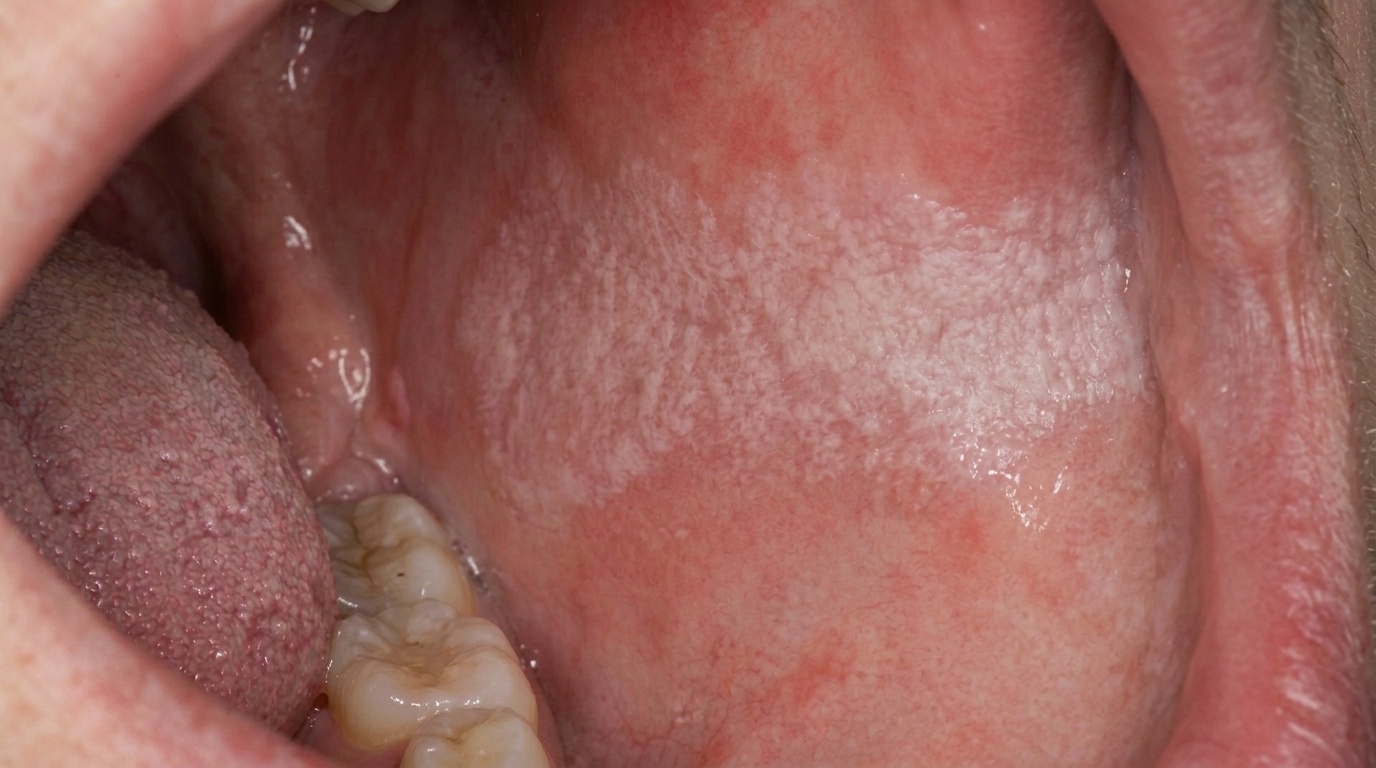
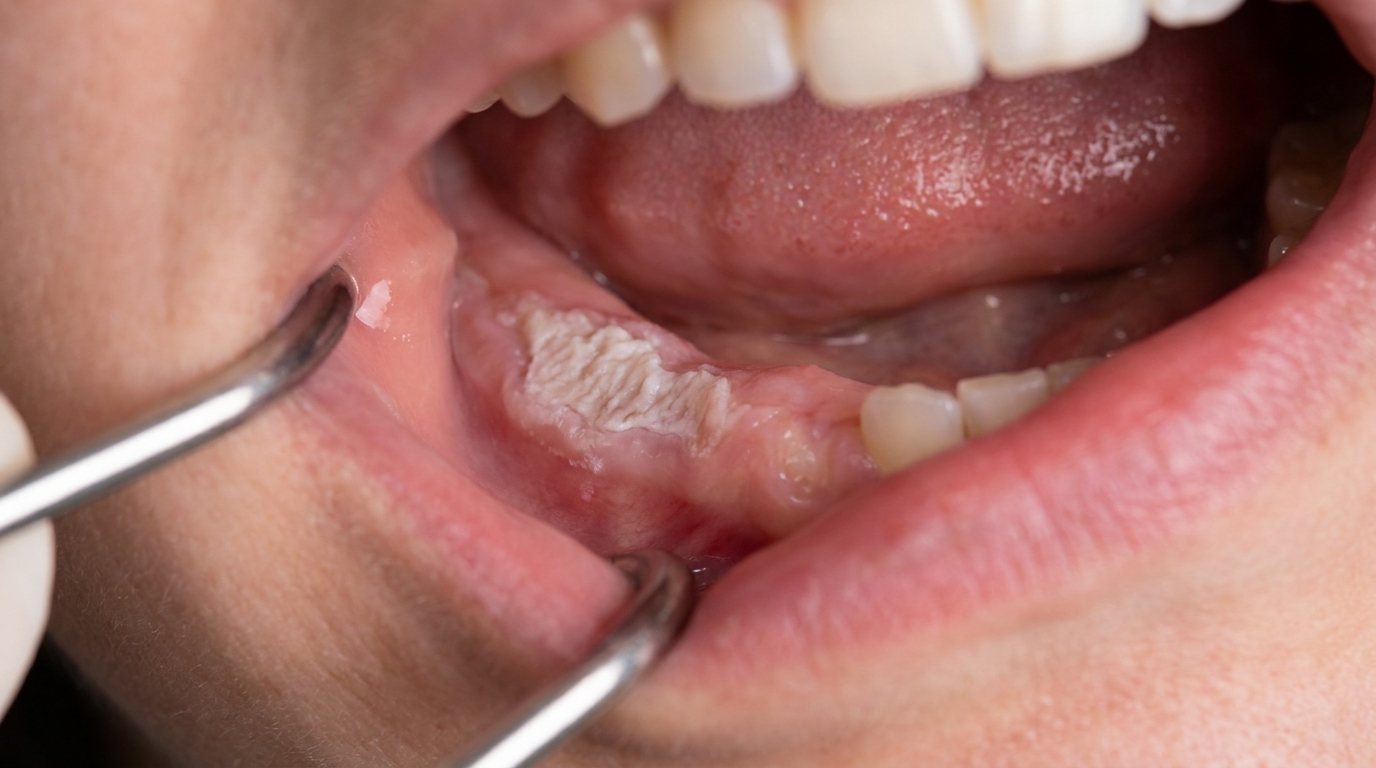
Frictional keratosis usually appears as a white or grey-white patch that is firmly attached to the underlying tissue. It cannot be wiped or scraped away with gauze, which separates it from things like thrush.
The edges of the patch are often poorly defined, it tends to blend gradually into the surrounding pink tissue rather than having a sharp border. The surface can be smooth and slightly opalescent (with a faint milky shine) in the early stages, or rough, shaggy, and ragged when chronic cheek or tongue biting is involved. Patches along the cheek bite line often run as a horizontal stripe, while those caused by a sharp tooth tend to sit directly opposite the offending edge.
What it feels like
Most frictional keratoses are completely painless. Patients are often surprised when a dentist points the patch out, because they had no idea it was there. Occasionally the area can feel slightly rough to the tongue, and if cheek biting is the cause, you may notice the shredded surface texture when running your tongue across the inside of the cheek.
What an X-ray might show
Frictional keratosis is a soft-tissue change only and does not show up on dental X-rays. Imaging is generally not needed for diagnosis, although your dentist may take routine X-rays to look for the underlying cause, for example, a fractured tooth or an over-erupted molar that is hitting the opposite gum.
What happens at the dentist?
The diagnosis of frictional keratosis is largely a clinical one, made by combining what your dentist sees in the mouth with what you describe about your habits and history.
A typical assessment includes:
A careful look at the patch itself, its location, size, surface texture, and whether it can be wiped off.
An examination of the tissue immediately opposite, to identify a sharp tooth, rough filling, or denture edge that lines up with the lesion.
Questions about chewing, biting, or tongue habits, denture wear, and how long the patch has been there.
A check of the rest of the mouth, since look-alike conditions such as lichen planus or leukoplakia often have features elsewhere.
If an obvious source of friction is identified, your dentist will usually recommend addressing it first and reviewing the patch in two to four weeks. A frictional keratosis should noticeably soften or resolve once the cause is removed. If the patch persists despite removal of the irritant, a biopsy may be recommended to be certain that nothing else is going on underneath.
Is this serious?
🟢 Usually harmless. A genuine frictional keratosis has no malignant potential, the surface is simply thickened, the underlying cells remain normal, and there is no documented evidence that ongoing minor trauma alone causes oral cancer.
That said, the appearance of frictional keratosis can closely mimic more serious white patches, including early leukoplakia. The reason a dental review matters is not that the keratosis itself is dangerous, but that confirming the diagnosis means ruling out the look-alikes.
If you have noticed a white patch in your mouth that has been there for more than two weeks, it is worth booking an assessment.
Could it be something else?
Several other conditions can produce a white patch that looks similar to frictional keratosis. Your dentist works through these differentials carefully before settling on a diagnosis.
Leukoplakia, a white plaque with no obvious cause that is considered potentially malignant. It looks similar because both appear as firmly attached white patches that cannot be wiped off. A dentist tells them apart by looking for an identifiable source of friction (present in keratosis, absent in leukoplakia) and by whether the patch resolves once that source is removed.
Oral lichen planus, an immune-related condition that often appears as a lacy white network on the cheeks. It can look similar where a reticular (net-like) pattern overlaps the bite line. A dentist tells them apart by the bilateral, symmetrical distribution of lichen planus and the presence of fine radiating white striae rather than a uniform thick patch.
Linea alba, a normal horizontal white line along the cheek where the teeth meet. It looks similar because both run along the bite plane. A dentist tells them apart by the symmetrical, narrow, smooth appearance of linea alba, which is considered a normal variant rather than a lesion.
Morsicatio buccarum (chronic cheek biting), a closely related condition caused by deliberate or habitual chewing of the cheek lining. It looks similar because both involve frictional thickening. A dentist tells them apart by the shredded, ragged, peeling surface of morsicatio compared with the smoother, more uniform plaque of frictional keratosis.
White sponge naevus, an inherited condition causing thick, folded, spongy white lesions in the mouth. It looks similar because both are persistent white plaques. A dentist tells them apart by the family history, early age of onset, bilateral and symmetrical distribution, and characteristic spongy folded texture of white sponge naevus.
Candidal leukoplakia (chronic hyperplastic candidiasis), a yeast-driven thickened white plaque, often at the corners of the mouth. It looks similar because both produce a stuck-on white area. A dentist tells them apart by examining for predisposing factors such as denture wear or smoking, and by the response of candidal lesions to antifungal therapy.
Hairy leukoplakia, a corrugated white patch on the side of the tongue associated with immune compromise, particularly HIV infection. It looks similar because both can affect the lateral tongue. A dentist tells them apart by the vertically corrugated, shaggy surface of hairy leukoplakia and the patient's broader medical context.
Leukoedema, a faint, milky, filmy greyish appearance of the cheek lining that is considered a normal variant. It looks similar because both produce a whitish overlay on the cheek. A dentist tells them apart by stretching the cheek, leukoedema disappears when the mucosa is pulled taut, while frictional keratosis does not.
Chemical burn, a white slough caused by something caustic held against the lining, classically aspirin tucked next to a sore tooth. It looks similar in the early stages because both produce a localised white patch. A dentist tells them apart by the history of chemical contact and the fact that a burn often peels or sloughs off, unlike a true keratosis.
Smokeless tobacco keratosis, a wrinkled, pouched white lesion in the area where chewing tobacco or snuff is held. It looks similar because both are caused by local irritation. A dentist tells them apart by the history of smokeless tobacco use and the characteristic location in the mucobuccal fold (the curve where the cheek meets the gum).
Squamous cell carcinoma or epithelial dysplasia, early oral cancer or pre-cancer, which can occasionally present as a persistent white area. It looks similar because both can appear as firm white patches. A dentist tells them apart by the absence of an identifiable cause, the failure to resolve after irritants are removed, and through biopsy if there is any doubt.
How is it treated?
The core principle of treatment is simple: find the cause, remove the cause, and the patch will usually look after itself. Treatment may include:
At home
Becoming aware of and reducing cheek, lip, or tongue biting habits, particularly during periods of stress.
Avoiding very hard or crusty foods if you have an edentulous ridge that is being traumatised by chewing.
Cleaning dentures thoroughly and removing them at night to give the tissues a rest.
At the dental clinic
Smoothing or reshaping a sharp tooth, broken cusp, or rough filling edge that is rubbing the lining.
Adjusting or relining a denture that is pressing on or rubbing one spot.
Replacing or recontouring a worn or fractured restoration.
Providing an occlusal splint or night guard if night-time clenching, grinding, or cheek biting is contributing.
Reviewing the lesion at a follow-up appointment, typically two to four weeks later, to confirm resolution.
If the patch does not improve once the source of friction has been addressed, a biopsy may be recommended. Referral to an oral medicine specialist is appropriate when the diagnosis remains uncertain after initial management, or when a biopsy raises any concern about the underlying tissue.
What's the long-term outlook?
The long-term outlook for frictional keratosis is excellent. Once the source of friction has been removed, most patches fade noticeably within a few weeks and resolve fully over a couple of months as the surface keratin sheds and is replaced by normal lining.
There is no intrinsic risk of malignant change from a true frictional keratosis. The patches can recur, however, if the underlying habit returns, for instance, if cheek biting starts up again during a stressful period, or if a denture begins to fit poorly as the underlying ridge changes shape over time. For this reason, regular dental check-ups are worth keeping up, both to monitor any persistent patches and to spot new sharp edges or denture issues before they cause problems, this kind of watchful, prevention-first review is part of how we look after long-standing patients.
If a patch is being attributed to friction but does not behave like a frictional keratosis, meaning it does not resolve once the cause is removed, that is the moment to investigate further with a biopsy, rather than to keep waiting and watching. Early reassessment is the key to making sure nothing else is being missed.
A note on this article
This article is for educational purposes only and does not constitute a clinical diagnosis. Please consult a registered dental practitioner for assessment and treatment advice.
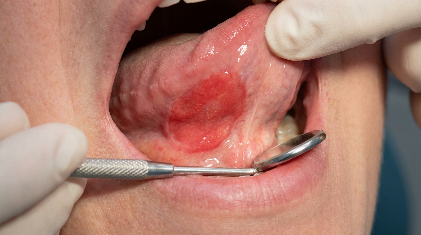
The cover image above is an AI-generated illustration based on the most common visible features of this condition described in clinical pathology references. It is not a photograph of a real case and should not be used to diagnose or rule out the condition in your own situation. If you are concerned about something you have noticed, please book an assessment with a registered dental practitioner.
References
Regezi, J. A., Sciubba, J. J., & Jordan, R. C. K. (2017). Oral pathology: Clinical pathologic correlations (7th ed.). Elsevier. Chapter 3, White Lesions: Focal (Frictional) Hyperkeratosis, pp. 83 to 84.
Neville, B. W., Damm, D. D., Allen, C. M., & Chi, A. C. (2023). Oral and maxillofacial pathology (5th ed.). Elsevier. Chapter 10, Epithelial Pathology: Frictional Keratosis and Alveolar Ridge Keratosis, pp. 383 to 384.
Cawson, R. A., & Odell, E. W. (2017). Cawson's essentials of oral pathology and oral medicine (8th ed.). Elsevier. Chapter 15, Benign Chronic White Mucosal Lesions: Frictional Keratosis and Cheek Biting, pp. 252 to 253.
Laskaris, G. Color atlas of oral diseases. Thieme. Chapter 4, Mechanical Injuries: Chronic Biting, pp. 46 to 47.