Compiled from clinical pathology references. Medically reviewed by Dr Cristian Dunker , Principal Dentist, ArtSmiles Cosmetic Dentistry.
Quick summary
Also called | Oral leukoedema |
How urgent? | 🟢 Not urgent, leukoedema is a benign, normal variation that requires no treatment |
Common or rare? | Very common, reported in 70-90% of black adults and a smaller, more variable proportion of white adults |
Who it affects | Adults of all backgrounds, with a much higher prevalence in people with darker mucosal pigmentation; also more common and more pronounced in smokers |
Who treats it | General dentist for diagnosis and reassurance; no treatment is required |
Based on | Neville, Cawson, with cross-references in Regezi |
What is it?
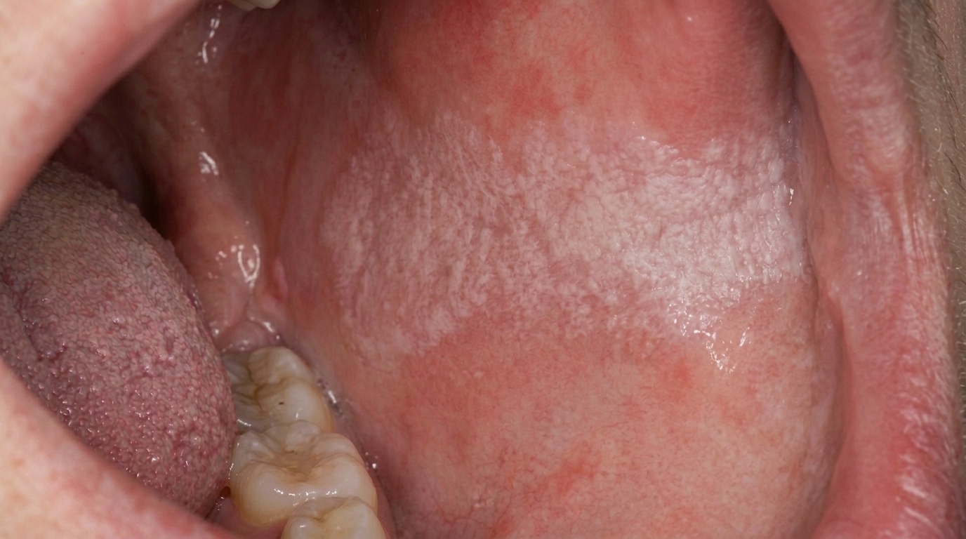
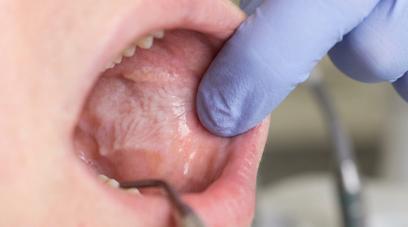
Leukoedema is a benign, very common change in the lining of the cheeks. The textbooks describe it as a bilateral, diffuse, milky-white or grey-white opalescent (with a faint milky shine) appearance of the inside of the cheeks. The "edema" in the name reflects its microscopic appearance, the surface cells of the cheek lining are gently swollen with fluid, scattering light and giving the area its faint white look. The hallmark feature is that the white appearance fades or disappears when the cheek is stretched, distinguishing leukoedema from most other white patches in the mouth. Because leukoedema is so common, the textbooks generally describe it as a variation of normal rather than a disease.
Who tends to get it?
The textbooks describe a fairly distinctive pattern of who tends to show leukoedema:
Adults of all ages, leukoedema is rare in children and develops gradually with age.
People of African and African-Caribbean ancestry, reported in 70-90% of black adults and 50% of black children. The change is more visible against the more pigmented background mucosa.
People of European ancestry, also affected, but the appearance is typically much milder and often hardly noticeable. Reported prevalence varies widely from less than 10% to more than 90% depending on diagnostic criteria.
Smokers, the textbooks specifically note that leukoedema is more common and more severe in smokers and tends to become less pronounced when smoking stops.
There is no strong sex predilection.
What causes it?
The exact cause is unknown, but the textbooks describe several relevant points:
Considered a variation of normal mucosa. Similar mild oedematous changes are also seen in the lining of the vagina and the larynx in many people, supporting the view that leukoedema reflects normal anatomical variability rather than disease.
Likely developmental. The change is not present in early childhood, develops slowly with age, and is influenced by background mucosal pigmentation.
Smoking aggravates the appearance. This is the only environmental factor consistently linked to leukoedema in the textbooks.
No infection, no allergy, no genetic syndrome is required for leukoedema to develop, and it is not contagious.
How does it develop?
Microscopically, leukoedema shows a slightly thickened cheek lining (acanthosis (thickening of the surface cell layer)) with a striking degree of intracellular oedema (fluid swelling inside the cells) in the spinous layer (the layer of prickle cells beneath the surface keratin), that is, the prickle cells of the epithelium are swollen with watery fluid. These vacuolated (swollen with fluid-filled spaces) cells scatter incoming light, giving the area its milky white appearance. There is no dysplasia (microscopic changes that can signal an increased cancer risk) and no inflammation. When the cheek is stretched, the swollen cells flatten out and the light scattering reduces, which is why the white appearance fades on stretching. Smoking thickens the surface keratin and exaggerates the white look; stopping smoking partially reverses this.
What might you notice?
What it looks like
The classic appearance is well described:
A bilateral, faint, milky-white or grey-white sheen inside both cheeks.
The change is diffuse rather than localised to one spot, and gradually fades into normal mucosa at the edges.
The surface may appear gently folded or wrinkled.
The most common location is the inside of the cheeks (buccal mucosa), sometimes extending forward onto the lip lining.
Unusually, it can also be seen on the floor of the mouth or the back of the throat.
The hallmark feature: the white appearance disappears or greatly reduces when the cheek is gently stretched between the fingers.
What it feels like
Leukoedema is asymptomatic. Most people are unaware of it until a dentist points it out.
No pain or soreness.
No taste change.
No bleeding or surface peeling.
No restriction of normal eating, drinking, or speaking.
What an X-ray might show
X-rays are not used for leukoedema, the diagnosis is made on clinical examination. Stretching the cheek to demonstrate fading is generally enough to confirm it.
What happens at the dentist?
Leukoedema is most often picked up at a routine dental check-up and clean at ArtSmiles when the dentist examines the inside of both cheeks. The typical assessment is short and reassuring:
Examine both cheeks under good light, looking for the diffuse milky-white change.
Stretch the cheek between fingers or with a mirror to confirm that the white appearance fades.
Ask about smoking habits, since smoking can exaggerate the change.
Document the appearance to set a baseline for future visits.
Reassure the patient that the change is benign and requires no treatment.
Recommend biopsy only in the rare case where the appearance is unusual or persists in a localised area that does not fade with stretching.
Is this serious?
🟢 Leukoedema is benign. The textbooks specifically classify it as a normal variation rather than a disease. There is no association with cancer, no risk to general health, and no risk to oral function. The most important reason to see a dentist is to be sure that what looks like leukoedema is actually leukoedema, and not one of the conditions it can mimic.
If you are unsure whether the white appearance inside your cheeks is leukoedema or something else, it is worth booking an assessment so the diagnosis can be confirmed with a simple clinical examination.
Could it be something else?
Several conditions can produce white patches in the mouth, and the stretch test is one of the most useful clinical tools for distinguishing them. The textbooks list these as the main differentials:
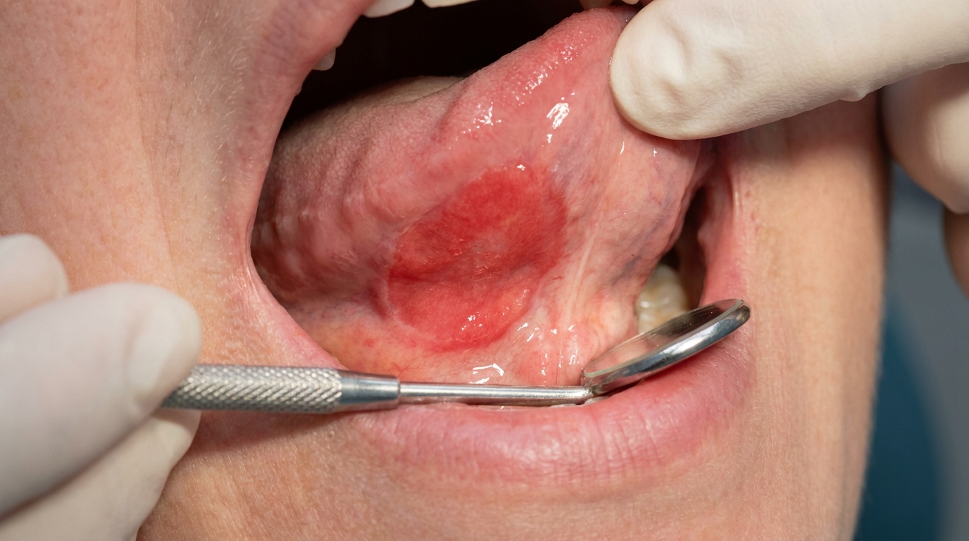
Idiopathic oral leukoplakia, a localised white patch that does not fade with stretching, and may carry a small risk of cancer change. Biopsy needed if unsure.
White sponge naevus, a hereditary, soft, spongy white change that is also bilateral but does not fade with stretching. Often a positive family history.
Pseudomembranous candidiasis (thrush), creamy-white plaque that wipes off, leaving red mucosa underneath.
Chronic hyperplastic candidiasis, a thicker, often nodular white plaque that does not wipe or stretch off; biopsy and antifungal trial may be needed.
Frictional hyperkeratosis (cheek-biting line), a localised white line where the teeth contact the cheek, with a clear cause.
Hairy leukoplakia, a corrugated white patch on the side of the tongue caused by Epstein-Barr virus, usually in the context of immunosuppression.
The fading-on-stretch sign is so characteristic that, in most cases, no other tests are needed.
How is it treated?
The textbooks all agree: leukoedema needs no specific treatment.
At-home measures and habits:
Continue normal oral hygiene, brushing twice a day with fluoride toothpaste and flossing daily.
Stop or reduce smoking. While smoking does not cause leukoedema, it makes the appearance more pronounced. Quitting often reduces the white sheen over weeks to months.
No need to avoid any food, drink or product because of the leukoedema itself.
Professional steps your dentist may consider:
Confirming the diagnosis by clinical examination and the stretch test.
Reassuring the patient about the benign nature of the change.
Documenting the appearance with photographs as a baseline.
Periodic check-ups as part of routine dental care.
Biopsy only in atypical cases where the appearance does not match the classical picture or where a localised area looks more solid than the surrounding leukoedema.
A patient-centred approach matters here too. People sometimes worry for years about a faint white change inside the cheeks. Calm, clear explanation of the diagnosis, the demonstration of fading on stretching, and the simple plan of "no treatment, just routine review" is itself part of effective care, values that sit at the heart of our clinical philosophy.
What's the long-term outlook?
The outlook is excellent. Leukoedema is a stable, harmless, lifelong appearance that does not progress to anything dangerous. There is no link to cancer, no link to systemic disease, and no need for special review beyond routine dental visits. For people who have wondered for years whether the white inside their cheeks meant something concerning, identifying leukoedema is usually the most reassuring step in the conversation.
A note on this article
This article is for educational purposes only and does not constitute a clinical diagnosis. Please consult a registered dental practitioner for assessment and treatment advice.
The cover image above is an AI-generated illustration based on the most common visible features of this condition described in clinical pathology references. It is not a photograph of a real case and should not be used to diagnose or rule out the condition in your own situation. If you are concerned about something you have noticed, please book an assessment with a registered dental practitioner.
References
Neville, B. W., Damm, D. D., Allen, C. M., & Chi, A. C. (2023). Oral and maxillofacial pathology (5th ed.). Elsevier. Chapter 1, Developmental Defects of the Oral and Maxillofacial Region: Leukoedema, with detailed clinical and histopathologic features and the characteristic fading-on-stretch sign, pp. 7 to 8.
Cawson, R. A., & Odell, E. W. (2017). Cawson's essentials of oral pathology and oral medicine (8th ed.). Elsevier. Chapter 15, Soft Tissue Disease: Leukoedema as a bilateral, diffuse, translucent greyish thickening representing a variation of normal, p. 252.
Regezi, J. A., Sciubba, J. J., & Jordan, R. C. K. (2017). Oral pathology: Clinical pathologic correlations (7th ed.). Elsevier. Chapter 3, White Lesions: Leukoedema as a hereditary mild opacification of the buccal mucosa.