Compiled from clinical pathology references. Medically reviewed by Dr Cristian Dunker , Principal Dentist, ArtSmiles Cosmetic Dentistry.
Quick summary
Also called | Oral hairy leukoplakia, HIV-associated hairy leukoplakia |
How urgent? | 🟡 Worth checking, the patch itself is harmless, but it is an important marker that the immune system may not be working properly |
Common or rare? | Uncommon overall, strongly associated with people who have HIV infection or other forms of immunosuppression (reduced immune function). Less common today thanks to modern antiretroviral therapy. |
Who it affects | Most often people living with HIV, particularly those with low CD4 counts; also seen after organ transplant, in some haematologic cancers, and with long-term corticosteroid use |
Who treats it | General dentist for diagnosis and biopsy, with referral to a GP, infectious diseases or HIV specialist for the underlying cause |
Based on | Cawson, Regezi, with cross-references in Neville |
What is it?
Hairy leukoplakia is a distinctive white patch that develops, almost always, on the side of the tongue. It is caused by replication of Epstein-Barr virus (EBV), the same virus responsible for glandular fever, within the surface cells of the tongue. The patch was first described in 1984, when an unusual corrugated white lesion was noticed in young men who later turned out to have HIV infection. Since then, hairy leukoplakia has been recognised as one of the more reliable oral signs that the immune system is significantly weakened. The patch itself is benign and does not turn into cancer, but it is an important pointer to an underlying systemic problem that needs attention.
Who tends to get it?
The textbooks describe a fairly tight clinical group:
People with HIV infection, particularly those with low CD4 counts and active virus levels. Up to 80% of untreated patients with hairy leukoplakia go on to develop AIDS, and the lesion is closely correlated with declining CD4 cells.
People who are immunosuppressed for other reasons, solid organ transplant recipients, people taking long-term high-dose corticosteroids (oral or topical), people receiving chemotherapy, and people with haematologic cancers such as leukaemia.
Adults rather than children, although it has been reported in children with HIV.
Predominantly men in the original series, reflecting the early recognition of HIV in men who have sex with men, although both sexes can develop the lesion.
The textbooks specifically note that hairy leukoplakia has declined in frequency in countries with widespread access to combination antiretroviral therapy, because well-controlled HIV no longer suppresses the immune system enough to allow the lesion to develop.
What causes it?
The cause is well established:
Epstein-Barr virus (EBV) replicating within the surface cells of the tongue. Most adults carry EBV silently after the original infection (often in childhood or adolescence), and the virus normally lives quietly in lymphocytes. In hairy leukoplakia, the virus moves into and actively multiplies within the keratinocytes (the surface skin and lining cells) of the tongue, something that does not happen in people with a healthy immune system.
Weakened cell-mediated immunity (the part of the immune system that uses T cells to fight virus-infected cells) is the key permissive factor. The textbooks describe a paucity of subepithelial (just beneath the surface lining) inflammation and very few Langerhans cells (immune cells that normally patrol the surface lining) in the lesion, suggesting that the local immune response is too quiet to suppress the virus.
Candida albicans is often a passenger in the surface keratin and may worsen the appearance, but it does not cause hairy leukoplakia on its own.
It is not understood why the lateral surface of the tongue is the favoured site. There is no link to oral hygiene, dental work, food, smoking or alcohol, although smoking can produce other white patches in the same area that need to be distinguished.
How does it develop?
Once active EBV replication begins in the tongue's epithelium, the surface keratin builds up and forms ridges and folds that give the lesion its corrugated, shaggy appearance. Microscopically, the upper layers of the epithelium contain "koilocyte-like" cells, pale, swollen cells with shrunken dark nuclei surrounded by a clear halo, full of viral particles. Surface candidal hyphae (long branching threads of the yeast) are often layered on top of the keratin without provoking the inflammatory reaction that would normally clear them, a reflection of the weakened local immunity. As the underlying immunosuppression worsens, the lesion typically becomes more extensive; with treatment of the underlying condition, it tends to regress.
What might you notice?
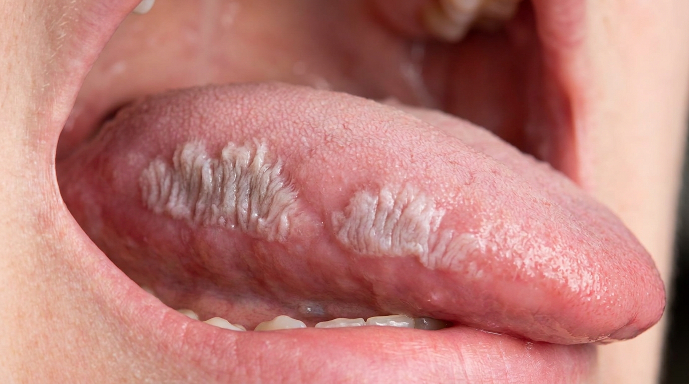
What it looks like
The classic clinical appearance is well described:
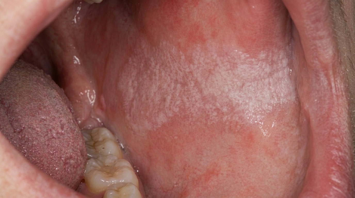
A well-demarcated white patch on the side of the tongue.
A vertically corrugated, ridged or "shaggy" surface, sometimes with hair-like extensions of keratin.
The patch may be flat and plaque-like, papillary or filiform in different cases.
The lesion may be unilateral or bilateral.
Most patches are on the lateral border of the tongue, occasionally extending onto the dorsum (top) or ventrum (underside).
Less commonly on the buccal (cheek) mucosa, floor of the mouth, or palate.
The patch does not wipe off with gauze, distinguishing it from oral thrush.
What it feels like
Hairy leukoplakia is usually painless. Many patients are not aware of it until a dentist or doctor points it out. When symptoms occur, they may include:
Mild discomfort on the side of the tongue, particularly with hot or spicy foods.
A rough or fuzzy sensation when the tongue moves against the teeth.
Soreness if there is a secondary candidal infection.
Cosmetic concern when the lesion is extensive enough to be visible on speaking or smiling.
What an X-ray might show
X-rays are not used for hairy leukoplakia, the diagnosis is made on clinical examination and confirmed on biopsy.
What happens at the dentist?
Hairy leukoplakia is often picked up at a routine dental check-up and clean at ArtSmiles, sometimes before the patient is aware they are immunosuppressed at all. The dentist will typically:
Examine the patch carefully, noting its location, size, surface pattern and whether it wipes off.
Take a careful medical history, including any recent illnesses, medications (especially corticosteroids and immunosuppressants), past organ transplant, history of HIV testing, or risk factors for HIV.
Arrange a biopsy in most cases. Confirmation of the diagnosis is essential, both to distinguish the lesion from other white patches and to give the patient a clear basis for further investigation.
Discuss further investigation of the immune system, typically through their GP. This usually involves an HIV test and, where indicated, other immune system tests.
Refer to an infectious diseases or HIV specialist when an underlying systemic cause is identified.
Reassure that the lesion itself is benign and treatable, and that addressing the underlying cause is the most important step.
Is this serious?
🟡 The patch itself is benign, it is not cancer and does not progress to cancer. The textbooks specifically note that hairy leukoplakia is not premalignant. However, it is an important sentinel sign (an early warning that something underlying needs attention) that the immune system is significantly weakened, and finding it should always prompt a careful check for an underlying cause. In people with HIV, the appearance of hairy leukoplakia historically marked a more rapid progression to AIDS in untreated patients. With modern antiretroviral therapy, that prognosis has improved markedly, but the lesion remains a meaningful clinical finding.
If you have a white patch on the side of your tongue that has not gone away after a couple of weeks, particularly one that is corrugated, shaggy or does not wipe off, it is worth booking an assessment so the right tests can be arranged.
Could it be something else?
Several conditions can produce a white patch on the side of the tongue. The textbooks list these as the main differentials:
Idiopathic oral leukoplakia, a white patch of unknown cause, sometimes premalignant. Usually flatter and less corrugated than hairy leukoplakia.
Frictional hyperkeratosis, a white thickening from chronic biting or rubbing of the tongue, typically with smoother edges and a clear cause from the patient's habits.
Tobacco-related leukoplakia, a white patch in a smoker, usually on the floor of the mouth, the cheek, or the gums rather than the lateral tongue.
Hyperplastic candidiasis (candidal leukoplakia), a thicker, often nodular white plaque that does not wipe off; biopsy and antifungal trial may be needed.
Pseudomembranous oral candidiasis (thrush), a creamy-white plaque that wipes off, leaving red mucosa underneath.
Oral lichen planus, usually has lacy white lines (Wickham striae) and may affect both cheeks and tongue.
Lupus erythematosus, can produce white patches with red atrophic centres on the inside of the cheeks and lips.
Hairy tongue, affects the central upper surface of the tongue rather than the side, and is caused by elongated filiform papillae rather than EBV.
How is it treated?
Treatment depends on whether the lesion is causing symptoms, on the patient's immune status, and on what is driving the immunosuppression.
At-home measures and habits:
Maintain excellent oral hygiene, brush the tongue gently and clean between teeth daily.
Manage candidal overgrowth if present, your dentist may recommend a topical antifungal mouthwash.
Limit alcohol and tobacco, both of which can aggravate any tongue lesion.
Avoid worrying about the lesion's appearance while you wait for biopsy results, it is benign in itself.
Professional steps your dentist may consider:
Biopsy to confirm the diagnosis on histopathology.
Investigation of the immune system, most often through your GP. Where HIV is suspected, an HIV antibody test is the most important first step.
Treating the underlying cause, well-controlled antiretroviral therapy in HIV, dose review of immunosuppressants after transplant, management of haematologic disease, is the most effective way to clear hairy leukoplakia. Lesions usually improve as the immune system improves.
Antiviral therapy with agents such as aciclovir, valaciclovir, ganciclovir, famciclovir, podophyllum or tretinoin can clear the lesion. The textbooks note, however, that lesions often return when the antiviral is stopped, and that prolonged use carries a risk of antiviral resistance which may be more serious for later, more severe infections. Treatment is therefore reserved for cases where the cosmetic or symptomatic burden is significant.
Addressing concurrent candidiasis with a topical or systemic antifungal where indicated.
Ongoing dental review, since people who develop hairy leukoplakia may also have a higher rate of other oral conditions (severe periodontal disease, oral candidiasis, Kaposi sarcoma) that warrant careful follow-up.
A patient-centred approach matters particularly here. A diagnosis that prompts an HIV test or other systemic investigation can be confronting. Calm, non-judgemental and unhurried discussion is itself part of effective care, values that sit at the heart of our clinical philosophy.
What's the long-term outlook?
The outlook for the lesion itself is excellent, hairy leukoplakia is benign, does not progress to cancer, and resolves with effective treatment of the underlying cause. The outlook for the underlying systemic condition depends entirely on what that condition is and how well it can be controlled. In people with HIV who start effective antiretroviral therapy, the immune system typically recovers, and hairy leukoplakia regresses or disappears completely. In people whose immunosuppression is necessary (for example, after organ transplant), a balance is struck between immune protection of the new organ and managing the lesion symptomatically. In every case, identifying the cause and acting on it is the single most important step.
A note on this article
This article is for educational purposes only and does not constitute a clinical diagnosis. Please consult a registered dental practitioner for assessment and treatment advice.
The cover image above is an AI-generated illustration based on the most common visible features of this condition described in clinical pathology references. It is not a photograph of a real case and should not be used to diagnose or rule out the condition in your own situation. If you are concerned about something you have noticed, please book an assessment with a registered dental practitioner.
References
Cawson, R. A., & Odell, E. W. (2017). Cawson's essentials of oral pathology and oral medicine (8th ed.). Elsevier. Chapter 15, Soft Tissue Disease: HIV-associated Hairy Leukoplakia, with EBV pathogenesis, koilocyte-like cells and management considerations including the risk of antiviral resistance, pp. 254 to 256.
Regezi, J. A., Sciubba, J. J., & Jordan, R. C. K. (2017). Oral pathology: Clinical pathologic correlations (7th ed.). Elsevier. Chapter 3, White Lesions: Hairy Leukoplakia, with first description in 1984, EBV replication, prevalence decline with antiretroviral therapy, AIDS development in untreated patients, and detailed differential diagnosis, pp. 87 to 89.
Neville, B. W., Damm, D. D., Allen, C. M., & Chi, A. C. (2023). Oral and maxillofacial pathology (5th ed.). Elsevier. Chapter on Bacterial, Viral and Fungal Infections: cross-reference to hairy leukoplakia in HIV-related lesions and as a key differential of hairy tongue.