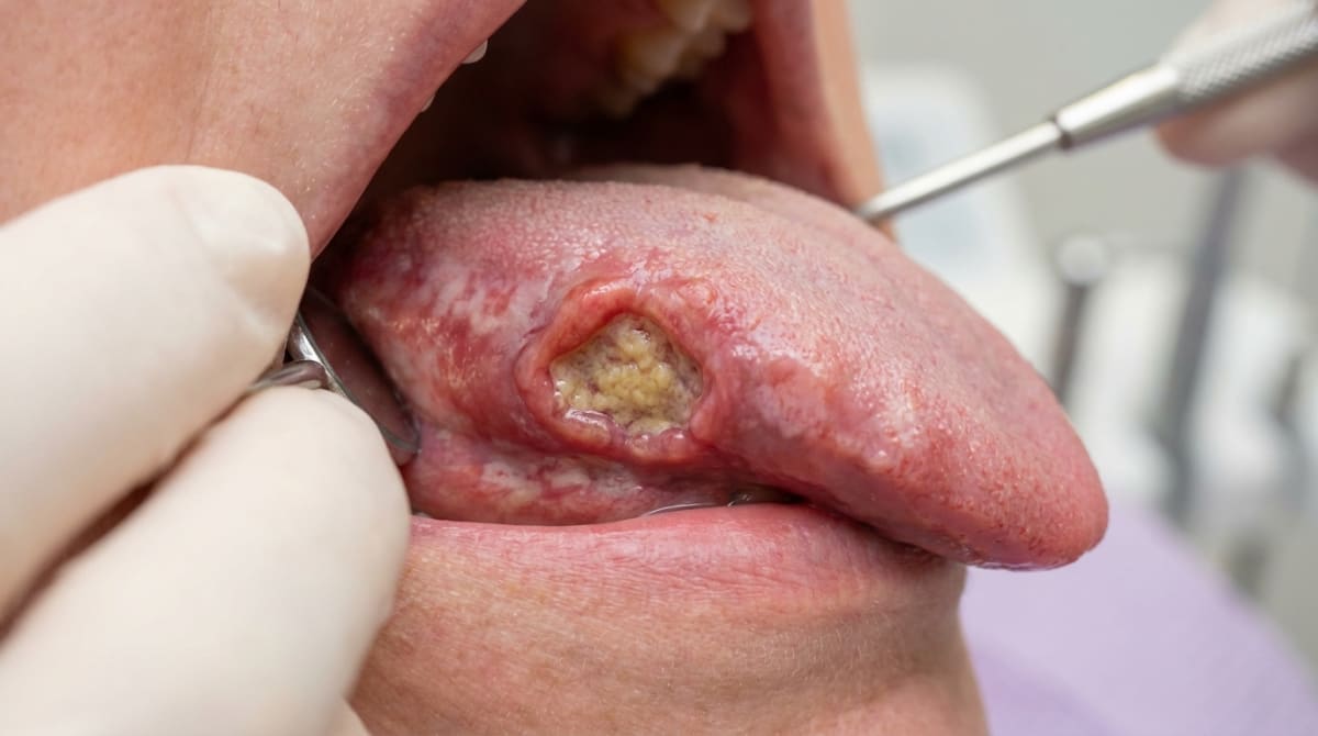
Compiled from clinical pathology references. Medically reviewed by Dr Cristian Dunker, Principal Dentist, ArtSmiles Cosmetic Dentistry.
Quick summary
Also called | Oral SCC, mouth cancer, oral cancer (when arising from the lining of the mouth) |
How urgent? | 🔴 See a dentist promptly, any mouth ulcer, lump, or red or white patch that has not healed within two weeks deserves an assessment. |
Common or rare? | Uncommon overall, but it is by far the most common cancer of the mouth, accounting for more than 90% of oral malignancies. |
Who it affects | Most often adults over 40, with a male predilection. Risk rises sharply with age. Younger adults can be affected, particularly with HPV-related disease. |
Who treats it | Detected by a general dentist; treated by a specialist team, usually oral and maxillofacial surgery, head and neck oncology, and radiation oncology. |
Based on | Regezi, Neville, Cawson |
What is it?
Oral squamous cell carcinoma is a cancer that develops in the thin layer of cells lining the mouth, the same kind of tissue that covers the tongue, floor of mouth, cheeks, gums, palate, and lips. It is the most common form of mouth cancer, making up more than 90% of oral malignancies.
It usually starts as a small, painless change in the mouth, a patch, an ulcer, or a lump, that does not go away. Because the early signs can look surprisingly innocent, regular dental check-ups are one of the most reliable ways it gets picked up early, when treatment outcomes are at their best.
Who tends to get it?
Oral squamous cell carcinoma is mainly a disease of older adults. Around 98% of patients are over the age of 40, and the incidence climbs steeply with age. Men have historically been affected more often than women, though that gap is narrowing in many Western countries.
In Australia and other developed nations, oral cancers account for roughly 2% to 3% of all cancers diagnosed each year. Globally, however, rates vary enormously, in parts of South Asia, mouth cancer can account for 40% or more of all cancers, largely because of betel quid chewing.
A newer pattern has also emerged. Cancers at the back of the mouth and throat (the oropharynx, base of tongue and tonsils) linked to human papillomavirus (HPV) are increasingly seen in younger and middle-aged adults, including many people who have never smoked.
What causes it?
The cause is multifactorial (many factors combine), usually a combination of long-term exposures rather than a single trigger. The well-established risk factors are:
Tobacco in any form. Cigarettes, cigars, pipes, and smokeless tobacco (chewing tobacco, snuff) are all linked to increased oral cancer risk. The risk is dose-dependent: the more someone smokes, and the longer they smoke, the higher the risk. Heavy smokers carry several times the risk of non-smokers.
Alcohol. Heavy drinking is an independent risk factor, and tobacco and alcohol together are far more dangerous than either alone, the combined risk can be 15 times higher or more.
Betel quid (paan). Common across South and South-East Asia, this chewing habit, often combining areca nut, slaked lime, betel leaf and tobacco, is one of the most carcinogenic mixtures known.
Sunlight (UV) exposure is the leading risk factor for cancer of the lower lip vermilion, particularly in fair-skinned outdoor workers, a real concern in the Australian climate.
Human papillomavirus (HPV), particularly HPV-16. HPV is now strongly linked to cancers of the tonsils and base of tongue. It plays a smaller role in cancers of the front of the mouth.
A weakened immune system, from medications after organ transplant, or long-standing HIV infection, modestly increases risk.
Pre-existing red or white patches in the mouth (erythroplakia and certain leukoplakias) can sometimes progress to cancer.
Iron deficiency in the rare Plummer-Vinson (Paterson-Kelly) syndrome (a combination of iron-deficiency anaemia, smooth tongue and difficulty swallowing).
Genetics generally play only a minor role. A few rare inherited conditions, such as Fanconi anaemia and dyskeratosis congenita (rare inherited bone-marrow disorders), increase risk significantly.
How does it develop?
Think of the lining of your mouth as a tightly organised wall of cells, replacing themselves in an orderly way. Every cell has built-in instructions telling it when to grow, when to stop, and when to be replaced.
Long-term exposure to carcinogens, chemicals in tobacco smoke, by-products of alcohol, UV light on the lip, gradually damages those instructions. Most damage gets repaired, but over years, mutations can build up in the cells' control genes. Eventually a clone of cells loses the ability to switch off its own growth, ignores the signals to die when it should, and starts spreading sideways and downward into the tissues beneath.
Many oral cancers pass through a recognisable in-between stage, a flat red or white patch (erythroplakia or leukoplakia), before becoming invasive. Others appear to develop in apparently normal-looking mucosa.
What might you notice?
Oral SCC has the unsettling habit of looking minor in its earliest, most curable stages. The four classic appearances are: a non-healing ulcer, a red patch, a white patch, or a mixed red-and-white patch.
What it looks like
Early lesions are usually small and painless. They may look like:
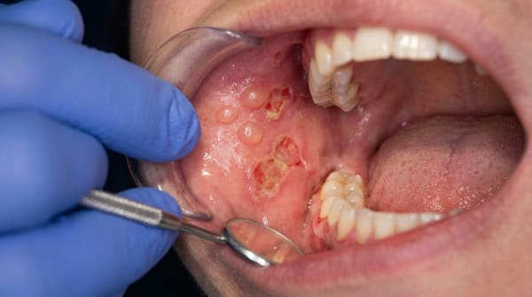
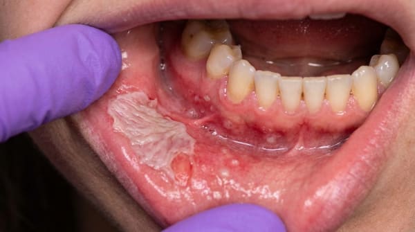
A persistent ulcer that does not heal after two weeks
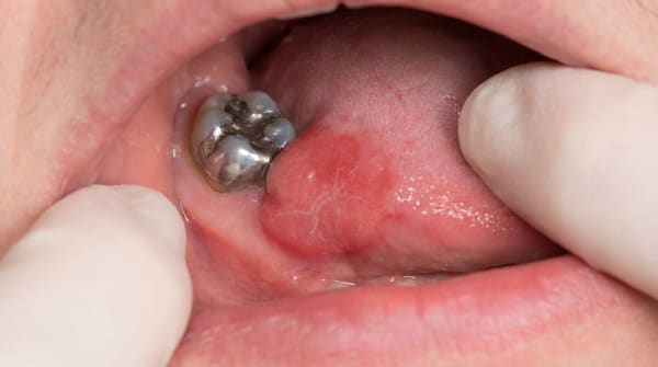
A red, velvety patch (erythroplakia), often the most concerning sign
A white patch (leukoplakia) that cannot be wiped away
A mixed red-and-white speckled patch
A lump, thickening, or warty growth that sits proud of the surface (sometimes called an exophytic growth)
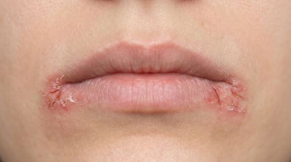
A crusted or scaly area on the lower lip
Later lesions tend to ulcerate and develop a raised, rolled, hard border around a central crater. The tissue often feels firm or hardened (the medical term is 'indurated') when pressed. There may be bleeding from minor trauma, or a tooth that becomes loose for no clear reason.
Over 70% of oral cancers form in a relatively small zone described as the "U-shaped cancer zone", the lateral and ventral surfaces of the tongue, the floor of mouth, and the soft palate complex extending back towards the oropharynx. This area accounts for only about 20% of the lining of the mouth, but the great majority of cancers concentrate there. The tongue alone accounts for more than half of intraoral cancers in many series.
What it feels like
In its earliest stages, oral SCC is often painless, which is part of why it can grow unnoticed. Pain, soreness, or burning when eating spicy or acidic foods tends to come later, once the lesion has ulcerated or invaded deeper structures. Other later symptoms can include:
A persistent sore throat or feeling that something is stuck
Difficulty or pain when swallowing
Numbness of the lip, tongue, or part of the jaw (from nerve involvement)
Loose teeth, a denture that suddenly feels different, or a changed bite
A lump in the neck, sometimes the first thing a patient notices
Voice changes, ear pain on one side, or unexplained weight loss
What an X-ray might show
A dental X-ray cannot diagnose mouth cancer, but it can show signs of advanced disease. Where a tumour has invaded the underlying jawbone, the X-ray may show an irregular, "moth-eaten" radiolucency with ragged edges, different from the clean outline of a benign cyst. CT, MRI, and PET scans are used by specialists to map the extent of disease and check for spread.
What happens at the dentist?
An experienced dentist at ArtSmiles screens the mouth at every routine check-up, looking systematically at the lips, tongue (including the under-surface and sides), floor of mouth, cheeks, gums, palate, and back of the throat, and feeling the neck for enlarged lymph nodes.
If something looks unusual, the assessment usually proceeds in steps:
A careful clinical examination, describing the lesion, palpating it, and checking the lymph nodes in the neck.
Watchful review, a clearly traumatic ulcer (for example, from a sharp tooth) may be reviewed after two weeks of removing the cause. Anything that does not heal in that time should not be given the benefit of the doubt.
Referral to a specialist, typically an oral medicine specialist or oral and maxillofacial surgeon, for any high-risk lesion.
Biopsy. This is the only definitive way to know whether a lesion is benign, dysplastic, or malignant. A small sample is taken under local anaesthetic and sent to a pathologist.
Imaging and staging, if cancer is confirmed, CT, MRI, and PET scans are used to assess size, depth, and any spread to lymph nodes or distant sites.
Dental practitioners are also encouraged to advise about prevention, discussing tobacco and alcohol with patients, and recommending lip protection for outdoor workers, and to follow up patients with known precancerous patches.
Is this serious?
🔴 Yes, oral squamous cell carcinoma is serious, and timely assessment matters. Overall five-year survival across all stages and sites of mouth and throat cancer sits around 50% to 67% in published series, but those figures hide an enormous range.
The single most important factor is stage at diagnosis. Small, early-stage cancers (still localised, less than 2 cm) are often curable with relatively conservative treatment, and survival rates are markedly better. Once the cancer has spread to lymph nodes in the neck, prognosis drops sharply. Cancers of the lip vermilion, picked up early, have an excellent outlook (around 90%+ five-year survival). Cancers of the posterior tongue and floor of mouth do worst, partly because they are harder to see and tend to be diagnosed later.
Untreated, oral SCC progresses by direct invasion of nearby tissues, muscle, bone, nerves, and by spread to lymph nodes in the neck, and eventually to the lungs, liver, or bones. So the message is not to panic, but not to wait either.
If you have noticed any of these signs for more than two weeks, it is worth booking an assessment.
Could it be something else?
Several mouth conditions can mimic oral SCC, especially in its early stages. Only a biopsy can give a definitive answer. Differentials your dentist will consider include:
Traumatic ulcer, caused by a sharp tooth, denture, or accidental bite. Looks like an ulcer, but heals within 10 to 14 days once the cause is removed; an ulcer that persists beyond that needs investigation.
Aphthous ulcer, recurrent, round, painful ulcers in healthy adults. They typically heal in 7 to 14 days; cancer is painless early and does not heal.
Leukoplakia, a white patch that cannot be wiped off. Some leukoplakias remain benign, but a proportion show dysplasia or hide an early carcinoma; biopsy distinguishes them.
Erythroplakia, a velvety red patch. The majority already show severe dysplasia or invasive cancer at biopsy, which is why any unexplained red patch is treated as urgent.
Oral lichen planus, chronic inflammatory condition with white striae and sometimes red erosions. Distinguished by its bilateral, lacy pattern and characteristic biopsy appearance.
Oral candidiasis (chronic hyperplastic), a white plaque caused by Candida that may not rub off. Responds to antifungal therapy, unlike a true neoplastic lesion.
Tuberculous or syphilitic ulcer, chronic infectious ulcers can clinically resemble cancer; biopsy and microbiological testing tell them apart.
Deep fungal infection, rare, but can present as a chronic non-healing ulcer; identified on biopsy.
Necrotising sialometaplasia, a self-resolving palatal ulcer that microscopically can mimic carcinoma; the history and biopsy distinguish it.
Pyogenic granuloma or epulis, a red, friable gum growth that bleeds easily. Usually distinguishable clinically, but biopsy is needed if features are atypical.
Verrucous carcinoma, a low-grade variant of SCC with a warty white surface; behaves less aggressively but still requires removal.
Keratoacanthoma, a rapidly growing, dome-shaped lip nodule that can look identical to SCC; many authorities now consider it a well-differentiated SCC variant and treat it as such.
How is it treated?
Treatment is highly individualised, it depends on where the cancer is, how big it is, whether lymph nodes are involved, the patient's general health, and HPV status (for cancers at the back of the mouth and throat).
What you can do at home, prevention and early detection:
Avoid tobacco in all forms; if you currently smoke or chew, quitting is the single most effective change you can make.
Keep alcohol intake moderate, and avoid combining heavy drinking with smoking.
Use SPF lip balm and a wide-brimmed hat outdoors, especially in the Australian sun.
Consider HPV vaccination, now approved as a preventive measure against HPV-related head and neck cancers as well.
Have regular dental check-ups, which include a soft-tissue mouth cancer screen.
Look in your own mouth from time to time. Anything that has not healed in two weeks deserves a professional opinion.
Professional treatment is led by a multidisciplinary head and neck cancer team (combining surgery, oncology, radiation oncology, dental and supportive care), and may include:
Surgery, usually the primary treatment for cancers of the front of the mouth. The aim is to remove the cancer with a clear margin of healthy tissue. This may also involve removing lymph nodes from the neck (a neck dissection) and reconstructive surgery to restore function and appearance.
Radiation therapy, used as the main treatment in some cases, or after surgery to reduce the chance of recurrence. Modern techniques (including intensity-modulated radiotherapy) aim to spare salivary glands and other healthy tissues.
Chemotherapy, often combined with radiation for advanced or higher-risk disease.
Targeted and immunotherapy agents, used in selected recurrent or advanced cases.
Pre-treatment dental care, extractions of unrestorable teeth, fluoride trays, and meticulous oral hygiene before radiotherapy reduce the risk of complications such as osteoradionecrosis (poor healing of irradiated bone). Your dentist plays an important role here.
Treatment is demanding. Side effects can include dry mouth, mucositis, taste changes, swallowing difficulty, and altered facial appearance, so support from speech pathologists, dietitians, and dental teams is part of standard care.
What's the long-term outlook?
Outcomes vary widely. Patients diagnosed at an early, localised stage, particularly with cancers of the lip, often do very well, with five-year survival above 80%. Patients diagnosed once the cancer has reached the neck nodes have a more guarded outlook, and outcomes drop further with distant spread.
Follow-up after treatment usually continues for at least five years. People who have had one oral cancer carry a lifelong, slightly increased risk of a second cancer of the mouth, throat, larynx, or lungs ("field cancerisation"), particularly if they continue to smoke or drink heavily. Quitting tobacco after diagnosis significantly reduces that risk.
For HPV-positive cancers at the back of the throat, the outlook is generally better than for HPV-negative tumours of the same stage, these tumours tend to respond well to treatment.
The single most powerful message about oral squamous cell carcinoma is this: early detection changes everything. A two-minute mouth check at your routine dental visit, and a willingness to investigate any sore that does not heal in a fortnight, are your best protection.
A note on this article
This article is for educational purposes only and does not constitute a clinical diagnosis. Please consult a registered dental practitioner for assessment and treatment advice.
The cover image above is an AI-generated illustration based on the most common visible features of this condition described in clinical pathology references. It is not a photograph of a real case and should not be used to diagnose or rule out the condition in your own situation. If you are concerned about something you have noticed, please book an assessment with a registered dental practitioner.
References
Regezi, J. A., Sciubba, J. J., & Jordan, R. C. K. (2017). Oral pathology: Clinical pathologic correlations (7th ed.). Elsevier. Chapter 2, Ulcerative Conditions: Squamous Cell Carcinoma of the Oral Cavity, pp. 52 to 75.
Neville, B. W., Damm, D. D., Allen, C. M., & Chi, A. C. (2023). Oral and maxillofacial pathology (5th ed.). Elsevier. Chapter 10, Epithelial Pathology: Squamous Cell Carcinoma, pp. 401 to 425.
Cawson, R. A., & Odell, E. W. (2017). Cawson's essentials of oral pathology and oral medicine (8th ed.). Elsevier. Chapter 17, Oral Cancer, pp. 277 to 289.
Frequently asked questions
What is oral squamous cell carcinoma?
Oral squamous cell carcinoma (OSCC) is the most common cancer of the mouth, arising from the squamous cells lining the cheeks, tongue, lips, gums, palate and floor of the mouth. It usually presents as a non-healing ulcer, a hard lump, a red or white patch, or an unexplained tooth mobility. Early detection dramatically improves outcomes.
What are the risk factors?
Tobacco (smoking, chewing, betel quid) and alcohol are the dominant risk factors, especially when combined. Other risk factors include high-risk HPV (particularly HPV-16 in oropharyngeal cancer), sun exposure (for lip cancer), poor oral hygiene, chronic irritation (sharp tooth, ill-fitting denture), nutritional deficiencies, immunosuppression, and family history. Around 5-10% of cases occur in people without classical risk factors.
What are the warning signs?
Any mouth ulcer that does not heal in three weeks, an unexplained lump, a persistent red or white patch (especially erythroplakia), unexplained tooth mobility, persistent throat pain, ear pain on one side, difficulty swallowing, a hoarse voice, or numbness of the lip should be investigated. Painless lesions are particularly concerning because they often present late.
How is OSCC diagnosed and treated?
Diagnosis is by biopsy. Imaging (CT, MRI, PET-CT) stages the disease (TNM). Treatment depends on stage — early cancers are usually treated with surgery alone with excellent outcomes (5-year survival 80%+). Advanced cancers need surgery plus radiotherapy and sometimes chemotherapy or immunotherapy, with lower survival. Lifelong follow-up is essential because risk of a second oral cancer is increased.