Compiled from clinical pathology references. Medically reviewed by Dr Cristian Dunker , Principal Dentist, ArtSmiles Cosmetic Dentistry.
Quick summary
Also called | Chronic atrophic candidiasis, denture sore mouth, denture-induced stomatitis, denture-associated candidiasis |
How urgent? | 🟡 Worth a check-up, usually painless but signals that the denture, the fit or the mouth itself needs attention |
Common or rare? | Very common, affects up to about 65% of people who wear a complete upper denture |
Who it affects | Adults who wear a removable upper denture or orthodontic plate, especially older adults |
Who treats it | General dentist (and prosthodontist if the denture needs remaking) |
Based on | Regezi, Neville, Cawson, Laskaris |
What is it?
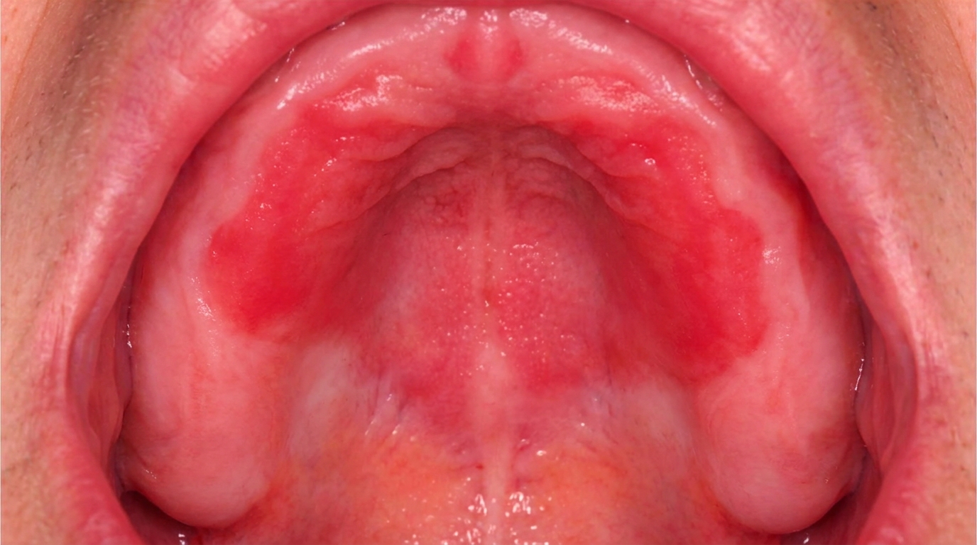
Denture stomatitis is a patch of red, sometimes slightly swollen mucosa (the soft tissue lining of the mouth) that sits underneath a removable denture, almost always an upper one. The redness mirrors the outline of the denture so neatly that it stops abruptly where the denture ends. Most people feel nothing at all; the change is usually picked up at a routine check-up when the denture is taken out.
The textbooks group it within the family of erythematous (red) oral candidiasis, and many authors use the term chronic atrophic candidiasis as a synonym. Despite the name, it is not always a true infection, it is usually a low-grade reaction to the yeast-rich biofilm that builds up on the denture's fitting surface.
Who tends to get it?
Denture stomatitis is one of the most common mucosal findings in older adults. Cawson notes that in the United Kingdom about 20% of people aged 55,64 are edentulous, and almost half of those over 65, so a very large group is at risk. Surveys of residential aged-care homes have found denture stomatitis in around 12% of residents.
Regezi reports that chronic erythematous candidiasis, the form linked to dentures, occurs in as many as 65% of older adults who wear a complete upper denture. Both sexes are affected, but it is particularly noticed in women, who are more likely to be edentulous in older age. Children and young adults are rarely affected because they rarely wear full dentures.
What causes it?
The condition is multifactorial. The textbooks consistently highlight the same group of contributing factors:
Wearing the denture continuously, sleeping in the denture is the single most important factor. The mucosa never gets a break from the prosthesis.
Poor denture hygiene, plaque and food debris accumulate on the fitting surface and harbour Candida albicans (the most common yeast that lives in the mouth) and other microorganisms.
A well-fitting (but airtight) upper denture, Cawson explains that an upper denture seals the underlying mucosa off from the protective washing action of saliva. The lower denture is more mobile and rarely shows the same change.
An ill-fitting or worn denture, chronic low-grade trauma, poor occlusion and porous old acrylic all worsen the problem.
Smoking, increases susceptibility to candidal colonisation.
Dry mouth (xerostomia), dry mouth, from medications, Sjögren's syndrome or radiotherapy.
Broad-spectrum antibiotics, disturb the normal oral flora and let yeasts overgrow.
Corticosteroid inhalers, particularly without a spacer or without rinsing afterwards.
Diabetes mellitus, raises the risk of any candidal infection.
Iron, folate or vitamin B12 deficiency, anaemia is repeatedly listed as a predisposing factor.
Immunosuppression, including HIV, chemotherapy, transplant medications and high-dose systemic steroids.
What the books make clear, and what is reassuring, is that allergy to the acrylic of the denture is not a real cause. Cawson is direct on this point: there is no foundation for the idea of a denture-base allergy, even though the myth persists.
How does it develop?
Think of the fitting surface of an upper denture as a covered, warm, slightly damp greenhouse pressed onto the roof of the mouth. Candida albicans is a yeast that lives quietly in most healthy mouths without causing trouble. Under a denture, three things shift in its favour: saliva can no longer wash the surface clean, the acrylic itself is microscopically porous and gives the yeast somewhere to hide, and any food or plaque left on the denture provides a steady food supply. The yeast multiplies into a sticky biofilm on the denture's fitting surface.
Neville and Regezi point out an important nuance, when the underlying mucosa is swabbed it often shows relatively few yeast cells, while the denture itself is heavily colonised. So in many cases the mucosa is not so much infected as reacting to the biofilm, the enzymes the yeast produces (such as phospholipases) and the chronic low-grade pressure of the denture. That is why the textbooks describe denture stomatitis as a spectrum that runs from a mild irritation through to a true candidal infection.
Clinicians often describe three stages, originally set out by Newton, that can progress one into the next if nothing is done:
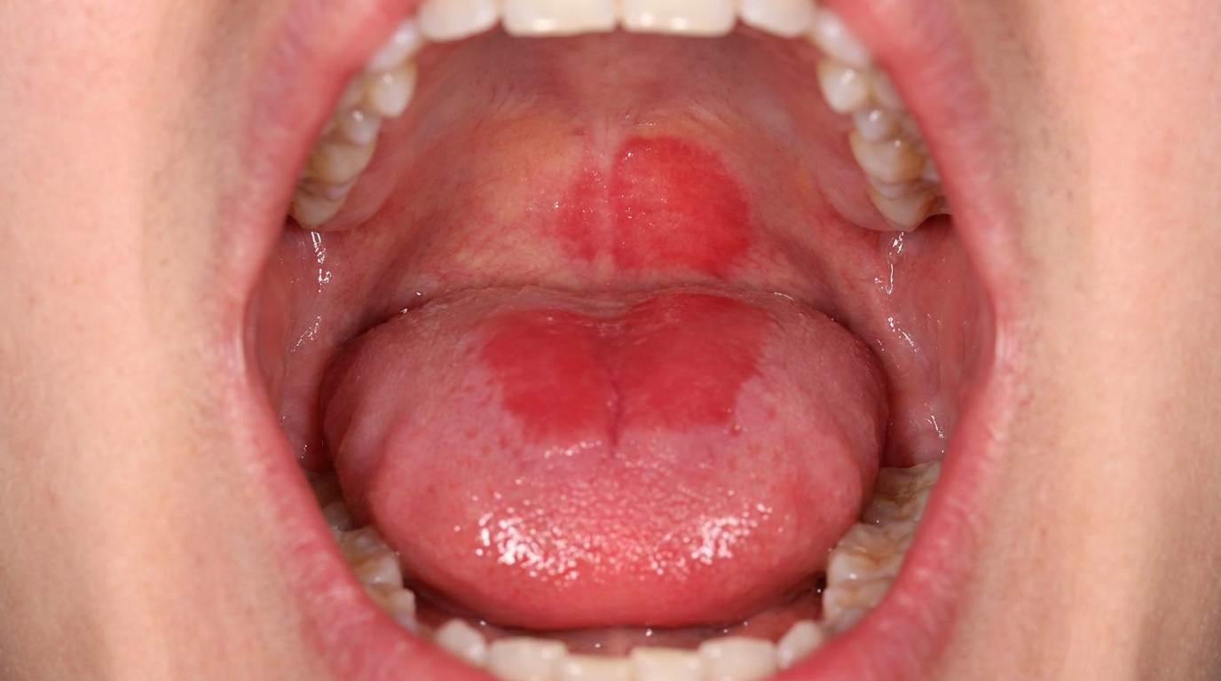
Type I, pinpoint red spots scattered across the palate.
Type II, a more diffuse, smooth red area covering most of the denture-bearing mucosa.
Type III, a nodular, lumpy ('papillary') hyperplasia, particularly in the centre of the palate, where the tissue itself starts to overgrow.
What might you notice?
What it looks like
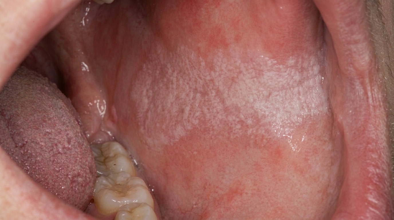
A bright red, sometimes slightly velvety or pebbly area on the roof of the mouth. The crucial clue is that the redness stops sharply at the edge of the denture, the mucosa beyond the denture margin looks completely normal. There may be tiny pinpoint red spots, a uniform red sheet, or in long-standing cases small nodular bumps in the centre of the palate. Sometimes there are little white flecks within the redness, which represent yeast colonies or food debris. The lower denture-bearing area almost always looks normal.
What it feels like
Surprisingly, the textbooks all stress the same point: denture stomatitis is usually painless. Some people describe a vague burning sensation (sometimes mistaken for burning mouth syndrome), a slight irritation or an altered taste, but most discover the condition only when the dentist takes the denture out and shows them. If there is significant pain, another cause (such as a traumatic ulcer, an ill-fitting flange or, rarely, something more serious) needs to be looked for.
A related sign that often appears at the same time is angular cheilitis, sore, cracked, red corners of the mouth. Saliva pools in the folds at the corners, especially when the bite has dropped, and the yeast colonises that warm, moist crease. Angular cheilitis is sometimes the complaint that brings someone in, and the denture-related cause is found on examination.
What an X-ray might show
Denture stomatitis is a soft-tissue change and does not show on X-rays. Imaging is not part of the diagnosis.
What happens at the dentist?
Diagnosis is almost always made on the clinical examination. At ArtSmiles, your dentist may:
Ask how long you have worn the denture, whether you sleep in it, how you clean it and whether you have noticed any change.
Take the denture out and examine both the mucosa and the fitting surface of the prosthesis under good light.
Check the fit, the occlusion and the vertical dimension (how much space your bite gives the lips and corners of the mouth).
Look for angular cheilitis at the corners of the mouth.
Ask about general health, diabetes, dry mouth, inhaled corticosteroids, antibiotics and any conditions that affect immunity.
Take a simple swab if the diagnosis is unclear; a Gram stain or PAS stain can confirm candidal hyphae (long branching threads of the yeast).
Order blood tests for iron, folate, vitamin B12 or blood sugar if the condition keeps coming back, as recommended by Cawson and Regezi.
Refer for further investigation if there is a persistent ulcer, a lesion that does not match the denture outline, or anything that looks unusual.
A biopsy is rarely needed for straightforward denture stomatitis. It would only be considered if a white or red patch failed to settle after appropriate treatment.
Is this serious?
🟡 Worth a check-up, but not an emergency. On its own, denture stomatitis is benign and does not turn into anything sinister. The textbooks are clear that, despite earlier suspicions, this form of candidiasis is not regarded as a precancerous lesion.
That said, it is a useful warning sign and is worth taking seriously for three reasons. First, if it is left, it can progress from a flat redness to nodular papillary hyperplasia (a pebbly thickening of the lining with many small bumps) of the palate, which is harder to reverse and may need surgery before a new denture can be made. Second, the underlying yeast can spread to the corners of the mouth (angular cheilitis) or, in immunocompromised people, become a more serious candidal infection. Third, and importantly, recurrent or unusually florid candidiasis can be the first clue to undiagnosed diabetes, anaemia or iron, folate or B12 deficiency, all of which deserve medical attention.
If the area under your denture looks red, or your denture has felt different for more than two weeks, it's worth booking an assessment.
Could it be something else?
Most cases are clear-cut, but a dentist will think about several look-alikes before settling on the diagnosis.
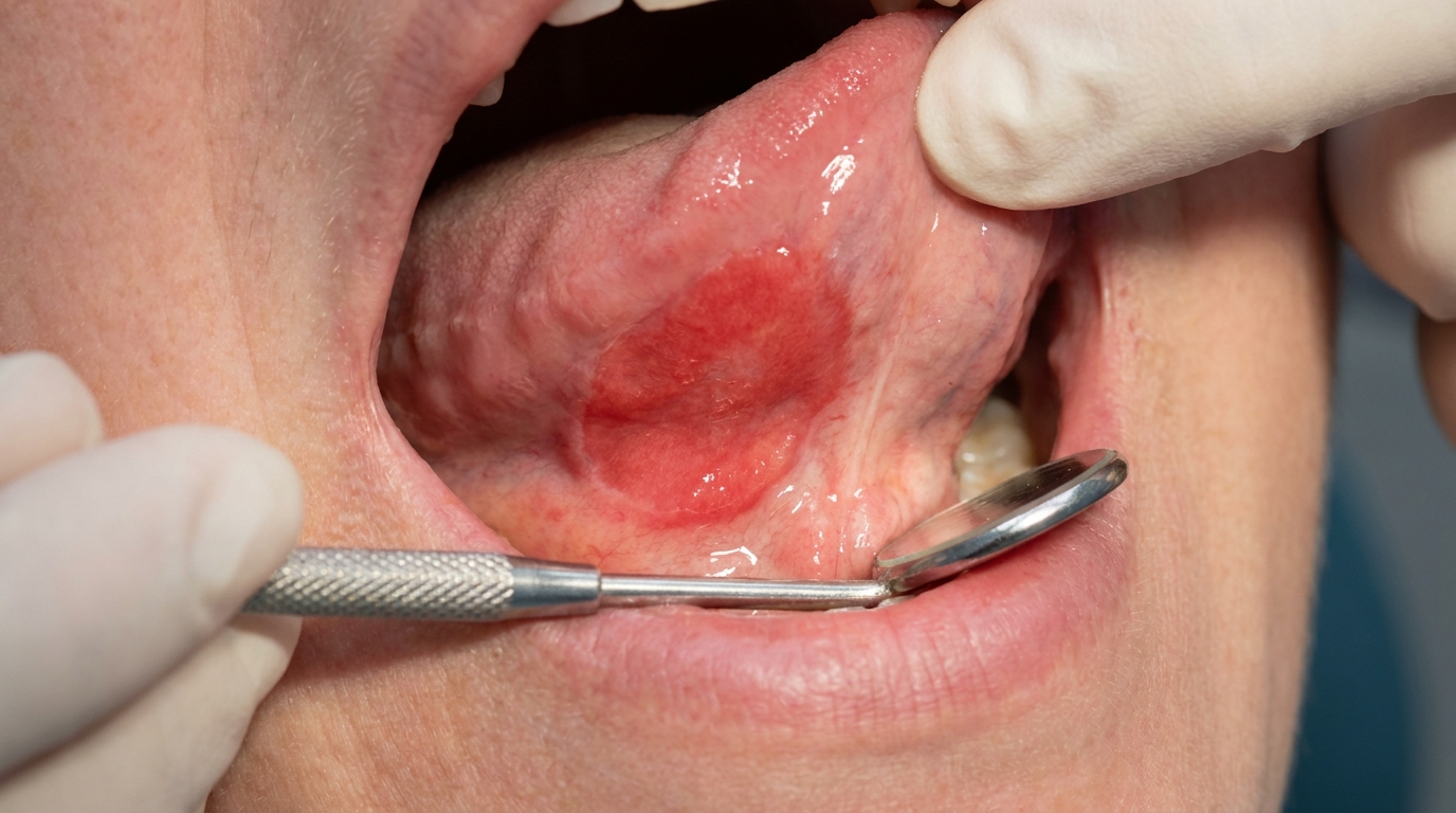
Traumatic ulcer or denture sore, a localised painful sore from an over-extended flange or a sharp edge. Unlike denture stomatitis, it hurts and is usually a single ulcer rather than a sheet of redness.
Papillary hyperplasia of the palate, described by Laskaris and Neville as a long-standing variant of denture stomatitis with multiple small nodules in the central palate. It overlaps with Newton's Type III and tends to need surgical removal.
Allergic contact stomatitis to acrylic, listed in Laskaris but considered very rare; Cawson notes the methylmethacrylate myth has little real basis.
Erythematous candidiasis unrelated to a denture, also red and atrophic, but the redness is not confined to the denture-bearing area; often seen on the dorsum of the tongue or palate in HIV-positive patients, after antibiotics or with dry mouth.
Median rhomboid glossitis (central papillary atrophy), a red, depapillated patch in the centre of the back of the tongue; can be a 'kissing' counterpart to a palatal (relating to the palate, the roof of the mouth) lesion in chronic multifocal candidiasis. Distinguished by location.
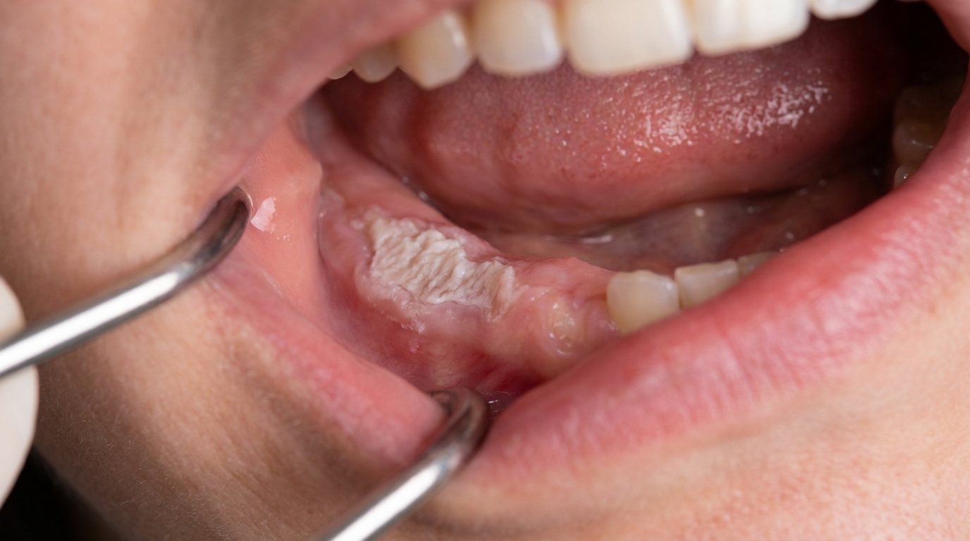
Erosive lichen planus, can cause red patches on the palate, but typically shows lacy white striae at the edges and affects sites beyond the denture outline.
Discoid lupus erythematosus, listed by Regezi as a differential for erythematous candidiasis; tends to have radiating fine white striae and is symmetrical.
Mucous membrane pemphigoid, produces erythema and erosions of the gingiva and palate, but usually painful and not confined to the denture-bearing area.
Erythroplakia or early carcinoma of the palate, uncommon, but any persistent red patch that does not respect the denture outline, or that fails to resolve after the denture and yeast are addressed, must be re-examined and biopsied.
How is it treated?
Treatment has two halves: changing the conditions that allowed the yeast to thrive, and (if needed) using an antifungal to settle the infection. Both halves matter, antifungals alone almost always fail if the denture habits do not change.
At home, what helps the most:
Take the denture out at night, every night. Cawson lists this as the single most important step.
Soak the denture overnight in dilute sodium hypochlorite (for fully acrylic dentures) or chlorhexidine mouthwash. This eliminates Candida from the porous acrylic, which Cawson identifies as the hidden reservoir that re-infects the mouth.
Brush the fitting surface daily with a soft denture brush and a non-abrasive denture cleaner.
Rinse the mouth out after using a corticosteroid inhaler, ideally with a spacer.
Keep on top of general oral hygiene and any natural teeth that remain.
What a dentist may recommend:
A topical antifungal such as nystatin suspension, miconazole oral gel or clotrimazole troches, applied for at least one to two weeks and continued for a week beyond resolution.
Application of miconazole gel directly to the fitting surface of the denture while it is being worn, three times a day, for one to two weeks.
Treatment of associated angular cheilitis with a topical antifungal, sometimes combined with fusidic acid if Staphylococcus aureus is also present.
Adjusting, relining (adding a new fitting surface to an existing denture so it sits properly) or remaking the denture if it is loose, worn, has lost vertical dimension, or has obvious porosity.
A systemic antifungal such as fluconazole or itraconazole if the condition is widespread, recurrent or in an immunocompromised patient.
Blood tests for anaemia, iron, folate, vitamin B12 or blood glucose if the condition keeps returning.
Surgical reduction (cryosurgery or excision) of papillary hyperplasia if Newton's Type III changes have developed before a new denture is made.
Resolution typically takes one to two weeks, but full settling of the mucosa may take longer if the denture is old or the underlying factors are stubborn.
What's the long-term outlook?
The outlook for denture stomatitis is excellent when the underlying habits are addressed. Regezi describes the prognosis for this form of candidiasis as excellent, and Cawson notes that the inflammation generally clears within one to two weeks of correct treatment.
The problem, in practice, is recurrence. If the denture goes back in 24 hours a day, if the fitting surface is not cleaned thoroughly, or if a predisposing factor (diabetes, dry mouth, an inhaler, an old porous denture) is not dealt with, the redness will return. Long-standing untreated cases can progress to papillary hyperplasia of the palate, which then needs surgical correction before a new prosthesis can be fitted comfortably.
The encouraging message from all four textbooks is the same: denture stomatitis is benign, it is not a marker for oral cancer, and it is largely preventable with simple denture hygiene and the discipline of taking the denture out at night. A regular dental review, even when there are no natural teeth left, gives the best chance of catching it early and keeping the mouth comfortable.
A note on this article
This article is for educational purposes only and does not constitute a clinical diagnosis. Please consult a registered dental practitioner for assessment and treatment advice.
The cover image above is an AI-generated illustration based on the most common visible features of this condition described in clinical pathology references. It is not a photograph of a real case and should not be used to diagnose or rule out the condition in your own situation. If you are concerned about something you have noticed, please book an assessment with a registered dental practitioner.
References
Regezi, J. A., Sciubba, J. J., & Jordan, R. C. K. (2017). Oral pathology: Clinical pathologic correlations (7th ed.). Elsevier. Chapter 3, White Lesions (Candidiasis), pp. 104 to 108; Chapter 4, Red-Blue Lesions, pp. 126 to 127.
Neville, B. W., Damm, D. D., Allen, C. M., & Chi, A. C. (2023). Oral and maxillofacial pathology (5th ed.). Elsevier. Chapter 6, Fungal and Protozoal Diseases (Erythematous Candidiasis / Denture Stomatitis), pp. 201 to 208.
Cawson, R. A., & Odell, E. W. (2017). Cawson's essentials of oral pathology and oral medicine (8th ed.). Elsevier. Chapter 12, Diseases of the Oral Mucosa: Mucosal Infections (Candidosis: Denture-induced Stomatitis), pp. 213 to 216; Appendix 12.1, Treatment of Candidosis.
Laskaris, G. Pocket atlas of oral diseases. Chapter 4, Mechanical Injuries (Denture Stomatitis), pp. 50 to 51; Chapter 18, Fungal Infections (Candida-associated Lesions), pp. 166 to 169.
Frequently asked questions
What causes denture stomatitis?
Denture stomatitis is a chronic redness of the palatal mucosa under a denture, caused mainly by Candida (yeast) growing on the denture surface and the mucosa. Wearing the denture continuously (especially at night), poor denture hygiene, ill-fitting dentures, and conditions that lower immunity (such as diabetes or certain medications) all contribute.
Is denture stomatitis painful?
Usually no. Most cases are surprisingly painless, despite the bright red appearance. The diagnosis is often made when the dentist notices the colour change at a routine review. Some patients notice a slight burning or unpleasant taste, particularly with denture food retention.
How is denture stomatitis treated?
Treatment combines denture hygiene (cleaning the denture thoroughly, soaking overnight in chlorhexidine or a mild bleach solution), taking the denture out at night to let the tissue rest, antifungal therapy (such as miconazole gel applied to the denture fit surface), and reviewing the denture fit. Underlying systemic factors like diabetes are also addressed.
Will denture stomatitis come back?
Recurrence is common if denture hygiene and night-time removal are not maintained. Long-term control combines consistent home care, regular dental review, and remaking or relining the denture if it no longer fits well. A new well-fitting denture significantly reduces recurrence risk.