Compiled from clinical pathology references. Medically reviewed by Dr Cristian Dunker, Principal Dentist, ArtSmiles Cosmetic Dentistry.
A bright red lump on the gum that appears within weeks and bleeds at the lightest touch is one of the more startling things a patient can find in their mouth. Most are not dangerous, but they do tend to grow, bleed, and refuse to settle without help. The medical name is pyogenic granuloma, and it is one of the most common reactive lesions of the mouth.
This article from the team at ArtSmiles, reviewed by Dr Cristian Dunker, explains what a pyogenic granuloma is, why it forms, and what to expect at a dental visit.
Quick summary
At a glance | Detail |
|---|---|
Also called | Lobular capillary haemangioma; pregnancy epulis or granuloma gravidarum when pregnancy-related |
How urgent? | 🟡 Worth assessing soon, the lump itself is benign but it bleeds easily and needs a quick diagnosis to exclude look-alikes |
Common or rare? | Common, one of the most frequent reactive lesions of the mouth |
Who it affects | Pregnant women, children and young adults, and anyone with local irritation in the mouth |
Who treats it | General dentist; sometimes a periodontist or oral surgeon for larger lesions |
Based on | Neville, Cawson, Regezi, Laskaris |
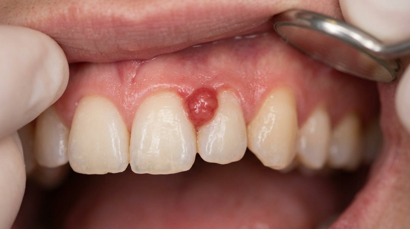
What is it?
Pyogenic granuloma is a misnomer (a misleading medical name), it is neither pus-filled (pyogenic) nor a true granuloma (a small ball of immune cells). The name has stuck for historical reasons. Today we understand it as a reactive vascular hyperplasia (an exuberant overgrowth of small blood vessels and connective tissue) formed in response to minor injury or local irritation.
In the mouth it usually appears as:
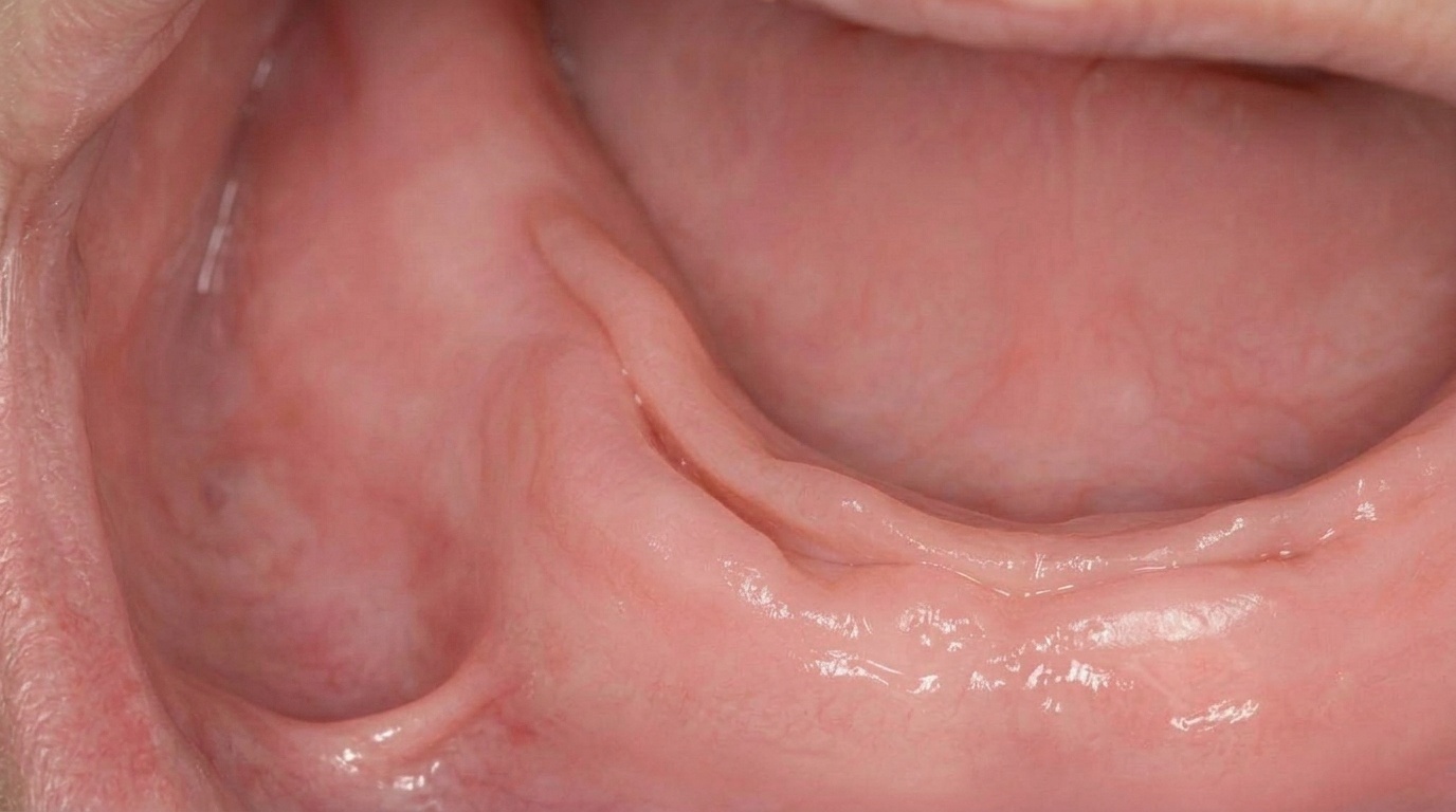
A smooth or lobulated, soft, exophytic lump (a lump that grows outward from the surface).
Deep red to purplish-red in colour.
Pedunculated (on a stalk) or sessile (on a broad base).
8 to 15 mm in size, sometimes larger.
Bleeds readily on light contact, often with a friable (easily broken), glistening surface.
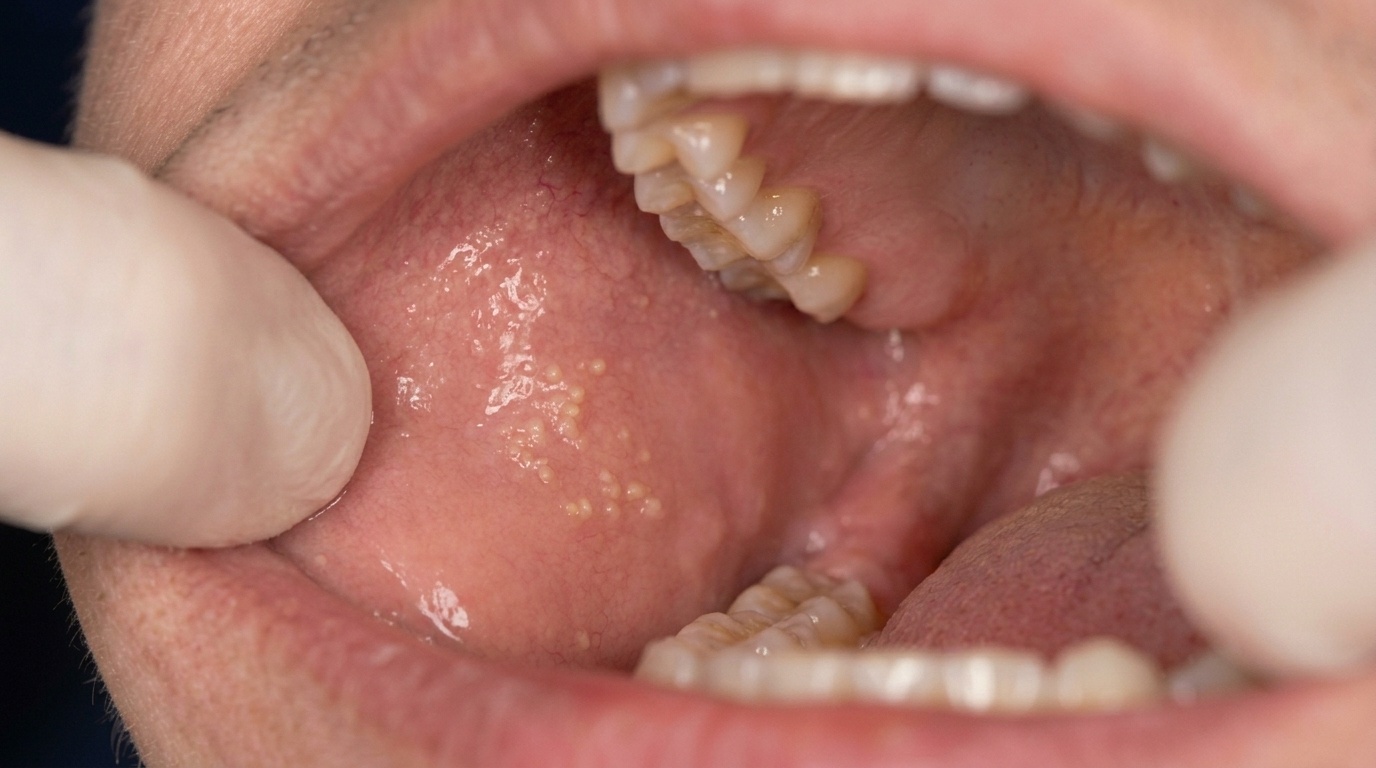
The most common site is the gum (gingiva), particularly between the front teeth, but it can also appear on the lips, tongue, cheek lining and palate.
Who tends to get it?
Pyogenic granuloma can affect anyone, but several groups are particularly prone:
Pregnant women. Around 5% of pregnancies feature a pyogenic granuloma on the gum, often called a pregnancy epulis or granuloma gravidarum. It typically appears in the second trimester and can shrink after delivery.
Children and young adults. It is one of the more common growths of the mouth in this age group.
People with persistent local irritation, calculus on the teeth, an overhanging filling, a sharp denture clasp, a partly erupted tooth, or a piece of food caught between teeth.
People taking certain medicines that affect blood vessels.
There is a slight female predominance overall, due in part to the link with pregnancy.
What causes it?
The trigger is usually a combination of:
Minor trauma or injury. A bite, a cut, a piece of food caught in the gum.
Local irritation. Plaque and calculus on the teeth, sharp edges of fillings, orthodontic wires.
Hormonal change. Pregnancy hormones (oestrogen and progesterone) make the gum more sensitive to plaque, so even small amounts of plaque produce an exaggerated response.
Immune and growth factors released by the irritated tissue, which encourage rapid growth of new blood vessels.
The lesion is not infectious and is not contagious.
How does it develop?
The classic pattern is rapid early growth followed by a stable phase:
A small red bump appears, often noticed first when it bleeds during brushing.
Over the next two to three weeks it grows, sometimes alarmingly quickly.
The surface becomes glossy and may ulcerate slightly.
After several weeks, growth slows and the lesion stabilises at its mature size.
With time, some lesions become firmer, paler and more fibrous (a fibroma-like maturation, becoming similar to a traumatic fibroma), but most stay red and friable until removed.
In pregnancy, the lesion typically shrinks after delivery as hormone levels normalise, and what is left can be removed easily.
What might you notice?
What it looks like
A deep red to purplish, soft, glistening lump, usually 8 to 15 mm, often on the gum between the front teeth. It may be on a stalk or on a broad base, with a smooth or slightly lobulated surface that bleeds when touched.
What it feels like
A rapidly growing red lump that has appeared within a few weeks.
Bleeding when brushing, flossing or eating crunchy foods.
A metallic taste from small amounts of blood throughout the day.
A lump that catches on the teeth or denture when chewing.
Tenderness rather than sharp pain, especially when something pushes on the lump.
Cosmetic concern when the lesion is at the front of the mouth.
The bleeding is usually small in volume but happens frequently, which is why patients feel as if it bleeds "all the time".
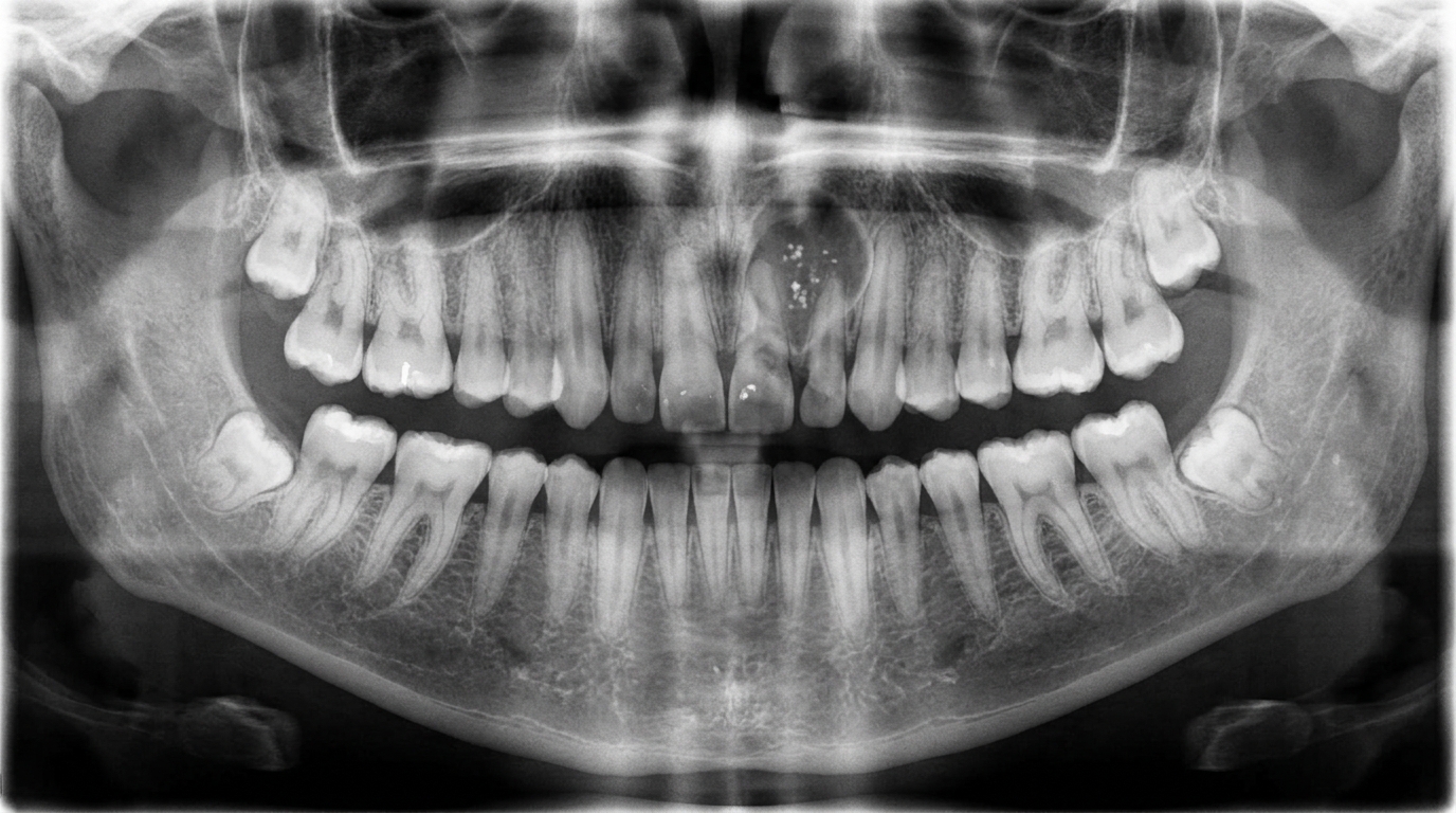
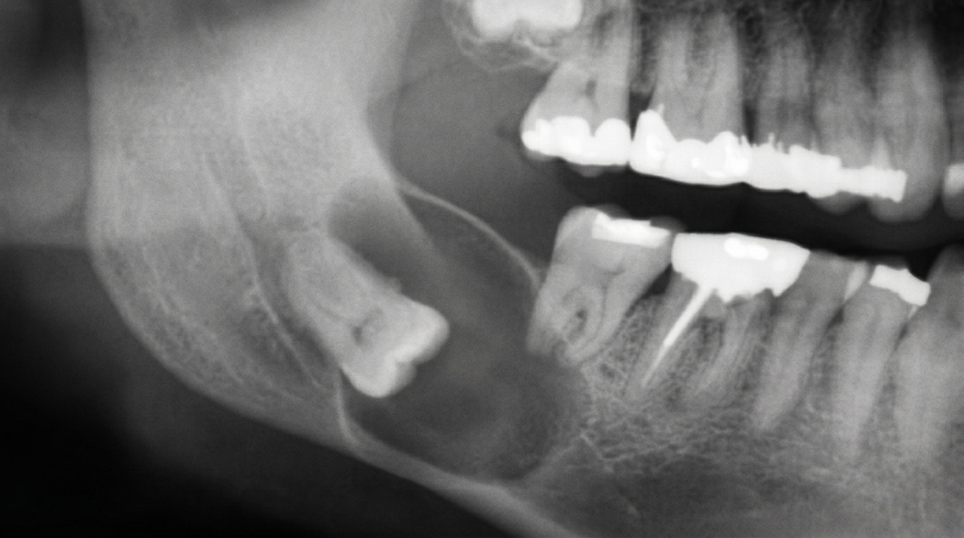
What an X-ray might show
Pyogenic granuloma is a soft-tissue lesion, so dental X-rays usually show no abnormality. The role of imaging is to check there is no underlying bone problem, such as a peripheral giant cell granuloma eroding the bone, that could explain the picture.
What happens at the dentist?
When you bring a bleeding lump to a check-up at ArtSmiles, the visit usually involves:
Listening to the story. When did it appear? How fast did it grow? Was there a recent injury, dental treatment or pregnancy?
A close clinical examination. We assess the size, shape, surface, base and texture, and identify any local source of irritation.
A check of the surrounding teeth. Plaque, calculus and any sharp edges of fillings or restorations are noted as likely contributors.
A photograph for the record.
A treatment plan. This usually combines removing the source of irritation with surgical excision of the lump. We will also explain what to expect afterwards.
Histopathology (examination of the removed tissue under the microscope). The removed tissue is sent to the laboratory to confirm the diagnosis. This step is important because several other lumps can look similar, and a definite answer reassures both clinician and patient.
If you are pregnant, treatment is often deferred until after delivery unless the lesion is a major problem, since lesions sometimes shrink or even resolve once hormone levels normalise.
Is this serious?
🟡 Pyogenic granuloma is a benign condition. It does not spread to other parts of the body, it is not cancer, and it does not turn into cancer. The reasons we still recommend treatment are:
Bleeding can be persistent and inconvenient.
Growth can interfere with chewing, brushing and aesthetics.
Diagnostic certainty matters, several other red lesions need to be excluded.
Recurrence is more likely if remnants are left or if the irritation is not addressed.
In short, the condition is not dangerous, but timely treatment is the most comfortable path forward.
Could it be something else?
Other lumps that can mimic a pyogenic granuloma include:
Peripheral giant cell granuloma, bluish-red, often on the gum, can erode underlying bone.
Peripheral ossifying fibroma, firm pink lump on the gum, often near a tooth.
Traumatic fibroma, firm, pale, painless lump from chronic minor trauma; sometimes the mature, fibrotic form of an old pyogenic granuloma.
Haemangioma or vascular malformation, generally present from earlier in life.
Kaposi sarcoma, a purplish red mucosal lesion seen in immunocompromised patients (see Oral Manifestations of HIV and AIDS).
Oral squamous cell carcinoma of the gum, usually firmer, more ulcerated and not following an obvious irritation pattern.
Metastatic tumours, rare causes of red gum lumps in patients with known cancer elsewhere.
This is why every excised pyogenic granuloma is sent to the laboratory.
How is it treated?
The standard treatment is surgical excision under local anaesthetic. The lesion is removed at its base, including a small surrounding margin, and the source of irritation is treated at the same visit. Common steps include:
Cleaning the affected area thoroughly to remove plaque and calculus.
Smoothing or replacing any sharp filling or restoration.
Adjusting an overhanging crown or denture.
In some cases, a small periodontal procedure to remove the entire base of the lesion.
Suturing the wound closed (or letting it heal as a small graze).
The tissue is sent for laboratory examination. Healing is usually complete within two weeks. Sometimes a laser or electrosurgery is used to control bleeding during removal, especially for larger lesions.
For pregnancy-related lesions, treatment is often delayed until after delivery, with careful oral hygiene and gentle cleaning in the meantime to limit growth.
Recurrence is possible, particularly if the source of irritation is not addressed, but uncommon when both steps (lump removal and trigger removal) are done well.
What's the long-term outlook?
The outlook is excellent. Once the lesion is excised and the local irritation removed, most patients have no further trouble. Pregnancy-related lesions may regress on their own after delivery. The piece of gum where the lump was usually heals smoothly with minimal scarring.
If you have a fast-growing red lump in your mouth that bleeds when you brush, please book an appointment rather than wait. We can usually offer a clear diagnosis and a treatment plan in a single visit.
A note on this article
This article is for educational purposes only and does not constitute a clinical diagnosis. Please consult a registered dental practitioner for assessment and treatment advice.
The cover image above is an AI-generated illustration based on the most common visible features of this condition described in clinical pathology references. It is not a photograph of a real case and should not be used to diagnose or rule out the condition in your own situation. If you are concerned about something you have noticed, please book an assessment with a registered dental practitioner.
References
Neville, B. W., Damm, D. D., Allen, C. M., & Chi, A. C. (2023). Oral and maxillofacial pathology (5th ed.). Elsevier. Chapter 8, Soft Tissue Tumors: Pyogenic Granuloma.
Cawson, R. A., & Odell, E. W. (2017). Cawson's essentials of oral pathology and oral medicine (8th ed.). Elsevier. Chapter 13, Reactive Lesions.
Regezi, J. A., Sciubba, J. J., & Jordan, R. C. K. (2017). Oral pathology: Clinical pathologic correlations (7th ed.). Elsevier. Chapter 5, Connective Tissue Lesions.
Laskaris, G. (2006). Pocket atlas of oral diseases (2nd ed.). Thieme. Pyogenic Granuloma.
Frequently asked questions
Is a pyogenic granuloma dangerous?
No. Despite the alarming name, a pyogenic granuloma is a benign, non-infectious overgrowth of small blood vessels and connective tissue in the gum or oral lining. It is not pus, not infection and not cancer. The main reasons to treat it are that it bleeds easily and can keep enlarging until removed.
Why do pyogenic granulomas bleed so easily?
They are made mostly of immature blood vessels with thin walls, packed close to the surface. Even gentle brushing, eating crusty food or accidentally touching the lump can break the surface and cause bleeding, sometimes well out of proportion to the size of the lesion.
Why do I get a pyogenic granuloma during pregnancy?
Hormonal changes in pregnancy, particularly elevated oestrogen and progesterone, exaggerate the gum's response to local irritants like plaque. The result is often a rapidly growing red lump on the gum, traditionally called a 'pregnancy tumour' or 'epulis gravidarum'. It is benign and usually shrinks after delivery, although it sometimes still needs removal.
Will a pyogenic granuloma come back after removal?
Recurrence happens in about 15-16% of cases, particularly if local irritants like plaque, sharp tooth edges or a foreign object in the gum are not addressed at the same time as surgery. Excising the lesion and then removing the local cause gives the best chance of a lasting result.