Compiled from clinical pathology references. Medically reviewed by Dr Cristian Dunker , Principal Dentist, ArtSmiles Cosmetic Dentistry.
Quick summary
Also called | Oral TB, tuberculous ulcer, secondary oral tuberculosis, orofacial tuberculosis |
How urgent? | 🔴 See a dentist or doctor promptly, a chronic, non-healing mouth ulcer may signal an underlying lung infection that requires medical workup |
Common or rare? | Rare in the mouth, even though tuberculosis is common globally; oral involvement is reported in roughly 0.5% to 5% of TB cases |
Who it affects | Most often middle-aged or older adults with active or undiagnosed pulmonary TB; also seen in people with reduced immunity, including those living with HIV |
Who treats it | General dentist (initial recognition and biopsy (a small tissue sample sent to the lab for testing) referral) plus a medical specialist (respiratory or infectious diseases) for systemic treatment |
Based on | Regezi, Neville, Cawson, Laskaris |
What is it?
Oral tuberculosis is a chronic mouth ulcer caused by the same bacterium that causes tuberculosis (TB) of the lungs, Mycobacterium tuberculosis. The mouth is rarely the first place TB shows up. In almost every case, the oral lesion is a secondary sign of an active lung infection that is shedding bacteria into saliva and sputum.
A tuberculous ulcer is typically painful, firm to the touch, and refuses to heal on its own over weeks. Spotting one matters far beyond the mouth, in roughly half of reported cases, the oral ulcer was the first clue that led to the diagnosis of underlying pulmonary TB.
In Australia, tuberculosis is a notifiable disease, which means cases must be reported to public health authorities so that contact tracing and treatment can be arranged.
Who tends to get it?
Globally, TB infects roughly one third of the world's population and remains the leading cause of death from a single infectious agent. In developed countries including Australia, rates fell sharply through the twentieth century but have not disappeared. Cases continue to occur, often in people born overseas in higher-prevalence regions, in people experiencing housing instability or overcrowding, and in those with compromised immunity.
Oral lesions specifically tend to develop in middle-aged and older adults with established pulmonary TB. The classic profile in the textbooks is an older man with a long-standing productive cough that has either gone unrecognised or been neglected. Primary oral TB, meaning the mouth is the first site of infection without lung involvement, is genuinely rare, though it is described slightly more often in children and adolescents.
People living with HIV represent a growing group affected by oral TB, partly because reduced immunity allows latent infection to reactivate, and partly because more than half of TB cases in this group involve sites outside the lungs.
What causes it?
The organism responsible is Mycobacterium tuberculosis, a slow-growing bacillus (rod-shaped bacterium) with a thick waxy coat that helps it resist immune attack. It spreads almost exclusively through the air, in tiny droplets coughed or sneezed out by someone with active pulmonary disease.
For an oral ulcer to develop, bacteria-laden sputum from the lungs needs to come into contact with the mouth lining. A small break in the mouth lining (mucosa), a denture rub, an extraction socket, a minor bite injury, gives the organism a foothold. Less commonly, the bacteria reach the mouth through the bloodstream or lymphatic system from a distant site.
Recognised risk factors include:
Active or undiagnosed pulmonary TB
Living or working in crowded conditions
HIV infection or other causes of weakened immunity
Diabetes, advanced age, and significant socioeconomic disadvantage
Immunosuppressive medications, including long-term corticosteroids
Travel to or origin from regions with high TB prevalence
Previous TB infection that may reactivate later in life
It is worth noting that only about 5% to 10% of people who become infected with M. tuberculosis go on to develop active disease, most contain the infection silently for years.
How does it develop?
Think of TB as a slow, drawn-out negotiation between the bacterium and the immune system. When droplets are inhaled, the bacteria settle in the lungs and are engulfed by immune cells called macrophages. In most healthy people, the immune system walls the organisms off inside small clusters called granulomas (small balls of immune cells), and the infection sits dormant, sometimes for life.
In a smaller group, the balance tips. The bacteria multiply, the granulomas break down in the centre into a soft, cheese-like material (a process called caseous necrosis), and the lung tissue is damaged. Bacteria are then coughed up in sputum.
When that infected sputum repeatedly washes across the mouth, the bacteria can seed into a tiny crack in the mucosa, most often on the dorsum of the tongue. The same granulomatous reaction now plays out locally: immune cells gather, the centre breaks down, and an ulcer forms. Because the immune system cannot easily clear the organism from the mouth either, the ulcer keeps deepening rather than healing.
What might you notice?
What it looks like
The classic tuberculous ulcer has a few recognisable features:
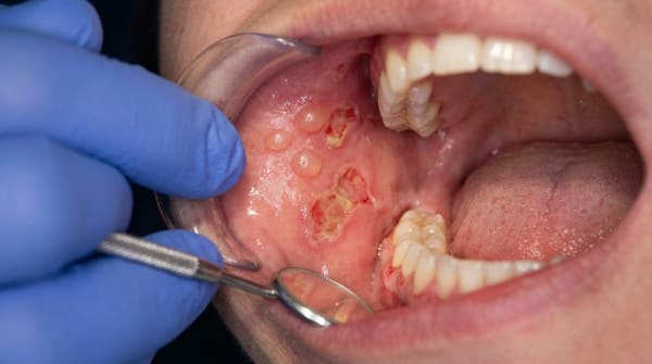
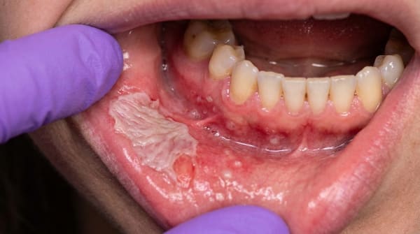
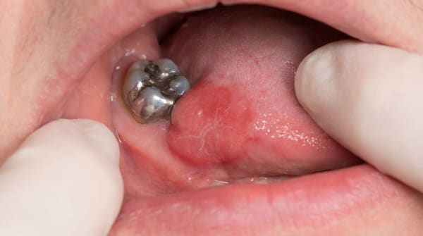
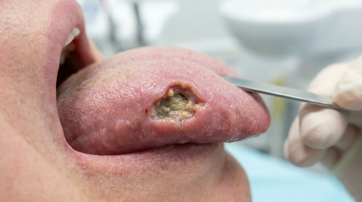
A single chronic ulcer, often on the mid-dorsum (top surface) of the tongue, but sometimes on the palate, lip, gum, or buccal mucosa
An angular, stellate (star-shaped), or irregular outline rather than a clean round shape
Overhanging, undermined edges and a pale, granular floor
Surrounding tissue that feels firm or indurated (firm and unyielding) when pressed
Size ranging from about one to five centimetres
A grey-yellow exudate may cover the surface
Other presentations described in the books include non-healing extraction sockets, areas of granular inflammation on the gum, swellings of the jaw, and enlarged neck lymph nodes that may eventually break down through the skin (a presentation called scrofula).
What it feels like
Descriptions vary across the source textbooks. Most authors describe the typical lesion as painful, particularly as it deepens, pain on chewing, swallowing, or speaking is common. Some texts note that early or shallow lesions can feel surprisingly mild or even painless, which is part of why people delay seeking care.
Pharyngeal involvement can cause difficulty swallowing, painful swallowing, and voice changes. Enlarged, firm neck lymph nodes are often felt at the same time.
Because oral TB is almost always secondary to lung disease, systemic symptoms are usually present in the background, a low-grade fever, night sweats, unexplained weight loss, fatigue, a persistent cough, and sometimes blood-streaked sputum.
What an X-ray might show
Dental X-rays are not the main diagnostic tool, but they may show two helpful clues:
A poorly defined area of bone destruction in the jaw if tuberculous osteomyelitis (bone infection) is present
Calcified cervical lymph nodes, sometimes mistaken for sialoliths (salivary stones), which point to past or current TB infection
A chest X-ray, ordered by a doctor, is usually the more revealing image, it commonly shows changes at the apex of the lungs in active disease.
What happens at the dentist?
The first job of the dentist at ArtSmiles is recognition. Any mouth ulcer that has not healed after two weeks deserves careful assessment, and one that is firm-edged, on the tongue, and accompanied by a chronic cough or weight loss raises the suspicion of TB.
A typical pathway might involve:
A detailed history covering travel, country of origin, prior TB exposure, immune status, cough, fever, night sweats, and weight loss
A clinical examination of the ulcer, the rest of the mouth, and the neck lymph nodes
Photographs and measurements to track the lesion over time
A biopsy of the ulcer edge, this is the key step. The tissue is sent for histopathology, which looks for caseating granulomas with multinucleated Langhans giant cells (a distinctive cell pattern with several nuclei found in TB-type infections)
Special staining of the sample using the Ziehl-Neelsen or Fite acid-fast stain (special lab stains that highlight TB-type bacteria) to look for the bacteria themselves
Mycobacterial culture or polymerase chain reaction (PCR) on the biopsy material, the gold standard for confirming the species
Onward referral to a medical practitioner for chest X-ray, sputum examination, an interferon-gamma release assay (IGRA, a blood test for TB exposure) or a tuberculin (Mantoux) skin test, and contact tracing
Newer rapid tests such as the Xpert MTB/RIF assay can detect M. tuberculosis DNA, and screen for rifampin resistance, within hours rather than the weeks needed for traditional culture.
The dentist's role is rarely to provide the cure for TB itself; it is to recognise the lesion, take a safe biopsy, and refer for systemic medical care. Strict infection control is followed during the appointment because active oral lesions can shed bacteria.
Is this serious?
🔴 Yes, this is a condition that needs urgent attention. Not because the mouth ulcer itself is immediately life-threatening, but because of what sits behind it.
Untreated pulmonary tuberculosis is a serious disease that can progress to widespread (miliary) infection, where the bacteria spread through the bloodstream to seed many organs, permanent lung damage, and death. It is also contagious, household members and close contacts can be exposed. In Australia, tuberculosis is a notifiable infectious disease, meaning the diagnosis triggers public health follow-up and contact tracing.
The positive news is that with proper diagnosis and a full course of medication, both the lung disease and the oral ulcer typically resolve well. The oral lesion itself is not the dangerous part, it is, in many cases, the visible warning sign of something the rest of the body has been quietly carrying for months.
If you've noticed a mouth ulcer that hasn't healed in more than two weeks, particularly one that feels firm, sits on your tongue, or comes with a long-standing cough, weight loss, or night sweats, it's worth booking an assessment.
Could it be something else?
The textbooks list a wide differential for chronic indurated oral ulcers. The most important are:
Squamous cell carcinoma, the most critical differential. Both can present as a chronic indurated ulcer with enlarged lymph nodes. A biopsy distinguishes them by showing malignant epithelial cells in cancer versus caseating granulomas with acid-fast bacilli in TB. The two can rarely coexist.
Primary syphilis (chancre), also produces a chronic ulcer with raised, indurated edges. Distinguished by serology (FTA-ABS, VDRL, RPR are blood tests for syphilis) and dark-ground microscopy showing Treponema pallidum (the spirochete bacterium that causes syphilis).
Major aphthous ulcer, can be deep, chronic, and painful. Distinguished by a history of repeated episodes, lack of granulomatous histology, and absence of acid-fast bacilli.
Chronic traumatic ulcer, looks similar but heals once the source of trauma (sharp tooth, denture flange, cheek-biting habit) is removed; biopsy shows non-specific inflammation.
Deep fungal infections (histoplasmosis, blastomycosis, coccidioidomycosis, paracoccidioidomycosis), also cause chronic granulomatous ulcers; distinguished by special stains and culture identifying the fungal organism.
Sarcoidosis (a chronic disease with non-caseating granulomas in many organs), produces granulomas, but they are non-caseating and acid-fast stains are negative; raised serum angiotensin-converting enzyme and bilateral hilar lymphadenopathy (swollen lymph nodes at the centre of the lungs on chest imaging) support the diagnosis.
Crohn's disease and orofacial granulomatosis, also produce non-caseating granulomas; distinguished by the broader gastrointestinal picture, lip swelling, cobblestone mucosa, and absence of acid-fast organisms.
Leprosy (Hansen disease), another mycobacterial infection; distinguished by skin nerve involvement, characteristic facial changes, and identification of Mycobacterium leprae.
Wegener's granulomatosis (granulomatosis with polyangiitis) (an autoimmune condition causing destructive midline ulcers), causes destructive midline ulcers; distinguished by ANCA (a blood marker for vasculitis) testing and renal involvement.
Lymphoma (cancer of the lymphatic system), can ulcerate the palate or tongue; distinguished by biopsy showing atypical lymphoid cells.
Actinomycosis (a chronic bacterial infection), chronic suppurating lesion that may discharge "sulfur granules"; distinguished by identification of Actinomyces colonies.
Cat-scratch disease (a bacterial infection caught from cats), causes granulomatous lymphadenitis; distinguished by history of animal exposure and identification of Bartonella henselae.
Eosinophilic ulcer (traumatic ulcerative granuloma), chronic tongue ulcer that can mimic TB; distinguished by characteristic deep eosinophil infiltrate on biopsy and absence of acid-fast bacilli.
How is it treated?
Treatment of oral tuberculosis is, in essence, treatment of the underlying systemic infection. There is no useful local-only therapy.
At home, supportive measures may include:
Gentle oral hygiene to reduce secondary infection
A soft diet to limit pain on chewing
Following the full medical treatment plan exactly, interrupted courses are the leading driver of drug resistance
Avoiding tobacco and alcohol, both of which slow mucosal healing
Attending all follow-up appointments and contact tracing meetings
Medical treatment centres on a long course of multi-drug antitubercular medication, prescribed and supervised by a respiratory or infectious diseases specialist. The standard regimen described in the source textbooks is the RIPE protocol:
Rifampin
Isoniazid
Pyrazinamide
Ethambutol
A typical schedule is an intensive 8-week phase using all four drugs, followed by a 16-week continuation phase of rifampin and isoniazid, roughly 6 months in total. Some patients require longer courses, and drug-resistant strains may need second-line agents over 9 to 24 months.
Directly observed therapy (DOT) is sometimes used to support adherence. Public health teams will follow up household and close contacts, who may need testing and preventive treatment of latent infection (silent infection that has not yet caused active disease).
The oral ulcer itself almost always heals once the systemic medication is taking effect, usually within a few weeks of starting therapy. A dentist may continue to monitor the mouth and review oral health while medical treatment progresses.
What's the long-term outlook?
With timely diagnosis and a complete course of treatment, the outlook for oral tuberculosis is generally good. The mouth ulcer resolves, lung function often recovers substantially, and most people return to normal life and work.
The main concerns are:
Late diagnosis, neglected pulmonary TB can cause permanent lung scarring before the oral lesion ever appears
Drug-resistant strains, multidrug-resistant TB is harder, longer, and more expensive to treat, with poorer outcomes
Coexisting HIV infection, alters treatment response and increases the risk of disease progression
Reactivation, even after successful treatment, latent organisms occasionally remain and can reactivate later in life if immunity drops
This is why the dental finding matters so much. A tongue ulcer that brings someone through the door of a clinic, and then through to a chest X-ray and a positive sputum test, can genuinely change the course of a hidden disease. The mouth, in this case, becomes a window onto the lungs.
A note on this article
This article is for educational purposes only and does not constitute a clinical diagnosis. Please consult a registered dental practitioner for assessment and treatment advice.
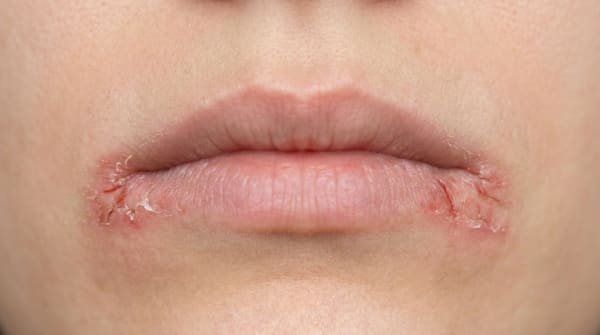
The cover image above is an AI-generated illustration based on the most common visible features of this condition described in clinical pathology references. It is not a photograph of a real case and should not be used to diagnose or rule out the condition in your own situation. If you are concerned about something you have noticed, please book an assessment with a registered dental practitioner.
References
Regezi, J. A., Sciubba, J. J., & Jordan, R. C. K. (2017). Oral pathology: Clinical pathologic correlations (7th ed.). Elsevier. Chapter 2, Ulcerative Conditions: Tuberculosis, pp. 31 to 34.
Neville, B. W., Damm, D. D., Allen, C. M., & Chi, A. C. (2023). Oral and maxillofacial pathology (5th ed.). Elsevier. Chapter 5, Bacterial Infections: Tuberculosis, pp. 186 to 189.
Cawson, R. A., & Odell, E. W. (2017). Cawson's essentials of oral pathology and oral medicine (8th ed.). Elsevier. Chapter 12, Diseases of the Oral Mucosa: Tuberculosis, pp. 211 to 212; Chapter 26, Cervical Lymphadenopathy: Tuberculous Cervical Lymphadenopathy, pp. 368 to 369.
Laskaris, G. (2003). Color atlas of oral diseases (3rd ed.). Thieme. Chapter 17, Bacterial Infections: Tuberculosis, pp. 158 to 160.
Frequently asked questions
What is oral tuberculosis?
Oral tuberculosis is an uncommon manifestation of TB caused by Mycobacterium tuberculosis. It usually appears as a chronic, painful, ragged ulcer with undermined edges, most often on the tongue or soft palate. Primary oral TB is rare; most cases are secondary to pulmonary TB and develop when infected sputum carries the organism into the mouth.
Who is at risk?
Risk factors mirror systemic TB risk — birth or long stays in TB-endemic countries, close contact with infected people, HIV infection, immunosuppression, diabetes, malnutrition, homelessness, and certain occupational exposures. Australia's TB rates are low but rising in specific groups, so awareness matters.
How is it diagnosed?
Diagnosis requires biopsy with Ziehl-Neelsen staining for acid-fast bacilli, culture and PCR for M. tuberculosis. Chest x-ray and sputum studies look for pulmonary disease. Interferon-gamma release assay (QuantiFERON-TB Gold) and tuberculin skin testing help. Differential diagnosis includes squamous cell carcinoma, deep fungal infection and syphilitic ulcer.
How is oral TB treated?
Treatment is the same as systemic TB — a 6-month course of combination antibiotics (isoniazid, rifampicin, pyrazinamide, ethambutol) supervised by infectious diseases or respiratory physicians. The oral ulcers usually heal within weeks of starting treatment. Notification, contact tracing and screening of close contacts are mandatory.