Compiled from clinical pathology references. Medically reviewed by Dr Cristian Dunker, Principal Dentist, ArtSmiles Cosmetic Dentistry.
Quick summary
Also called | Invasive mycoses, deep mycoses, systemic mycotic infections of the mouth |
How urgent? | 🔴 See a dentist or doctor promptly, these are uncommon but serious infections, particularly in immunocompromised patients |
Common or rare? | Rare in healthy adults; uncommon overall, more often seen in immunocompromised patients |
Who it affects | Mostly immunocompromised adults (HIV/AIDS, organ transplant, chemotherapy, poorly controlled diabetes); occasionally healthy people after heavy environmental exposure |
Who treats it | Specialist team, infectious disease physicians and oral and maxillofacial surgeons, with the dentist supporting early recognition |
Based on | Regezi, Neville, Cawson |
Most fungal infections of the mouth that we see are oral thrush, a creamy white film caused by Candida that responds to a course of antifungal medicine. Far less common, but important to recognise, is a group of conditions called deep fungal infections, invasive infections caused by environmental fungi that can produce persistent ulcers, deep tissue destruction and, in some cases, life-threatening illness in patients with reduced immunity.
This article from the team at ArtSmiles, reviewed by Dr Cristian Dunker, explains what deep fungal infections are, who is at risk, and why early recognition matters.
What is it?
A deep fungal infection is one that has spread from a surface (skin or mouth lining), mucosa into deeper tissues, invading muscle, blood vessels or bone. In the mouth, the conditions most often involved include:
Histoplasmosis, caused by Histoplasma capsulatum, often acquired from soil contaminated with bird or bat droppings.
Blastomycosis, caused by Blastomyces dermatitidis, found in moist soil and decaying wood.
Coccidioidomycosis ("valley fever"), caused by Coccidioides immitis, common in the southwestern United States, parts of Mexico, and Central and South America.
Paracoccidioidomycosis, caused by Paracoccidioides brasiliensis, prevalent in Latin America.
Mucormycosis ("zygomycosis", "phycomycosis"), caused by Mucor, Rhizopus and related fungi, an aggressive infection particularly seen in patients with poorly controlled diabetes or marked immune suppression.
Aspergillosis, caused by Aspergillus, most often involving the sinuses with possible spread into the palate.
These infections behave differently from each other but share the feature of being deep, invasive and potentially serious.
Who tends to get it?
Deep fungal infections are most often seen in:
Patients with HIV/AIDS with low CD4 counts (a measure of immune-cell levels).
Patients on long-term immunosuppressive therapy for autoimmune disease or after organ transplant.
Patients receiving cancer chemotherapy.
Patients with poorly controlled diabetes (especially for mucormycosis).
Patients on long-term high-dose corticosteroids.
Travellers and residents of endemic regions (places where the disease is naturally found).
Otherwise healthy people occasionally, particularly with histoplasmosis after heavy exposure to bat or bird droppings (cave exploration, demolition work).
In Australia, deep fungal infections are uncommon but are seen in patients who have travelled to or lived in endemic regions, in immunocompromised patients, and in poorly controlled diabetes (particularly mucormycosis after periods of severe illness).
What causes it?
Each deep fungal infection has its own ecological niche, but some general principles apply:
Inhalation of fungal spores from soil, dust or bird/bat droppings is the usual entry point.
The lungs are infected first; the mouth is involved by spread through the bloodstream or directly from the sinuses.
Reduced immunity allows the fungus to spread beyond a contained early infection.
Tissue acidosis (higher acid levels in the body) and high blood sugar in poorly controlled diabetes provide ideal conditions for Mucor and related organisms to invade blood vessels and tissues quickly.
These infections are not transmitted person to person in everyday contact.
How does it develop?
The course depends on the organism:
Histoplasmosis and similar. A primary lung infection, often mild and self-limiting. In immunocompromised hosts, the infection spreads through the bloodstream and may produce a chronic, granulomatous (with small clusters of immune cells) oral ulcer weeks to months later.
Mucormycosis. Often acute and rapidly progressive. Begins in the sinuses, spreads to the orbit and brain in days, and can cause palatal tissue death (necrosis) with a characteristic black eschar. A medical emergency.
Aspergillosis. Usually starts in the sinuses; chronic forms can erode into the palate.
In all cases, the deep nature of the infection means that ordinary surface treatments (mouthwashes, topical antifungals) are not sufficient.
What might you notice?
What it looks like
Common features include:
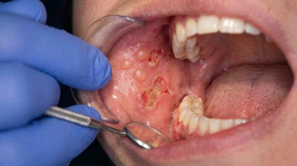
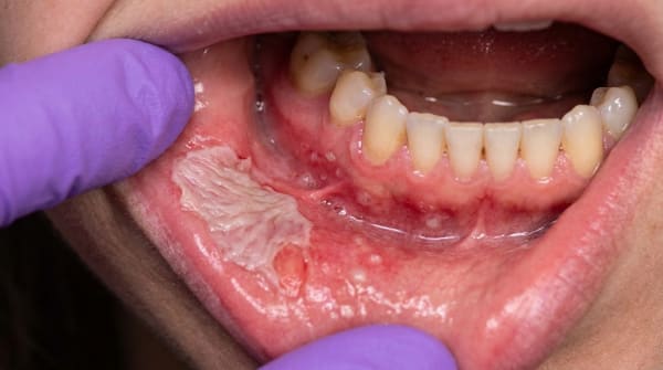
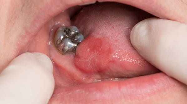
A persistent, painful ulcer in the mouth, often on the palate, gum or tongue, that has not healed despite weeks of conservative treatment.
Raised, granular or warty edges to the ulcer.
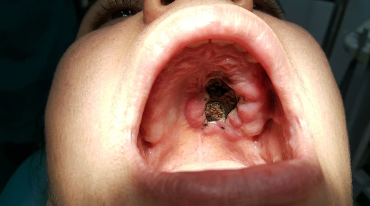
Black or grey patches of dead tissue (eschar), particularly in mucormycosis.
A hole through the palate between mouth and nose in advanced mucormycosis.
Loose teeth in the affected area.
Facial swelling, fever and feeling unwell, especially in invasive infections.
Eye, nose and brain symptoms in advanced mucormycosis (a medical emergency).
In immunocompromised patients, even subtle ulcers should be taken seriously.
What it feels like
Pain at the ulcer is usually the most prominent symptom, often localised to the palate, gum or tongue and worsening over weeks rather than improving. Eating, brushing and speaking can all aggravate the soreness. As deeper tissue is destroyed, you may notice a foul taste or bad breath from the affected area, loose teeth in advanced disease, and general unwellness with fever or fatigue when the infection is invasive.
In severe mucormycosis, eye, nose and brain symptoms can develop within days, alongside swelling and pain across the cheek and forehead. That combination is a medical emergency and warrants same-day hospital assessment, not a wait-and-see approach.
What an X-ray might show
Dental X-rays cannot diagnose deep fungal infections, but they may show advanced changes. In mucormycosis or aspergillosis spreading from the sinuses, an X-ray can reveal cloudiness of the maxillary sinus, erosion of the sinus floor, or moth-eaten bone destruction in the palate. CT and MRI scans, ordered by a specialist, are far more useful for mapping the extent of disease.
What happens at the dentist?
When a non-healing oral ulcer is encountered at ArtSmiles, the visit usually involves:
A thorough history with attention to immune status, current medicines, recent illness, diabetes control, travel history and any environmental exposure.
A careful examination of the ulcer, the surrounding tissues, lymph nodes and the rest of the mouth.
Imaging when needed, panoramic X-ray, sinus X-ray, CT or MRI in suspected sinus or bone involvement.
Urgent referral to an oral and maxillofacial surgeon or oral medicine specialist for biopsy and tissue culture. Special fungal stains (PAS and Grocott methenamine silver, special lab stains for fungi) and culture identify the organism.
Coordination with infectious disease and other specialists depending on the picture.
Supportive dental care while specialist diagnosis and treatment are organised.
A persistent oral ulcer in an immunocompromised patient is treated as urgent until proven otherwise.
Is this serious?
Yes, deep fungal infections are serious because:
Mucormycosis can be life-threatening within days if untreated.
Other invasive fungi can produce significant tissue destruction over weeks to months.
Systemic spread affects lungs, brain and other organs.
Underlying immune compromise complicates both diagnosis and treatment.
Mimics of more common conditions (oral cancer, tuberculosis, syphilitic ulcer) mean that misdiagnosis can delay essential antifungal therapy.
For these reasons, prompt biopsy is the most important step in any persistent oral ulcer in an at-risk patient.
If you've noticed any of these signs for more than two weeks, it's worth booking an assessment.
Could it be something else?
Several conditions can mimic deep fungal infections:
Oral squamous cell carcinoma. A persistent ulcer with raised firm edges in any patient, particularly with smoking or alcohol history.
Tuberculous ulcer of the mouth.
Syphilitic ulcer (chancre and gumma, syphilis ulcers at different stages).
Necrotising periodontal disease in immunocompromised hosts.
Lymphoma of the oral cavity.
Aphthous-like ulcers in HIV infection.
Wegener granulomatosis (granulomatosis with polyangiitis).
Trauma-related ulcer that has not been allowed to heal.
Biopsy with appropriate stains and cultures is the only reliable way to tell these apart.
How is it treated?
Treatment is tailored to the organism, the extent of disease and the patient's immune status. Common elements include:
Systemic antifungal medicines. Liposomal amphotericin B for mucormycosis; itraconazole or other azoles for many others.
Surgical debridement. Particularly for mucormycosis, where dead tissue is removed urgently to limit spread.
Treatment of the underlying immune problem. Better diabetes control, antiretroviral therapy (HIV treatment) in HIV, dose adjustment of immunosuppressants where possible.
Reconstruction. Defects after surgical debridement may need later reconstruction with prosthetics or grafts.
Long-term follow-up to confirm resolution and prevent relapse.
Treatment is led by infectious disease specialists working with oral and maxillofacial surgeons. The dentist's role is early recognition, urgent referral and supportive care.
What's the long-term outlook?
The outlook depends heavily on:
The fungus involved (histoplasmosis better than mucormycosis).
How early treatment is started.
The patient's underlying immune status.
The extent of tissue destruction at diagnosis.
With timely diagnosis and appropriate treatment, many patients recover well. Those with significant immune suppression and advanced disease have a more guarded outlook, which is why prompt referral matters so much.
If you are an immunocompromised patient with a sore in your mouth that has not healed in two weeks, please book an urgent appointment. We will give priority to your case and work with your medical team to get you the right diagnosis quickly.
A note on this article
This article is for educational purposes only and does not constitute a clinical diagnosis. Please consult a registered dental practitioner for assessment and treatment advice.
The cover image above is an AI-generated illustration based on the most common visible features of this condition described in clinical pathology references. It is not a photograph of a real case and should not be used to diagnose or rule out the condition in your own situation. If you are concerned about something you have noticed, please book an assessment with a registered dental practitioner.
References
Neville, B. W., Damm, D. D., Allen, C. M., & Chi, A. C. (2016). Oral and maxillofacial pathology (4th ed., Ch. 6: Fungal and Protozoal Diseases, Histoplasmosis, Blastomycosis, Mucormycosis). Elsevier.
Cawson, R. A., & Odell, E. W. (2017). Cawson's essentials of oral pathology and oral medicine (8th ed., Ch. 17: Mycotic Infections). Elsevier.
Regezi, J. A., Sciubba, J. J., & Jordan, R. C. K. (2017). Oral pathology: clinical pathologic correlations (7th ed., Ch. 2: Ulcerative Conditions). Elsevier.
Frequently asked questions
What are deep fungal infections of the mouth?
Deep (also called systemic or invasive) fungal infections involve the mouth as part of a wider body infection. They include histoplasmosis, blastomycosis, coccidioidomycosis, paracoccidioidomycosis, cryptococcosis, mucormycosis and aspergillosis. They usually present as a chronic non-healing ulcer or a hard nodule, often mimicking oral cancer.
Who gets these infections?
Deep fungal infections are uncommon in healthy people in Australia but are seen more often in travellers returning from endemic areas (Americas for histoplasmosis, blastomycosis, paracoccidioidomycosis), immunocompromised patients (HIV/AIDS, transplant recipients, chemotherapy), and people with poorly controlled diabetes (especially mucormycosis). Aspergillosis can occur after invasive dental procedures in immunocompromised patients.
How are they diagnosed?
Biopsy is essential. Tissue is sent for routine histology (showing characteristic fungal forms), special stains (PAS, Grocott's methenamine silver), fungal culture and sometimes PCR. Imaging (CT, MRI) defines the extent. Investigation of the immune status and search for primary lung or other infection are needed.
How are they treated?
Treatment requires systemic antifungal therapy in collaboration with infectious diseases specialists — amphotericin B, voriconazole, posaconazole, itraconazole or fluconazole, depending on the organism. Mucormycosis often needs aggressive surgical debridement in addition to antifungal therapy. Treating the underlying immunosuppression is critical.