Compiled from clinical pathology references. Medically reviewed by Dr Cristian Dunker, Principal Dentist at ArtSmiles Cosmetic Dentistry.
A denture that has been comfortable for years can quietly start to rub the gum. At first nothing more than a tender spot, but over time, in some patients, the constant pressure produces a soft, folded ridge of tissue right where the denture sits. That ridge is epulis fissuratum.
The name sounds dramatic, but the condition itself is benign and treatable. This article from the team at ArtSmiles, reviewed by Dr Cristian Dunker, explains why it happens, how it is treated, and how to prevent it.
What is it?
Epulis fissuratum (also called denture-induced fibrous hyperplasia or denture epulis) is overgrown soft tissue that develops along the edge of an old or ill-fitting denture. It is the mouth's slow response to long-term mechanical irritation, much like the way the skin produces a callus where shoes rub the foot.
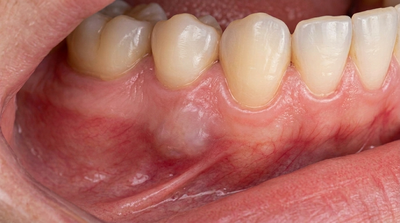
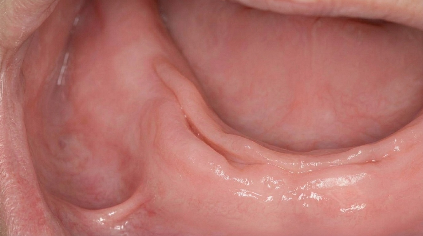
It typically forms a firm, pink, painless fold of tissue running parallel to the denture flange (the edge of the denture that sits in the gum vestibule). The fold often has a fissured groove down its middle, into which the denture flange sits. Sometimes there are several folds stacked on top of each other.
It is not a tumour, not a cancer, and not an infection.
Who tends to get it?
Epulis fissuratum is mostly seen in:
Adults wearing full or partial dentures, particularly long-standing ones.
Older adults, in whom the gum and underlying bone shrink slowly over time, making old dentures progressively looser.
People who continue to wear a denture day and night without giving the tissues a rest.
People who have not had a denture review for several years, when small adjustments could have stopped the rubbing.
It can occur in either jaw and is most common along the front of the upper jaw and the buccal (cheek-side) edges.
What causes it?
The single cause is chronic mechanical irritation from a denture flange that no longer fits. As bone resorbs (shrinks) under a denture over the years, the denture base sits lower and lower, and the flange digs into a slightly different spot. The tissue at that spot:
Is squeezed during chewing.
Is rubbed back and forth as the denture moves during speech.
Slowly develops thickened, scarred connective tissue in response.
The resulting fold of tissue is the body's protective adaptation, but it also keeps the denture in the wrong position and continues the cycle of rubbing.
How does it develop?
The progression is gradual:
A denture that has fit well for years starts to feel loose.
The flange begins to rub against the gum vestibule, often without the patient feeling pain because the rubbing is constant rather than sharp.
The body lays down more fibrous tissue in the irritated area to protect itself.
Over months to years, this tissue builds into a fold or several folds. The flange sits in the groove between folds.
Without intervention, the folds continue to enlarge and the denture fits worse and worse.
Because the tissue is fibrous and collagen-rich, it is firm to touch and cannot simply disappear with denture adjustment alone once it is well established.
What might you notice?
Common features of epulis fissuratum include:
A firm, pink fold of tissue along the edge of an upper or lower denture.
A groove down the middle of the fold, where the denture sits.
Multiple parallel folds, in long-standing cases.
Mild discomfort with chewing, but often no pain at all.
An ulcer in the groove where the flange digs in. This may be tender and slightly red, but it heals quickly when the denture pressure is removed.
A denture that feels loose, rocks or "pops" out during speech or eating.
Bleeding while brushing the gum if the flange crosses the area.
Because the tissue is benign and grows slowly, many patients simply tolerate it for a long time before mentioning it.
What happens at the dentist?
When you mention a lump or fold near your denture at a check-up, a typical visit involves:
A careful look at the lump and the denture together. We see whether the fold sits exactly along the flange and how mobile and snug the denture is.
A check for any unusual features. Most epulis fissuratum is straightforward, but any lump that bleeds easily, has a colour change, ulcerates outside the obvious flange line or grows quickly is investigated more carefully.
A discussion about denture history, how old, how often worn, how often relined, and whether you wear it overnight.
A treatment plan, usually a combination of denture adjustment, possible reline or remake, and (in established cases) minor surgical removal of the folded tissue.
Sometimes a biopsy. If anything about the appearance is unusual or if the lump does not fit the typical pattern, we may remove a small sample for the laboratory to confirm the diagnosis.
We will never tell you to "just live with it" if it is worth treating. A simple denture review can change how comfortable your mouth feels every day.
Is this serious?
Epulis fissuratum is not serious in the medical sense, it is not cancer and it does not spread. The reasons it still deserves attention are:
Comfort. Folded tissue and a loose denture make eating less enjoyable.
Denture stability. A denture that no longer fits properly compromises chewing, speaking and confidence in social settings.
Confirming the diagnosis. Several other lumps in the same area can look similar at first glance, including some that do need urgent attention. A careful examination and, when needed, a biopsy gives certainty.
Bone preservation. A poorly fitting denture continues to cause faster bone loss in the jaw, which makes future denture or implant treatment harder.
Could it be something else?
Lumps in the gum area near a denture may also include:
Pyogenic granuloma. A bright red, soft, easily bleeding lump that grows quickly and is usually triggered by a minor injury or pregnancy.
Peripheral giant cell granuloma. A bluish-red lump on the gum that can erode underlying bone.
Peripheral ossifying fibroma. A firm pink lump on the gum, often near a tooth or denture flange.
Squamous cell carcinoma of the gum. Rare but serious; usually painful, ulcerated, firmer than expected, and not following the line of the denture.
Salivary gland tumour. Rare, but can present as a firm lump in the floor of the mouth or palate.
Mucocele. A bluish, soft, painless cyst from a blocked salivary gland duct, more common on the inner lip.
The reason for this list is to explain why we examine, photograph and sometimes biopsy lumps rather than just trim them off.
How is it treated?
Treatment is a combination of two steps that work together:
Surgical excision of the folded tissue under local anaesthetic. This is usually a quick procedure with sutures, healing over a couple of weeks. In most cases the gum heals smoothly and the original shape of the vestibule is restored.
Adjustment or remake of the denture. Removing the lump without fixing the underlying loose denture means the problem will simply return. Options include relining the existing denture, remaking it, or, in some patients, transitioning to an implant-retained denture for a more stable long-term result.
Smaller, early lesions may regress with denture adjustment alone. Larger, well-established folds nearly always need surgery as well.
After healing, regular reviews, at least once a year, keep dentures fitting well and prevent recurrence.
What's the long-term outlook?
The outlook for epulis fissuratum is excellent. Once the denture is no longer rubbing, the tissue does not come back. With well-maintained dentures, regular reviews and good oral hygiene, you can expect to enjoy comfortable chewing and speaking again. If your denture has not been reviewed for some time and you have noticed a fold of tissue along its edge, please book a visit. We can usually offer a clear plan in a single appointment.
A note on this article
This article is for educational purposes only and does not constitute a clinical diagnosis. Please consult a registered dental practitioner for assessment and treatment advice.
The cover image above is an AI-generated illustration based on the most common visible features of this condition described in clinical pathology references. It is not a photograph of a real case and should not be used to diagnose or rule out the condition in your own situation. If you are concerned about something you have noticed, please book an assessment with a registered dental practitioner.
References
Neville, B. W., Damm, D. D., Allen, C. M., & Chi, A. C. (2016). Oral and maxillofacial pathology (4th ed., Ch. 8: Soft Tissue Tumors, Inflammatory Hyperplasia). Elsevier.
Cawson, R. A., & Odell, E. W. (2017). Cawson's essentials of oral pathology and oral medicine (8th ed., Ch. 13: Reactive Lesions). Elsevier.
Regezi, J. A., Sciubba, J. J., & Jordan, R. C. K. (2017). Oral pathology: clinical pathologic correlations (7th ed., Ch. 5: Connective Tissue Lesions). Elsevier.
Laskaris, G. (2006). Pocket atlas of oral diseases (2nd ed., Denture-Induced Hyperplasia). Thieme.
Frequently asked questions
What causes epulis fissuratum?
It is caused by long-term mechanical rubbing from a denture flange that no longer fits. As the bone under the denture shrinks over the years, the flange digs into a slightly different spot of gum and the body lays down extra fibrous tissue to protect itself, eventually forming a fold.
Is epulis fissuratum cancer?
No. Epulis fissuratum is a benign reactive overgrowth of tissue, not a tumour or cancer. It does not spread. Other lumps in the same area can occasionally look similar, so a careful examination and sometimes a small biopsy is used to be certain of the diagnosis.
Can it heal without surgery?
Small, early folds can sometimes settle once the denture is relined or adjusted so the flange no longer rubs. Larger, well-established folds usually need a small surgical excision under local anaesthetic, combined with denture adjustment, to fix the underlying cause.
Will it come back after treatment?
Not if the denture is properly adjusted, relined or remade after the lump is removed. The condition keeps coming back if the same loose, rubbing denture is worn again. Regular denture reviews, at least once a year, keep the fit good and prevent recurrence.