Compiled from clinical pathology references. Medically reviewed by Dr Cristian Dunker , Principal Dentist, ArtSmiles Cosmetic Dentistry.
Quick summary
Also called | Perlèche, angular stomatitis, angular cheilosis, commissural cheilitis |
How urgent? | 🟡 Worth a check-up, usually settles with the right treatment, but persistent cases can point to an underlying issue worth investigating |
Common or rare? | Common, especially in older adults and denture wearers |
Who it affects | Most often older adults, denture wearers, people with diabetes, immunosuppression, or iron, B12 or folate deficiency; also seen in children who lick their lips habitually |
Who treats it | General dentist, sometimes alongside a GP if a nutritional or medical cause is suspected |
Based on | Regezi, Neville, Cawson, Laskaris |
What is it?
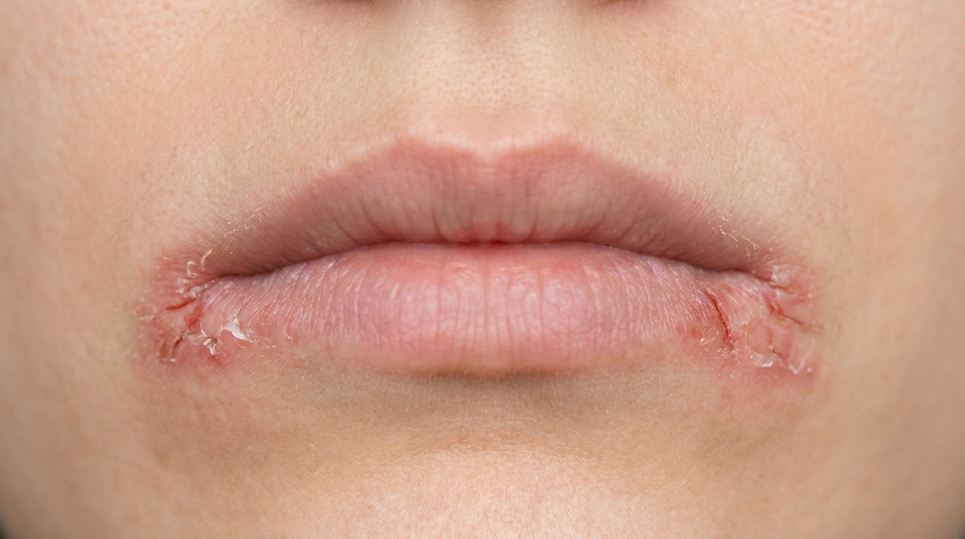
Angular cheilitis is a sore, red, cracked patch at one or both corners of the mouth. The skin in these creases becomes inflamed, cracked (fissured), and sometimes crusted or scaly. It is often a mixed infection, usually involving the yeast Candida albicans together with bacteria such as Staphylococcus aureus, that takes hold in skin folds where saliva tends to pool.
Who tends to get it?
Angular cheilitis is most common in older adults, particularly those who wear complete dentures. The textbooks describe it as especially prevalent in people who have deep folds at the corners of the mouth as a result of mandibular over-closure, that is, when the lower jaw sits a little too close to the upper jaw because the natural height of the bite has been lost.
It also turns up frequently in:
People with iron-deficiency anaemia, vitamin B12 or folate deficiency, or riboflavin (B2) deficiency.
People with diabetes, especially when blood sugar is poorly controlled.
People who are immunosuppressed, including those living with HIV.
Children and adults who habitually lick their lips, suck their thumb, or apply petroleum-based ointments around the mouth.
Patients with Plummer-Vinson syndrome (a rare condition combining iron-deficiency anaemia, glossitis, and difficulty swallowing).
It can affect either sex but is reported slightly more often in women, partly because iron deficiency (low body iron stores) is more common in women of reproductive age.
What causes it?
The sore corners are usually caused by a mixed infection. Microbiological studies cited in the textbooks suggest that around 20% of cases are caused by the yeast Candida albicans alone, around 60% are a mixed infection of C. albicans plus the bacterium Staphylococcus aureus, and around 20% are caused by Staphylococcus aureus on its own. Streptococci can also be involved.
For those organisms to take hold, something usually has to set the stage. The main predisposing factors documented in the source texts include:
Reduced vertical dimension of the bite, in plain language, the lower jaw sits a little too close to the upper jaw because tooth height has been lost (worn-down or missing teeth, or ill-fitting / aged dentures). This deepens the skin folds at the corners of the mouth, where saliva pools.
Denture-related candidal infection, angular cheilitis frequently sits alongside denture stomatitis (a red, inflamed patch on the palate beneath an upper denture). The denture acts as a reservoir for Candida, which then leaks out at the corners with saliva.
Nutritional deficiencies, iron, vitamin B12, folate, and riboflavin (B2) deficiencies are all classically linked to angular cheilitis.
Immunosuppression, uncontrolled diabetes, HIV infection, chemotherapy, long-term corticosteroids (including steroid inhalers), and broad-spectrum antibiotics that disturb the normal oral flora.
Dry mouth (xerostomia), for example from medications, radiotherapy, or Sjögren's syndrome (an autoimmune condition that causes dry mouth and dry eyes).
Lip-licking, thumb-sucking, or constant application of greasy lip balms or petroleum jelly, these keep the corner skin moist and macerated (softened from being constantly wet), which favours yeast growth.
Mechanical trauma, smoking, and chronic exposure to cold or windy weather.
How does it develop?
The corners of the mouth are a naturally tricky neighbourhood. They sit at a fold where two surfaces meet, and saliva can collect and seep in.
If the bite has lost height, say, from worn teeth, missing back teeth, or a denture that has gradually become too short, the upper and lower lips press into each other a little too snugly. That deepens the crease at the corner of the mouth and turns it into a small, warm, damp pocket. Think of it like a poorly drained gutter: water (or in this case, saliva) sits there instead of running away, and the surface underneath softens and breaks down.
Once the skin softens, Candida yeast, which lives harmlessly in many people's mouths, and bacteria such as Staphylococcus aureus find a perfect environment to multiply. The skin reacts with redness, fissuring, and sometimes a yellowish crust. Because the underlying conditions (the deep fold, the denture, the nutritional deficiency) usually persist, the lesion tends to wax and wane unless those drivers are addressed.
What might you notice?
What it looks like
A red, sore, sometimes scaly patch at one or both corners of the mouth. The skin often looks cracked or fissured, and there may be a yellowish crust or small whitish plaques. In long-standing cases, the inflammation can spread a little along the skin folds away from the mouth. When candidal infection extends further onto the surrounding skin (often because someone has been smearing on petroleum jelly), the whole area can look red and crusted, a pattern sometimes called cheilothrush (candida spreading onto the skin around the mouth).
What it feels like
The corners often feel sore, raw, or burning, particularly when opening wide to eat or yawn. Some people describe a sense of dryness or tightness. The lesions tend to come and go in severity rather than disappear completely. There may be discomfort with acidic or salty foods.
Where angular cheilitis sits alongside denture stomatitis, the palate under the upper denture can also look red, although that part is usually not painful.
What an X-ray might show
Angular cheilitis is a soft-tissue condition of the skin around the corners of the mouth, so an X-ray is not used to diagnose it. Imaging may be taken for other reasons during your visit, for example to check the fit of a denture or look at the back teeth that influence your bite height.
What happens at the dentist?
A dentist at ArtSmiles will usually start with a careful look at the corners of the mouth, the lips, the tongue, and the rest of the oral mucosa. They will also check the fit and condition of any dentures and assess the bite height.
Depending on what they find, the assessment may include:
A clinical exam of the rest of the mouth to check for related signs, such as denture stomatitis on the palate, central papillary atrophy on the tongue (a smooth red patch), or thrush (white plaques that wipe off).
A swab or smear of the corner of the mouth and the fitting surface of any denture, to confirm Candida and identify whether Staphylococcus is also involved.
A review of medications and medical history, particularly steroid inhalers, long courses of antibiotics, diabetes control, and any conditions associated with dry mouth.
A referral to a GP for blood tests if the lesion is recurrent or unusually persistent. The textbooks specifically recommend testing for iron deficiency, ferritin, vitamin B12, folate, and a screen for diabetes.
A denture review or remake if loss of vertical dimension is contributing, see the practice's denture-discomfort guidance for related information.
A biopsy is rarely needed for angular cheilitis itself but may be considered if a lesion looks unusual or fails to respond to standard treatment.
Is this serious?
🟡 Worth a check-up. Angular cheilitis itself is not dangerous, but it is uncomfortable and can be persistent. More importantly, it can be a visible signpost to something else worth attending to, a candidal infection elsewhere in the mouth, a vitamin or iron deficiency, poorly controlled diabetes, or an issue with denture fit.
Left untreated, the textbooks note that angular cheilitis can last for a long time, with periods of improvement and flare-up. The cracks can become deeper and more painful, and recurring infection in the same spot is common. In rare cases, persistent angular cheilitis in an adult with no obvious local cause can be one of several signs that prompt screening for HIV infection or another underlying immune problem.
If you've noticed any of these signs for more than two weeks, it's worth booking an assessment.
Could it be something else?
Several other conditions can look similar at the corners of the mouth, and a dentist will usually want to rule them out before settling on the diagnosis.
Herpes labialis (cold sores), can also produce sore, crusted lesions near the lip border. The textbooks describe cold sores as starting with tingling and small clustered blisters that crust over within a few days, whereas angular cheilitis is a more persistent, fissured patch fixed at the corner of the mouth (commissure).
Median lip fissure, a deep vertical crack in the middle of the upper or lower lip, often with secondary candidal or bacterial infection. It looks similar but sits in the centre of the lip, not at the corners.
Actinic cheilitis, chronic sun damage of the lower lip in older adults, often with dryness, scaling, and atrophy. Distinguished by its location across the vermilion (the red border of the lip) of the lower lip rather than at the corners, and by a history of long-term sun exposure.
Exfoliative cheilitis, persistent scaling and crusting of the lip vermilion, typically in younger women and often linked to lip-picking or atopy. Usually affects the whole lip border rather than the angles.
Contact (allergic or irritant) cheilitis, a reaction to lipsticks, toothpastes, foods, or topical products. Tends to follow the area of contact and improves once the offending product is removed.
Plummer-Vinson syndrome, angular cheilitis here is severe and accompanied by a smooth, red tongue, difficulty swallowing, and iron-deficiency anaemia. Diagnosis is confirmed by blood tests and endoscopy.
Cheilocandidiasis, a more extensive candidal infection of the perioral skin, often in someone applying petroleum jelly to the corners of the mouth. Distinguished by its broader spread beyond the commissure.
Riboflavin (B2) and other B-vitamin deficiencies, can produce angular stomatitis with glossitis (inflammation of the tongue). Distinguished by blood tests and the presence of other deficiency signs.
Crohn's disease and orofacial granulomatosis (persistent lip and facial swelling), can occasionally produce persistent fissuring or swelling of the lips and angles. Distinguished by other oral and gastrointestinal features and by biopsy.
Squamous cell carcinoma of the lip or commissure, rare, but worth ruling out in any non-healing lesion at the corner of the mouth in an older patient. A dentist can recognise this by its firmness, induration, and failure to settle with antifungal treatment, and will refer for biopsy.
How is it treated?
Treatment usually has two parts: settling the infection in the skin, and dealing with whatever is keeping it coming back.
At home, helpful measures may include:
Keeping the corners of the mouth gently clean and dry through the day.
Avoiding heavy use of petroleum jelly or thick balms on the affected area, since these can trap moisture and feed the yeast.
Limiting lip-licking.
For denture wearers, taking the denture out at night, brushing it carefully, and soaking it in a recommended cleanser (the textbooks suggest dilute sodium hypochlorite or chlorhexidine overnight for Candida-affected dentures). Other evidences show success leaving the dentures dry and clean in a container at night.
At the dentist or GP, treatment may include:
A topical antifungal cream such as miconazole, applied to the corners of the mouth several times a day. The textbooks note that miconazole and clotrimazole have the advantage of also acting against some bacteria.
A combined antifungal,antibacterial approach when Staphylococcus aureus is also involved. Cawson specifically describes the use of fusidic acid cream alongside antifungal therapy, while Neville mentions iodoquinol-with-corticosteroid creams as another option.
Treatment of any associated intra-oral candidal infection at the same time, for example with nystatin or amphotericin lozenges, or miconazole gel. The textbooks emphasise that angular cheilitis won't fully clear if denture stomatitis or thrush elsewhere in the mouth is left untreated.
Adjusting or remaking dentures to restore proper vertical dimension where over-closure is contributing. This won't always eliminate the deep folds completely, but it often reduces them.
Investigating and correcting nutritional deficiencies (iron, B12, folate, riboflavin) through a GP, which can resolve cases that otherwise keep recurring.
Optimising diabetes control or reviewing steroid inhaler technique (using a spacer and rinsing the mouth after use) where these are relevant.
Treatment typically takes one to two weeks of regular topical application, sometimes longer if an underlying cause needs to be addressed. Resistant or unusually severe cases may need a systemic antifungal such as fluconazole or itraconazole, prescribed by a doctor.
What's the long-term outlook?
For most people, angular cheilitis settles well once the right combination of antifungal and antibacterial treatment is used and the underlying driver is addressed. The texts describe the prognosis for routine candidal-related angular cheilitis as excellent.
The key to keeping it away is dealing with what set it up in the first place. If a denture has lost height, the corners will keep getting sore until that is corrected. If iron levels are low, the skin will remain vulnerable. If a steroid inhaler is being used without a spacer, Candida will keep being seeded back into the mouth. Patients whose underlying factors are addressed, better-fitting dentures, corrected nutrition, controlled diabetes, improved inhaler technique, generally see the lesions stay away.
Where there is an ongoing immune problem, such as HIV infection or chronic mucocutaneous candidiasis (a rare condition with persistent yeast infection of the skin and mucous membranes), angular cheilitis can be more stubborn and may need long-term management alongside the underlying condition.
A note on this article
This article is for educational purposes only and does not constitute a clinical diagnosis. Please consult a registered dental practitioner for assessment and treatment advice.
The cover image above is an AI-generated illustration based on the most common visible features of this condition described in clinical pathology references. It is not a photograph of a real case and should not be used to diagnose or rule out the condition in your own situation. If you are concerned about something you have noticed, please book an assessment with a registered dental practitioner.
References
Regezi, J. A., Sciubba, J. J., & Jordan, R. C. K. (2017). Oral pathology: Clinical pathologic correlations (7th ed.). Elsevier. Chapter 3, White Lesions: Candidiasis (Angular Cheilitis form), pp. 104 to 108.
Neville, B. W., Damm, D. D., Allen, C. M., & Chi, A. C. (2023). Oral and maxillofacial pathology (5th ed.). Elsevier. Chapter 6, Fungal and Protozoal Diseases: Candidiasis, Angular Cheilitis, pp. 201 to 212; and Chapter 17, Oral Manifestations of Systemic Diseases: Iron-Deficiency Anemia and Plummer-Vinson Syndrome, pp. 829 to 831.
Cawson, R. A., & Odell, E. W. (2017). Cawson's essentials of oral pathology and oral medicine (8th ed.). Elsevier. Chapter 12, Diseases of the Oral Mucosa: Candidosis and Angular Stomatitis, pp. 213 to 217; and Chapter 22, Anaemias, Leukaemias and Lymphomas, pp. 336 to 337.
Laskaris, G. (2003). Color atlas of oral diseases (3rd ed.). Thieme. Chapter 13, Diseases of the Lips: Angular Cheilitis, pp. 102 to 103; and Chapter 18, Fungal Infections: Candida-associated Lesions, pp. 166 to 167.