Compiled from clinical pathology references. Medically reviewed by Dr Cristian Dunker , Principal Dentist, ArtSmiles Cosmetic Dentistry.
Quick summary
Also called | Stomatitis medicamentosa, medication-induced oral ulcers, drug-related stomatitis, caused by medical treatment (iatrogenic) oral ulcer formation, mouth lining (mucosa)l drug reaction |
How urgent? | 🟡 Worth a check-up, most heal once the medication is identified, but persistent or large ulcers (especially on the tongue or palate) need assessment |
Common or rare? | Common, adverse drug reactions are increasingly frequent as more people take long-term medications |
Who it affects | Adults, especially those on multiple medications; risk rises sharply once a person is taking five or more drugs |
Who treats it | General dentist working in liaison with the prescribing doctor; specialist referral if ulcers persist |
Based on | Regezi, Neville, Cawson |
What is it?
Drug-induced oral ulceration is the umbrella term for mouth sores that appear as a side effect of a medication. The medical name is stomatitis medicamentosa, which simply means inflammation of the mouth caused by a drug.
The ulcers can look like ordinary aphthous (canker) sores, or like the lacy white patches and erosions of lichen planus (a chronic immune skin and mouth condition), or they can be larger, deeper, and more stubborn. What links them all is that the medication, not an infection or trauma, is the trigger.
Who tends to get it?
Mucosal reactions to systemic medications are very common, and the risk grows with the number of drugs a person takes. Published estimates suggest that taking two medications carries about a 6% risk of an adverse reaction; with five drugs the figure rises to roughly 50%, and with eight or more medications it approaches 100%, in other words, almost everyone on lots of long-term medication will get some kind of mucosal reaction at some point.
Older adults are therefore the most affected group, simply because they are most likely to be on long-term combinations for blood pressure, heart disease, arthritis, diabetes, or mental health. People taking newer biologic and immune-modulating drugs (such as checkpoint inhibitors used in cancer care) are also recognised as a growing group.
Women are diagnosed with chronic drug-related mucosal lesions slightly more often than men, although this likely reflects healthcare-seeking behaviour as much as biology.
What causes it?
Many classes of medication have been linked to oral ulceration. The textbooks divide them by the pattern of reaction they tend to produce.
Drugs that cause non-specific ulcers and erosions:
Nicorandil, a potassium-channel activator used for angina; well known for causing large, painful, persistent ulcers
Methotrexate, a disease-modifying drug for rheumatoid arthritis and some skin conditions
Mycophenolate, used after organ transplants
Nonsteroidal anti-inflammatory drugs (NSAIDs) including aspirin, ibuprofen and naproxen
Cancer chemotherapy drugs, especially methotrexate, 5-fluorouracil, cyclophosphamide and many others
Drugs that produce a lichen planus-like (lichenoid) pattern of striae and erosions:
ACE inhibitors, common blood-pressure medications (captopril, enalapril)
Beta blockers
Thiazide diuretics and furosemide
Allopurinol (used for gout)
Antimalarials
Gold salts and penicillamine (older arthritis treatments)
Methyldopa (an older antihypertensive)
Oral hypoglycaemic (blood-sugar lowering) medications used in diabetes
Some statins (lipid-lowering medications)
Tumour necrosis factor (TNF) antagonists, a type of injectable medicine used for autoimmune conditions, and newer immune checkpoint inhibitors (a class of cancer immunotherapy drugs) (such as pembrolizumab, nivolumab, ipilimumab)
Drugs that produce a fixed drug eruption, a recurring sore that appears in the same spot each time the medication is taken, include barbiturates, sulphonamides, tetracycline, naproxen, paracetamol, gold salts and penicillamine.
Drugs that can cause severe widespread ulceration through erythema multiforme or marrow suppression include sulphonamides, barbiturates, anticonvulsants, antithyroid agents and propylthiouracil (a medication used to treat overactive thyroid).
A detailed medication history is therefore essential whenever an unexplained ulcer turns up.
How does it develop?
Drug reactions in the mouth follow two broad mechanisms.
The first is immune-mediated. Part of the drug acts as a hapten, a small chemical that latches onto a protein in the mucosa and is then mistakenly recognised by the immune system as foreign. Lymphocytes (immune cells) gather at the junction between the surface lining and the deeper tissue, attack the basal cells (the bottom layer of the surface lining), and an erosion or ulcer develops. This is the mechanism behind lichenoid reactions, lupus-like reactions and the pemphigus- and pemphigoid-like patterns described in the textbooks.
The second is direct toxicity. Some drugs (or their breakdown products) damage the rapidly dividing cells of the mouth lining without involving the immune system at all. Cancer chemotherapy mucositis (soreness and ulcers in the mouth from cancer treatment) is the clearest example, the drug kills cells that are turning over quickly, and the mouth, which renews its lining every week or so, takes a heavy hit. Aspirin held against the cheek also burns the mucosa directly.
Think of the lining of the mouth as a high-traffic surface that is constantly resurfacing itself. A drug reaction is like roadworks: either the immune system has dug up a section of road by mistake, or the resurfacing crew has been laid off and the surface starts to break down.
What might you notice?
What it looks like
The appearance varies widely. The textbooks describe patterns ranging from:
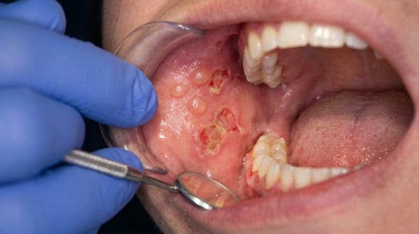
Single or multiple round ulcers with a yellow-grey base and a red rim, very similar in appearance to common mouth ulcers
Larger, irregular, persistent ulcers, most typical of nicorandil, which tends to produce big sores on the side of the tongue, the inside of the cheek or the back of the mouth
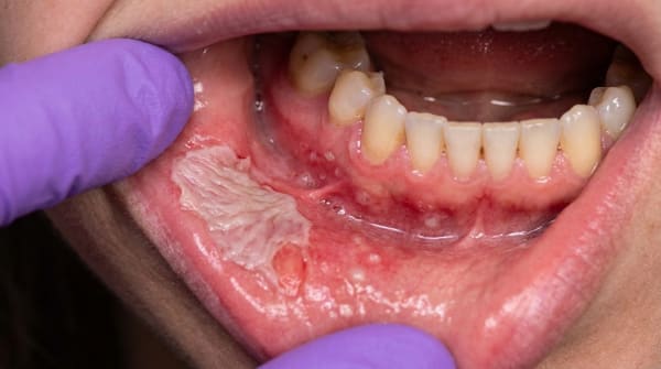
Lacy white striae, atrophic red patches, or shallow erosions resembling lichen planus, often on the inside of the cheek and the side of the tongue
Localised areas of redness or vesicles in the same spot each time a particular medication is taken (the fixed drug eruption pattern)
Widespread erosions and sloughing of the lining in severe immune-mediated reactions
Lesions are often (but not always) bilateral and roughly symmetrical. The most commonly affected sites are the inside of the cheeks, the side of the tongue and the lower lip, the same places where lichen planus usually shows up.
What it feels like
Most drug-induced ulcers are sore, particularly when eating, drinking acidic or spicy foods, or brushing nearby teeth. Burning, stinging or roughness are common. Larger ulcers (especially those caused by nicorandil) can be persistently painful for weeks or months. Striae alone, the lacy white pattern of a lichenoid reaction, may be painless and only noticed by chance.
What an X-ray might show
Drug-induced mouth ulcers are a soft-tissue problem and do not show up on a routine dental X-ray. Imaging is generally not needed for diagnosis.
What happens at the dentist?
The diagnosis is built on history more than on any single test. At ArtSmiles, a dentist will typically:
Take a careful medical and medication history, including prescription drugs, over-the-counter medicines, herbal preparations and recent medication changes. The textbooks emphasise that recently introduced medications are more often the culprit, and that any drug started in the months leading up to the ulcer should be considered.
Examine the mouth in detail, looking at the size, shape, distribution and pattern of the ulcers, and whether they look more like aphthae, lichen planus, a fixed drug eruption, or something else.
Consider liaising with the prescribing doctor, because the most reliable diagnostic step is identifying and, where possible, withdrawing the suspected medication. This is never done by the dentist alone; a substitute drug usually has to be agreed with the original prescriber.
Recommend a biopsy (a small tissue sample sent to the lab for testing) if the ulcer is unusual or persistent, especially if it has not healed after two weeks following drug withdrawal, or if there is any feature that raises concern about another cause. A biopsy in drug-induced ulceration usually shows non-specific inflammatory features, its main role is to rule out other diagnoses.
Refer to an oral medicine specialist if the diagnosis is unclear, if the ulceration is widespread or severe, or if the suspected drug cannot easily be changed.
For lupus-like drug reactions, blood tests for antinuclear antibodies and anti-histone antibodies (blood markers used to look for lupus-like reactions) may be requested. For chronic lichenoid reactions, immunofluorescence testing (a special lab stain that highlights immune-system markers) of a biopsy sometimes helps separate a drug reaction from genuine lichen planus.
Is this serious?
🟡 Worth a check-up. Most drug-induced ulcers are uncomfortable rather than dangerous, and they resolve once the offending medication is identified and changed. However, three points are worth taking seriously:
A few drug reactions (toxic epidermal necrolysis [a severe skin-shedding reaction], severe erythema multiforme, agranulocytosis) can be life-threatening. The mouth is sometimes the first place these show up.
Large, persistent ulcers, particularly those caused by nicorandil, can interfere with eating, sleeping and quality of life for months until the cause is recognised.
Any ulcer that does not heal within roughly two weeks needs assessment regardless of the suspected cause, because mouth cancer can occasionally look similar to a stubborn drug reaction.
If you've noticed any of these signs for more than two weeks, it's worth booking an assessment.
Could it be something else?
Many other conditions can produce ulcers that look a lot like a drug reaction. The textbooks list the following differentials.
Recurrent aphthous stomatitis (mouth ulcers), common round, painful ulcers with a yellow base and red rim. Tend to recur in the same person but not in the same site, and resolve in 7,14 days; drug-induced ulcers may be larger, more persistent, and linked in time to a new medication.
Oral lichen planus, a chronic immune-driven inflammation producing white striae, red patches or erosions, often bilateral. Lichenoid drug reactions look almost identical clinically and on biopsy; the distinguishing feature is the temporal link to a medication and improvement on withdrawing it.
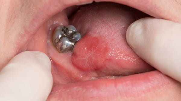
Lichenoid reaction to dental restorations, looks like lichen planus but localised to mucosa in direct contact with an amalgam or composite restoration. Distinguished by its strict relationship to the restoration and improvement after the restoration is removed.
Traumatic ulcer, caused by a sharp tooth, ill-fitting denture or accidental bite. Usually has an obvious mechanical cause and heals once the trauma is removed.
Aspirin or chemical burn, a white, slough-covered area where a tablet, gel or mouthwash has been held against the mucosa. Distinguished by the patient's account of placing the substance against the sore area.
Behçet's disease, recurrent oral ulcers combined with genital ulcers, eye inflammation and skin lesions. Distinguished by the involvement of multiple body sites and a typical demographic pattern.
Erythema multiforme and Stevens-Johnson syndrome (a severe drug reaction with widespread blistering), widespread, severe blistering and ulceration of mouth, lips and skin, often triggered by a drug or by herpes simplex. Distinguished by the rapid onset, target-shaped skin lesions and systemic illness.
Pemphigus vulgaris and mucous membrane pemphigoid, autoimmune blistering diseases producing fragile blisters and erosions. Distinguished on biopsy with direct immunofluorescence; some drugs can trigger pemphigus- or pemphigoid-like reactions, blurring the line.
Lupus erythematosus, can produce oral erosions, striae and red patches very similar to a lupus-like drug reaction. Distinguished by serum antibodies (anti-double-stranded DNA in true lupus) and by the systemic features of lupus.
HIV-associated oral ulcers, large, persistent ulcers in immunocompromised patients. Distinguished by HIV testing and the broader clinical picture.
Necrotising ulcerative gingivitis (and agranulocytosis-related ulceration), painful gum margin ulceration, sometimes triggered by drugs that suppress the bone marrow (agranulocytosis means the white-cell count drops dangerously low). Distinguished by a blood count showing very low white cells.
Cancer therapy-related mucositis, widespread painful ulceration of the non-keratinised mucosa (the soft, unprotected lining inside the cheeks and lips) during chemotherapy or head and neck radiotherapy. Distinguished by the obvious treatment context.
Squamous cell carcinoma (mouth cancer), a persistent ulcer with rolled, indurated (firm and raised) edges. Always needs to be excluded with a biopsy if a suspected drug-induced ulcer fails to heal after withdrawing the medication.
Chronic ulcerative stomatitis and eosinophilic ulcer, rarer ulcerative conditions distinguished by their distinctive biopsy and immunofluorescence findings.
How is it treated?
The single most important step is identifying the drug responsible and, where possible, withdrawing or substituting it, always in consultation with the doctor who prescribed it. Once the medication is stopped, most ulcers heal within days to a few weeks, although some chronic immune-mediated reactions may take months and occasionally need short courses of immune-suppressing medication to settle.
Beyond that, treatment may include:
Symptom relief at home, soft, bland foods; avoiding spicy, salty or acidic items; warm salty mouth rinses; and over-the-counter protective gels or sprays.
Topical anaesthetic gels or sprays for short-term pain relief before meals.
Topical corticosteroid preparations prescribed by a dentist for localised lichenoid or erosive lesions, to dampen the inflammation.
Antifungal preparations if a secondary thrush infection complicates the picture (which can happen with topical steroids).
Systemic corticosteroids or other immune-modulating therapy, used by a specialist for severe or persistent reactions, particularly when the offending drug genuinely cannot be stopped.
Specialist referral to oral medicine, dermatology or the patient's treating physician for complex cases, severe drug reactions, or when the diagnosis remains uncertain.
For chemotherapy-related mucositis, supportive measures (oral hygiene, ice chips during certain infusions, pain relief, infection prevention) form the mainstay; the cancer treatment itself usually cannot be stopped.
What's the long-term outlook?
The outlook is generally good. Most drug-induced ulcers heal completely once the medication is identified and changed, and they leave no scarring. Nicorandil ulcers, for example, typically heal within a few weeks of stopping the drug, even after months of trouble.
Lichenoid drug reactions are slower to settle. The textbooks note that mucosal lesions can persist for months, and occasionally years, after the drug has been stopped, particularly with older agents such as gold salts. A small number of patients need ongoing topical treatment to keep the lesions comfortable.
Reintroducing the drug usually causes the ulcer to come back, which is why a substitute medication is preferred wherever possible. With the right liaison between dentist, doctor and (if needed) specialist, the great majority of people make a full recovery.
A note on this article
This article is for educational purposes only and does not constitute a clinical diagnosis. Please consult a registered dental practitioner for assessment and treatment advice.
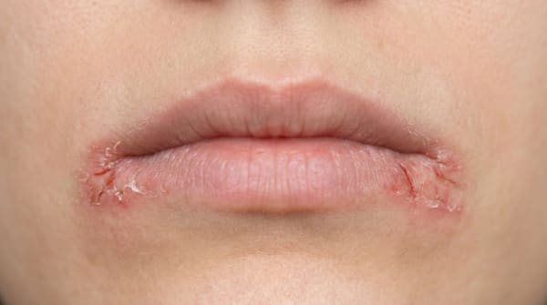
The cover image above is an AI-generated illustration based on the most common visible features of this condition described in clinical pathology references. It is not a photograph of a real case and should not be used to diagnose or rule out the condition in your own situation. If you are concerned about something you have noticed, please book an assessment with a registered dental practitioner.
References
Regezi, J. A., Sciubba, J. J., & Jordan, R. C. K. (2017). Oral pathology: Clinical pathologic correlations (7th ed.). Elsevier. Chapter 2, Ulcerative Conditions: Drug Reactions and Contact Allergies, pp. 47,50.
Neville, B. W., Damm, D. D., Allen, C. M., & Chi, A. C. (2023). Oral and maxillofacial pathology (5th ed.). Elsevier. Chapter 9, Allergies and Immunologic Diseases: Mucosal Reactions to Systemic Drug Administration, pp. 337,342; Chapter 8, Physical and Chemical Injuries: Chemical Injuries of the Oral Mucosa and Cancer Therapy-Related Mucositis, pp. 278,281.
Cawson, R. A., & Odell, E. W. (2017). Cawson's essentials of oral pathology and oral medicine (8th ed.). Elsevier. Chapter 13, Diseases of the Oral Mucosa: Nicorandil-Induced Oral Ulceration and Lichenoid Reactions, pp. 225,228; Chapter 35, Complications of Systemic Drug Treatment, pp. 443,446.