Compiled from clinical pathology references. Medically reviewed by Dr Cristian Dunker , Principal Dentist, ArtSmiles Cosmetic Dentistry.
Quick summary
Also called | Oral findings in HIV, HIV-related oral lesions |
How urgent? | 🔴 Important , many of these lesions can be the first clue to undiagnosed HIV infection or to disease progression in someone already known to have HIV |
Common or rare? | Common in advanced or untreated HIV infection. Modern antiretroviral therapy (ART, the daily medication combination that suppresses HIV) (ART) has dramatically reduced their frequency. |
Who it affects | People living with HIV, particularly those with low CD4 counts (a blood measure of the number of these immune cells) or who are not receiving effective treatment |
Who treats it | General dentist for diagnosis and oral care, in close coordination with the patient's HIV physician or GP |
Based on | Neville, Cawson, with cross-references in Regezi |
What is it?
"Oral manifestations of HIV and AIDS" is the term clinicians use for the cluster of mouth conditions that develop when the immune system has been weakened by Human Immunodeficiency Virus (HIV) infection. The textbooks describe a remarkably distinctive set of oral findings , some of which are so closely tied to HIV that they were among the earliest clues recognised in the AIDS epidemic of the 1980s. With modern combination antiretroviral therapy (ART), most of these lesions have become much less common, but they remain important because they can be the first sign that an undiagnosed person might have HIV, or that a person already known to have HIV may need treatment review.
Who tends to get it?
The textbooks describe a fairly clear risk profile:
People with established HIV infection, particularly those with CD4 counts below about 200 cells per microlitre (the threshold for AIDS).
People not receiving effective antiretroviral therapy , by far the most important risk factor.
People with a delayed diagnosis of HIV , sometimes the dental finding is what prompts the test.
People with treatment failure or poor adherence to ART.
People with co-existing risk factors , heavy smoking, poor nutrition, severe systemic illness, other infections.
Children with vertical transmission , a smaller but important group historically.
What causes it?
HIV itself does not cause most of the oral lesions directly. Instead, by gradually destroying the immune system's CD4 helper T cells (white blood cells that coordinate the immune response, the main target of HIV), HIV allows other organisms , fungi, viruses, and bacteria , to overgrow or reactivate. The textbooks group the typical oral findings as follows:
Fungal infections , particularly oral candidiasis (thrush) caused by Candida albicans.
Viral infections , including oral hairy leukoplakia (Epstein-Barr virus), recurrent herpes simplex, shingles (varicella-zoster virus), severe cytomegalovirus and HPV-related lesions.
Vascular tumours , particularly Kaposi sarcoma (a cancer of blood vessel cells caused by human herpesvirus 8) (caused by human herpesvirus 8, HHV-8).
Lymphomas , particularly non-Hodgkin lymphoma, often in the palate.
Bacterial gum disease , including necrotising periodontal diseases that progress unusually rapidly.
Salivary gland disease , diffuse infiltrative lymphocytosis syndrome (a condition where immune cells crowd into glands, often the saliva glands) causing parotid swelling and dry mouth.
Aphthous-like ulcers , often unusually large, deep and slow to heal.
How does it develop?
Once the CD4 count drops below a critical level, the body can no longer hold these other organisms in check. Erythematous (red) candidiasis typically appears below 400 cells/μL, while the more obvious pseudomembranous (white plaque) candidiasis usually develops below 200 cells/μL. Hairy leukoplakia appears as Epstein-Barr virus replicates unchecked in the lateral tongue. Kaposi sarcoma develops where HHV-8 has lingered in the body for years and finally has the opportunity to multiply in the now-permissive environment. Each lesion has its own mechanism, but all share the underlying story of weakened immune surveillance allowing previously controlled organisms to flourish.
What might you notice?
What it looks like
The textbooks describe a recognisable spectrum of findings:
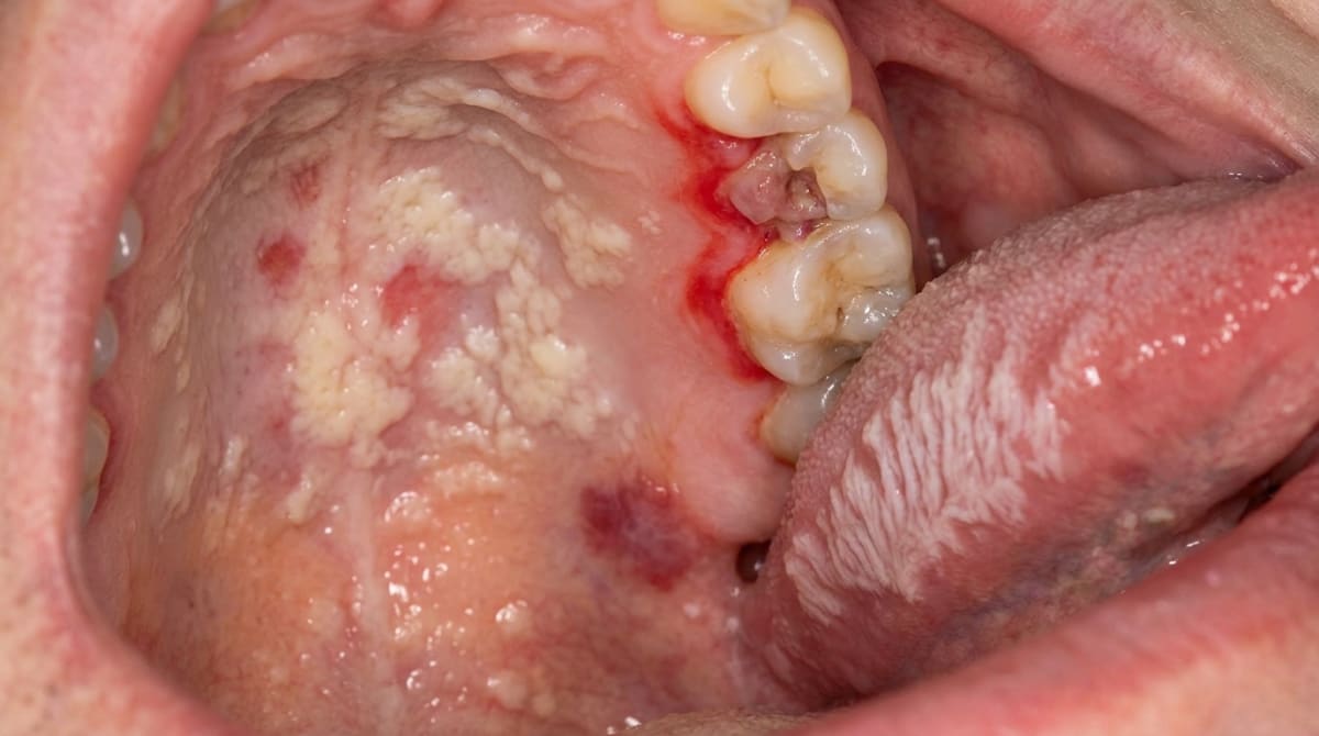
Pseudomembranous candidiasis (the form of oral thrush with creamy white plaques) (thrush) , creamy white plaques on the cheeks, palate or tongue that wipe off, leaving a red base.
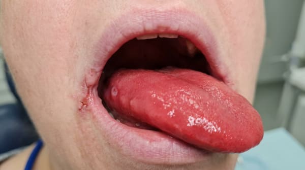
Erythematous (red, smooth) candidiasis , diffuse smooth red areas on the palate or back of the tongue, often without obvious white component.
Angular cheilitis , cracking, redness and soreness at the corners of the mouth.
Oral hairy leukoplakia (a white, corrugated tongue patch driven by Epstein-Barr virus) , vertical white corrugations or shaggy patches on the side of the tongue that do not wipe off.
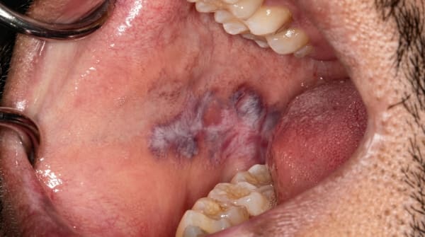
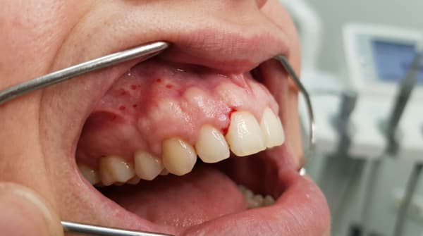
Kaposi sarcoma , purple, brown or red flat or raised patches, most often on the hard palate or gingiva.
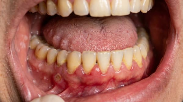
Severe linear gingival erythema (a thin red band along the gum margin) , a bright red band along the gum margin out of proportion to plaque.
Necrotising ulcerative gingivitis or periodontitis (rapidly destructive gum and bone disease with painful tissue death) , rapidly destructive gum disease with painful necrosis and bone loss.
Recurrent or unusually severe herpes , large, persistent or atypically located herpetic ulcers.
Aphthous-like ulcers , often larger than typical aphthae, slow to heal, sometimes recurrent.
Salivary gland enlargement , bilateral parotid swelling with a dry mouth.
Lymphomatous masses , firm, slowly enlarging swellings in the palate or floor of the mouth.
What it feels like
Symptoms depend on the lesion:
Burning, soreness or altered taste with candidiasis.
Pain on eating with severe ulceration or gum disease.
Cracking and pain at the corners of the mouth with angular cheilitis.
Generally no pain with hairy leukoplakia.
Pain, bleeding or tooth mobility in advanced Kaposi sarcoma involving the palate or gingiva.
Dry mouth with parotid enlargement.
Severe gum pain and odour in necrotising periodontal disease.
What an X-ray might show
X-rays may show:
Rapid bone loss in necrotising periodontal disease.
Bone destruction under invasive Kaposi sarcoma or non-Hodgkin lymphoma.
No specific findings in most other manifestations , these are soft-tissue conditions.
What happens at the dentist?
A dentist at ArtSmiles , typically as part of a dental check-up and clean , may sometimes be the first to notice changes that prompt HIV testing. The dentist will typically:
Examine every part of the mouth carefully, recording photographs and detailed notes.
Take a sensitive history about general health, recent illnesses, weight changes, and any known immunosuppression.
Ask about past HIV testing in a non-judgemental way when oral findings suggest it.
Take a fungal swab and tissue biopsy as appropriate to confirm specific diagnoses.
Refer to a GP, infectious diseases physician or HIV specialist when HIV is suspected or known and the oral findings need broader management.
Treat the oral conditions themselves , antifungals, periodontal care, biopsy of suspicious masses, and referral for cancer therapy when required.
Coordinate with the patient's HIV team so that dental care is timed safely around any active HIV-related illness.
Is this serious?
🔴 The lesions themselves vary in seriousness , from completely treatable candidiasis to life-threatening Kaposi sarcoma or lymphoma. The bigger picture is that finding these lesions is often a flag that the underlying HIV is poorly controlled or undiagnosed. The most important action is therefore to confirm or exclude HIV, optimise antiretroviral therapy, and treat the specific oral conditions in parallel. With effective ART, most patients see their oral findings improve dramatically over weeks to months.
If you have noticed unfamiliar mouth lesions and you are not sure what is causing them, it is worth booking an assessment so the right tests can be arranged and care coordinated with your GP.
Could it be something else?
Many of these oral findings can occur in people without HIV. The textbooks emphasise that the same lesions can be caused by other forms of immunosuppression, including:
Diabetes-associated candidiasis , particularly when blood glucose control is poor.
Drug-induced immunosuppression , organ transplant medications, long-term corticosteroids, chemotherapy.
Inhaler-related candidiasis in asthma and COPD.
Smoking-related leukoplakia , distinguished from hairy leukoplakia by location and clinical pattern.
Allergic or contact stomatitis , distinguishable by history and biopsy.
The clinical pattern, history and laboratory tests together usually allow distinction, but biopsy and HIV testing have low thresholds in any unfamiliar oral finding.
How is it treated?
Treatment combines management of the oral lesion and management of the underlying HIV.
At-home measures and habits:
Maintain excellent oral hygiene , gentle brushing twice daily and flossing.
Use a soft-bristled toothbrush and a non-irritating toothpaste.
Adhere strictly to prescribed ART regimens , consistent treatment is the most powerful single change.
Stop smoking , smoking aggravates almost every HIV-related oral condition.
Maintain good general nutrition and hydration.
Attend regular dental check-ups and notify your dentist of any new mouth changes.
Professional steps your dentist may consider:
Antifungal therapy , fluconazole orally for 1 to 2 weeks (often the first choice in HIV-infected patients), or topical agents in milder cases.
Antiviral therapy for severe herpes infections.
Specific therapy for Kaposi sarcoma , initiation of ART often induces regression; locoregional therapies for symptomatic lesions; systemic chemotherapy for advanced disease.
Specialist treatment of non-Hodgkin lymphoma with combination chemotherapy and ART.
Aggressive management of necrotising periodontal disease with debridement, antibiotics (typically metronidazole), oral hygiene support and pain control.
Targeted treatment of hairy leukoplakia with antiviral agents only when symptoms or appearance warrant it; many cases regress on ART alone.
Routine dental care including fillings, scaling and protection of the dentition, with attention to bleeding risk if platelet counts are low.
Coordination of dental procedures with the HIV team to time them around stable phases of treatment.
A patient-centred approach is particularly important here. People with HIV often face stigma, anxiety and complex medical care, and their dental experience should feel respectful, calm and confidential. Honest, unhurried conversation about findings, tests and treatment is itself part of effective care, values that sit at the heart of our clinical philosophy.
What's the long-term outlook?
The outlook for the oral manifestations of HIV has been transformed by modern antiretroviral therapy. Most lesions improve substantially or resolve completely once ART is well established and the immune system recovers. Aggressive conditions such as Kaposi sarcoma and lymphoma still need specialist care, but their incidence has fallen dramatically. The single most important factor in a good long-term outcome is consistent engagement with the HIV treatment team and continuing dental care alongside the medical care.
A note on this article
This article is for educational purposes only and does not constitute a clinical diagnosis. Please consult a registered dental practitioner for assessment and treatment advice.
The cover image above is an AI-generated illustration based on the most common visible features of this condition described in clinical pathology references. It is not a photograph of a real case and should not be used to diagnose or rule out the condition in your own situation. If you are concerned about something you have noticed, please book an assessment with a registered dental practitioner.
References
Neville, B. W., Damm, D. D., Allen, C. M., & Chi, A. C. (2023). Oral and maxillofacial pathology (5th ed.). Elsevier. Chapter 7 , Viral Infections: HIV Infection oral manifestations including candidiasis, oral hairy leukoplakia, Kaposi sarcoma, non-Hodgkin lymphoma, persistent generalised lymphadenopathy and the impact of antiretroviral therapy, pp. 252 to 258.
Cawson, R. A., & Odell, E. W. (2017). Cawson's essentials of oral pathology and oral medicine (8th ed.). Elsevier. Chapter 24 , HIV and AIDS: cross-reference for oral signs and dental management.
Regezi, J. A., Sciubba, J. J., & Jordan, R. C. K. (2017). Oral pathology: Clinical pathologic correlations (7th ed.). Elsevier. Chapter on HIV-related oral lesions.