Compiled from clinical pathology references. Medically reviewed by Dr Cristian Dunker , Principal Dentist, ArtSmiles Cosmetic Dentistry.
Quick summary
Also called | Central papillary atrophy of the tongue, posterior lingual papillitis |
How urgent? | 🟢 Not dangerous, benign and stable; treated mainly to confirm the diagnosis and clear any fungal component |
Common or rare? | Affects roughly 0.01-1% of adults |
Who it affects | Adults; uncommon in children; more common in smokers, denture wearers, people with dry mouth and people on inhaled corticosteroids |
Who treats it | General dentist for diagnosis and antifungal management |
Based on | Neville, Cawson, with cross-references in Regezi |
What is it?
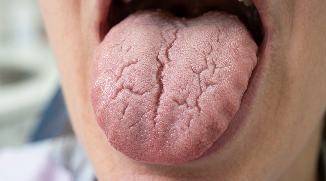
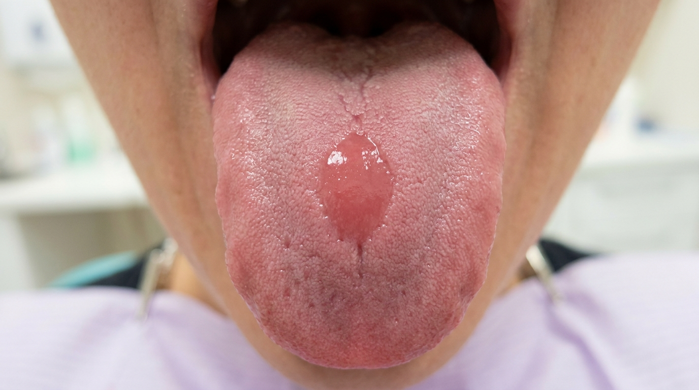
Median rhomboid glossitis is a smooth, well-defined red patch on the central back of the tongue, just in front of the V-shaped row of larger taste papillae (the circumvallate papillae). The textbooks describe it formally as central papillary atrophy of the tongue, a name that more accurately reflects what is happening, since the redness is caused by loss of the small surface "hairs" (filiform papillae, the tiny hair-like projections on the tongue surface) in that area. Originally believed to be a developmental defect, the textbooks now describe it as a form of chronic erythematous (atrophic) candidiasis, a long-standing, often asymptomatic Candida infection of that part of the tongue. It is benign, harmless and usually responds to antifungal therapy.
Who tends to get it?
The textbooks describe a fairly recognisable profile:
Adults, most often middle-aged or older. The lesion is rare in children, which is part of the evidence that it is not truly developmental.
No strong sex predilection.
More common in smokers, particularly heavy long-term smokers.
More common in denture wearers, especially when dentures are worn at night or are poorly cleaned.
More common in people with dry mouth (xerostomia, a dry mouth) from any cause.
More common in people using inhaled corticosteroids for asthma or COPD without rinsing the mouth afterwards.
Sometimes seen with diabetes mellitus or other immunocompromising conditions.
The reported prevalence in adults is around 0.01-1%, with substantial variation depending on the population studied and the criteria used.
What causes it?
The textbooks describe a clear shift in understanding over the past few decades:
Originally thought to be a developmental defect, failure of the embryonic tuberculum impar (a small midline mound of tissue in the developing tongue) to be properly covered by the lateral parts of the developing tongue.
Now widely accepted as a chronic candidal infection of that area. Several lines of evidence support this:
The lesion is rare in children, which would be surprising if it were truly developmental.
Candida albicans is consistently identified in the affected tissue.
Lesions improve with antifungal therapy in most cases.
Similar lesions can be induced experimentally on the tongues of laboratory animals by inoculating them with Candida.
So while a small developmental contribution may exist, the practical view is that median rhomboid glossitis is a long-standing, low-grade Candida infection that should usually be treated as such.
How does it develop?
A small population of Candida albicans yeast cells, normally present in the mouth, settles in the central back of the tongue, a relatively sheltered area where saliva flow is lower. With contributing factors such as smoking, dry mouth or inhaled steroids, the yeast multiplies enough to cause chronic, low-grade inflammation. Over months and years, the inflammation gradually destroys the filiform papillae in that area, leaving a smooth, red, well-demarcated patch. Under the microscope, the textbooks describe a psoriasis-like inflammation pattern with small clusters of immune cells and long, branching yeast threads working their way into the upper layers of the surface lining.
What might you notice?
What it looks like
The classic appearance is well described:
A well-demarcated, smooth, red patch on the central posterior dorsal tongue, just in front of the row of large papillae at the back.
The patch is often lozenge-shaped or oval, hence the historical name "rhomboid".
The colour ranges from bright red to dusky pink.
The surface may be smooth or lobulated, sometimes slightly raised.
The patch is symmetrical and stable in shape.
A matching red patch may appear on the palate where the back of the tongue rests at rest, the "kissing lesion" of chronic multifocal candidiasis.
What it feels like
Most people are unaware of median rhomboid glossitis, since:
It is typically asymptomatic.
Some patients describe a mild burning sensation or sensitivity to hot or spicy foods.
A rough or fuzzy feeling can be reported when the lesion is examined with the tongue.
Severe pain or ulceration is not part of this condition and should prompt other investigation.
What an X-ray might show
Median rhomboid glossitis is confined to the surface of the tongue and does not show on X-rays.
What happens at the dentist?
Median rhomboid glossitis is most often picked up at a routine dental check-up and clean at ArtSmiles when the dentist examines the tongue. The dentist will typically:
Examine the tongue carefully, with the patient sticking the tongue out as far as possible to expose the central posterior surface.
Note the size, shape and stability of the patch and compare with any past records or photographs.
Look for the "kissing lesion" on the palate, which together with the tongue patch points to chronic multifocal candidiasis.
Take a careful history of smoking, denture wear, inhaler use, dry mouth and other contributing factors.
Take a fungal swab from the affected area in selected cases.
Recommend a course of antifungal treatment as both diagnostic and therapeutic, since most lesions improve with antifungal therapy.
Recommend biopsy only when the diagnosis is uncertain, for example, if the lesion is enlarging, ulcerated or has features unusual for median rhomboid glossitis.
Is this serious?
🟢 Median rhomboid glossitis is benign. The textbooks specifically note that carcinoma virtually never develops at this site. The classic textbook concern, historically, was that the appearance of the lesion under the microscope could be mistaken for a carcinoma by a pathologist not familiar with the condition, a concern about misdiagnosis, not about cancer risk. With modern pathology review and an understanding of the candidal nature of the lesion, this is rarely an issue.
If you have noticed a smooth red patch on the central back of your tongue that has been present for some weeks, it is worth booking an assessment so the diagnosis can be confirmed and any antifungal treatment arranged.
Could it be something else?
Several other conditions can produce a red patch on the tongue. The textbooks list these as the main differentials:
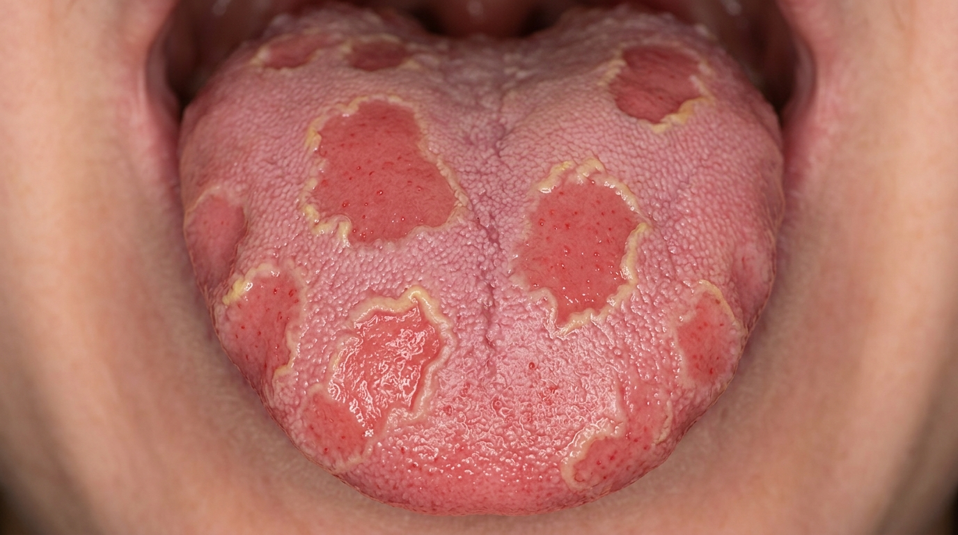
Geographic tongue, produces multiple migrating red patches with serpentine white borders, mainly on the front two-thirds of the tongue, rather than a single central patch.
Chronic hyperplastic candidiasis, can produce thicker white/red plaques rather than a smooth red patch; biopsy distinguishes them.
Atrophic glossitis from anaemia or vitamin deficiency, usually affects the entire tongue surface rather than a central patch.
Erythroplakia, a high-risk red patch in the mouth that needs biopsy; usually irregular and on the floor of the mouth or under the tongue rather than the central dorsum.
Squamous cell carcinoma, uncommon at this site but considered if the lesion is enlarging, ulcerated or has irregular features.
Oral lichen planus atrophic form, can produce red atrophic areas, but usually with white striae and a more bilateral distribution.
How is it treated?
Because the lesion is now widely understood as a candidal infection, antifungal therapy is generally the treatment of choice.
At-home measures and habits:
Stop smoking, the single most important change.
Clean dentures thoroughly every day if you wear them, and avoid wearing them overnight.
Rinse the mouth with water after every dose of an inhaled corticosteroid.
Stay well hydrated and address dry mouth with saliva substitutes if needed.
Maintain excellent oral hygiene including gentle tongue brushing.
Professional steps your dentist may consider:
A course of antifungal therapy, most often topical (such as a miconazole gel or nystatin oral suspension) for 2-4 weeks, or systemic fluconazole in selected cases. Many lesions resolve completely; some only partially.
Treating any associated angular cheilitis with a combined antifungal-corticosteroid cream.
Reviewing predisposing factors, smoking, inhalers, dentures, dry mouth, and addressing them where possible.
Biopsy in atypical cases.
Periodic review, even after a successful course of antifungal therapy, the lesion can recur if the underlying factors are not addressed.
A patient-centred approach matters here too. Many patients are surprised that a "tongue patch" they have had for years is treatable. Calm, clear explanation of the candidal basis, the role of factors like smoking and inhalers, and what to expect from a course of antifungals is itself part of effective care, values that sit at the heart of our clinical philosophy.
What's the long-term outlook?
The outlook is excellent. Median rhomboid glossitis is benign and does not progress to anything dangerous. Most lesions improve with antifungal therapy and management of the underlying factors. Some retain a mild residual change in the central tongue surface, which is harmless. Recurrence is possible if smoking continues, dentures are not cleaned properly or inhaler technique is not improved. Across all situations, regular dental review and consistent management of the contributing factors are the keys to a good long-term outcome.
A note on this article
This article is for educational purposes only and does not constitute a clinical diagnosis. Please consult a registered dental practitioner for assessment and treatment advice.
The cover image above is an AI-generated illustration based on the most common visible features of this condition described in clinical pathology references. It is not a photograph of a real case and should not be used to diagnose or rule out the condition in your own situation. If you are concerned about something you have noticed, please book an assessment with a registered dental practitioner.
References
Neville, B. W., Damm, D. D., Allen, C. M., & Chi, A. C. (2023). Oral and maxillofacial pathology (5th ed.). Elsevier. Chapter 6, Fungal and Protozoal Diseases: Erythematous Candidiasis (Central Papillary Atrophy of the Tongue / Median Rhomboid Glossitis), with chronic multifocal candidiasis "kissing lesion", pp. 203-205.
Cawson, R. A., & Odell, E. W. (2017). Cawson's essentials of oral pathology and oral medicine (8th ed.). Elsevier. Chapter 14, Soft Tissue Disease: Median Rhomboid Glossitis, with the note that carcinoma virtually never develops at this site, p. 249.
Regezi, J. A., Sciubba, J. J., & Jordan, R. C. K. (2017). Oral pathology: Clinical pathologic correlations (7th ed.). Elsevier. Chapter on Red and Blue Lesions: cross-reference for median rhomboid glossitis.
Frequently asked questions
What is median rhomboid glossitis?
Median rhomboid glossitis is a smooth, red, slightly raised rhombus-shaped patch in the middle of the back of the tongue, just in front of the V-shaped row of large papillae. It represents an area where the small papillae are missing and is now recognised as a form of chronic atrophic oral candidiasis.
What causes it?
It was once thought to be a developmental defect, but is now considered a chronic localised Candida infection. Risk factors include smoking, denture wearing, broad-spectrum antibiotics, inhaled corticosteroids, diabetes, immune suppression and dry mouth. A 'kissing' lesion may appear on the palate where the affected area of the tongue touches it.
Is median rhomboid glossitis serious?
No, it is benign and not a precancerous condition. Persistent or sudden enlargement does warrant biopsy because oral squamous cell carcinoma can rarely occur in the same area. The lesion's main clinical interest is as a clue to candida overgrowth and to investigate the underlying risk factors.
How is it treated?
Treatment is by addressing predisposing factors (smoking, denture hygiene, controlling diabetes, rinsing after inhalers) and using topical antifungals such as nystatin suspension or miconazole gel for two to four weeks. The patch may persist after treatment because the missing papillae do not regrow, but the redness and any burning should improve.