Compiled from clinical pathology references. Medically reviewed by Dr Cristian Dunker , Principal Dentist, ArtSmiles Cosmetic Dentistry.
Quick summary
Also called | Mottled enamel, fluorosed enamel, enamel fluorosis |
How urgent? | 🟢 Not urgent, mild dental fluorosis is harmless and primarily a cosmetic concern; severe cases need restoration |
Common or rare? | Common, about 40% of US adolescents show some degree of fluorosis; the figure has risen over the past decades |
Who it affects | Children whose teeth formed during a period of higher-than-recommended fluoride intake from drinking water, fluoride supplements, infant formula made up with fluoridated water, or swallowed toothpaste |
Who treats it | General dentist for assessment and cosmetic restoration where appropriate |
Based on | Neville, with cross-references in Cawson and Regezi |
What is it?
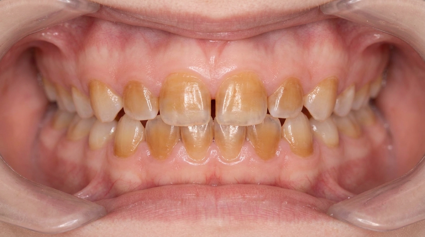
Dental fluorosis is a developmental change in the enamel caused by higher-than-recommended fluoride intake during the time the teeth were forming. The textbooks describe it as one of the recognised forms of enamel hypoplasia, distinguished by its specific cause, fluoride, and its characteristic appearance of lusterless white opacities, sometimes with yellow-brown mottling. Mild fluorosis is harmless and often unnoticed. Moderate to severe fluorosis can produce more visible mottling and, in extreme cases, pitted or chipped enamel. Importantly, fluorosed teeth are more resistant to decay than normally formed enamel, which sits within the wider story of why fluoride remains a fundamental tool in dentistry: most fluoride exposure prevents disease, while excessive exposure during a narrow window of childhood produces the cosmetic changes of fluorosis.
Who tends to get it?
The textbooks describe a fairly recognisable risk profile:
Children whose enamel was forming during a period of higher fluoride intake, most critically the first 3 years of life for the upper front teeth, with the relevant window for back teeth running through to about age 8.
Children given fluoride supplements in areas where the water was already fluoridated, or in addition to other fluoride sources.
Children whose infant formula was reconstituted with fluoridated water during the first year.
Children who swallowed adult toothpaste before they learned to spit it out, the textbooks describe ingestion of fluoride dentifrice as responsible for about two-thirds of fluorosis cases in the United States.
People who grew up in areas with naturally high fluoride water, including parts of East Africa, some areas of India and parts of South-East Asia.
Both sexes equally affected.
The textbooks specifically note that the prevalence of mild fluorosis has risen from about 22.6% of US adolescents in 1986-87 to about 40.7% in 1999-2004, reflecting wider use of fluoride in toothpaste, drinking water and other products.
What causes it?
The cause is well established:
Excess fluoride during enamel formation, particularly in the first 3 years of life when the cosmetically important upper front teeth are being made.
Fluoride retention causes incomplete maturation of the enamel proteins (amelogenins, proteins that organise enamel formation), producing a more porous, hypomineralised (with less mineral than normal, making it weaker) enamel that scatters light and looks chalky white.
Sources of excess intake include:
Drinking water with naturally or artificially high fluoride (above the recommended 0.7 ppm).
Fluoride toothpaste swallowed by young children, the dominant cause in many countries.
Fluoride supplements prescribed where they were not needed, or stacked on top of existing fluoridated water.
Infant formula reconstituted with fluoridated tap water for the entire first year.
Some bottled drinks and processed foods made with fluoridated water.
The dose-response is well documented: more fluoride during the critical window means more severe fluorosis. There is also a genetic component, children with similar fluoride exposures can show different severity of fluorosis, which the textbooks attribute to inherited differences in enamel formation.
How does it develop?
Fluoride incorporated into the developing enamel binds to calcium and changes how the enamel proteins are removed during maturation. The enamel ends up more porous than normal, with retained organic material between the calcium crystals. This porosity scatters incoming light, giving the enamel its characteristic chalky-white appearance. In more severe cases, the porous areas can pick up dietary pigments and discolour over time, producing the brown spots that worry patients. In the most severe cases, the enamel is so under-mineralised that it can chip or pit after the tooth has erupted.
What might you notice?
What it looks like
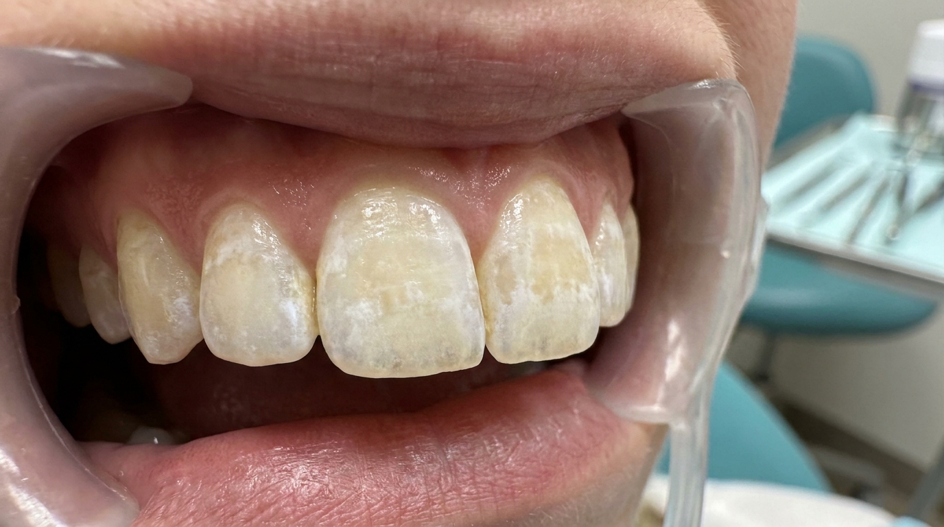
The textbooks describe a recognisable spectrum:
Very mild: small, lusterless paper-white areas on less than 25% of the tooth surface, often only noticeable when the tooth is dried.
Mild: larger but still small white opacities on less than 50% of the surface.
Moderate: most of the enamel surface is involved; may show patchy brown staining.
Severe: enamel is rough and pitted, with widespread brown staining and confluent pits.
The pattern is typically symmetrical and bilateral because both sides of the mouth are exposed to the same fluoride.
The deciduous (baby) teeth are usually less affected because their enamel forms before fluoride dentifrice exposure begins.
What it feels like
Dental fluorosis is generally asymptomatic (causing no symptoms):
No pain or sensitivity in mild and moderate cases.
Mild sensitivity to cold in severe cases where pits or chipped areas expose the underlying dentine (the softer layer beneath the enamel).
Aesthetic concern is the most common reason patients seek treatment.
Lower risk of decay in the affected teeth, fluorosed enamel is more resistant to acid demineralisation (loss of mineral content from enamel) than normal enamel.
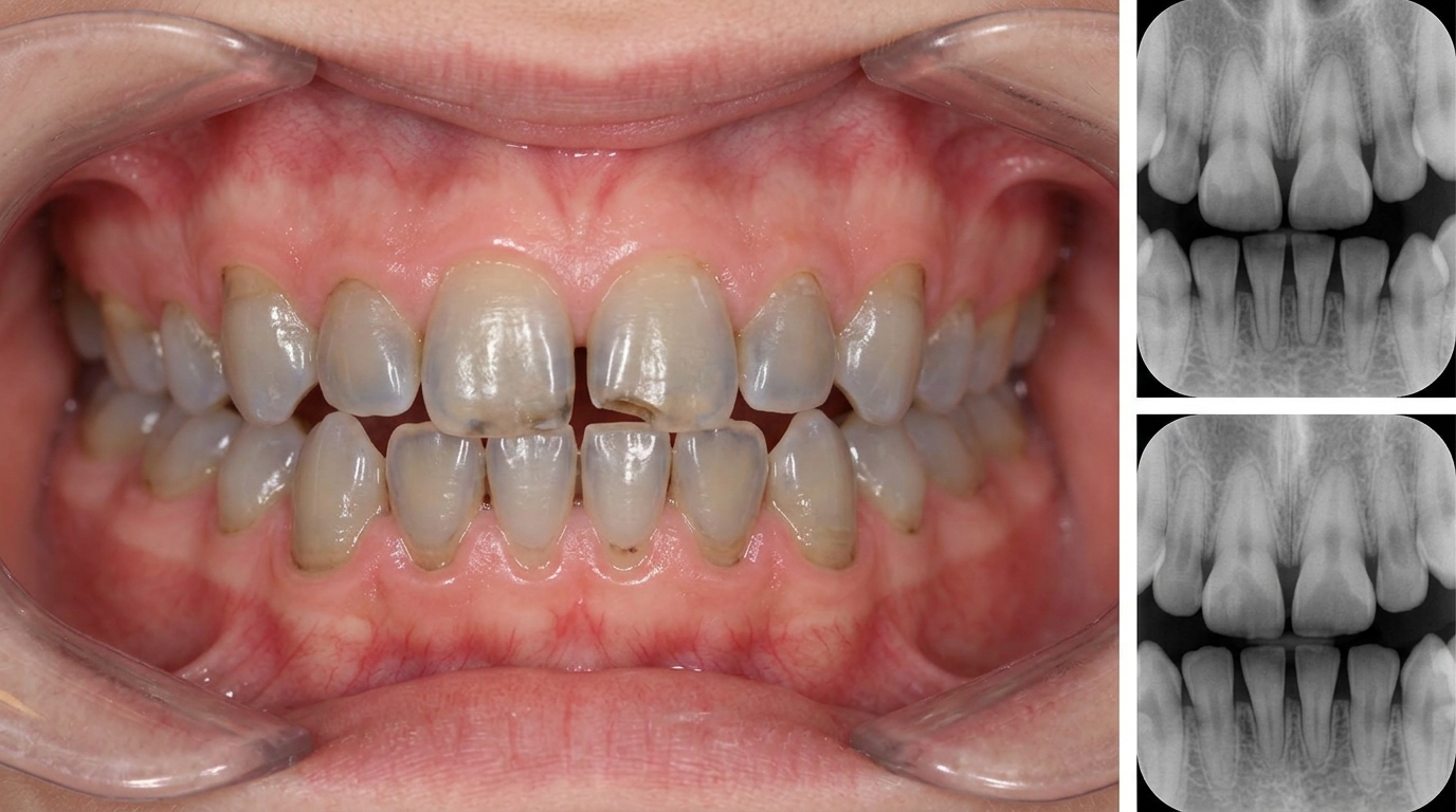
What an X-ray might show
X-rays do not usually show dental fluorosis directly. In severe cases with pitting, the enamel surface may appear less smooth on a high-resolution radiograph.
What happens at the dentist?
Dental fluorosis is most often picked up at a routine dental check-up and clean at ArtSmiles when the dentist examines the teeth. The dentist will typically:
Examine the teeth carefully under good light and after gentle drying, many milder cases are only visible when the enamel is dry.
Take a careful history of childhood drinking water, area of upbringing, infant formula, fluoride supplements and toothpaste habits.
Look for the characteristic bilateral, symmetrical pattern that distinguishes fluorosis from other enamel defects.
Discuss whether treatment is needed based on the severity and the patient's aesthetic concern.
Document the appearance with photographs and clinical records.
Is this serious?
🟢 Mild and moderate fluorosis is benign and primarily a cosmetic concern. Severe fluorosis can produce sensitivity, chipping and a more demanding appearance to manage, but is far less common. The textbooks specifically note that fluorosed teeth are more resistant to decay than normal teeth, a useful balance to keep in mind when discussing the condition. The most important practical message is that dental fluorosis is preventable through careful management of total fluoride intake during the first few years of life.
If you have noticed white or brown mottling on your or your child's teeth that has been there since the teeth came through, it is worth booking an assessment so the diagnosis can be confirmed and aesthetic options discussed if needed.
Could it be something else?
Several conditions can produce mottled or opaque enamel. The textbooks list these as the main differentials:
Environmental enamel hypoplasia from systemic illness or trauma, usually shows pits or grooves rather than the lusterless white opacities of fluorosis.
Amelogenesis imperfecta, hereditary defect of enamel formation, typically affecting all teeth and present in family members.
Molar-incisor hypomineralization (MIH), demarcated white, yellow or brown opacities, usually asymmetrical and involving first permanent molars.
White-spot lesions from early caries, typically near the gum line in plaque-prone areas, with a different distribution from fluorosis.
Tetracycline staining, yellow, grey or brown banding from medication taken during enamel formation.
The combination of bilateral symmetrical distribution, lusterless white opacities and a history of relevant fluoride exposure is the strongest clue toward dental fluorosis.
How is it treated?
Treatment depends on the severity and the patient's wishes.
At-home measures and habits:
Continue using fluoride toothpaste in adult life, it does not cause fluorosis at this point, since enamel formation is complete, and it helps prevent decay.
Use a low-abrasivity toothpaste if affected teeth are pitted or sensitive.
Avoid heavy at-home bleaching without dental advice, bleaching white-spot lesions can sometimes accentuate them.
Photograph the appearance if you want to monitor for any change over time.
Professional steps your dentist may consider:
Microabrasion (gentle removal of a very thin surface layer of enamel), a gentle technique that removes a very thin layer of mottled outer enamel using a mildly acidic abrasive paste; particularly effective for surface white opacities.
External bleaching, alone or after microabrasion, to lighten the surrounding enamel and reduce contrast with the opaque areas.
Resin infiltration (a tooth-coloured resin that fills the microscopic gaps in chalky enamel), a low-viscosity light-cured resin that infiltrates porous enamel and masks white opacities without significant tooth removal. The textbooks specifically note that this approach has been a useful addition to the toolkit.
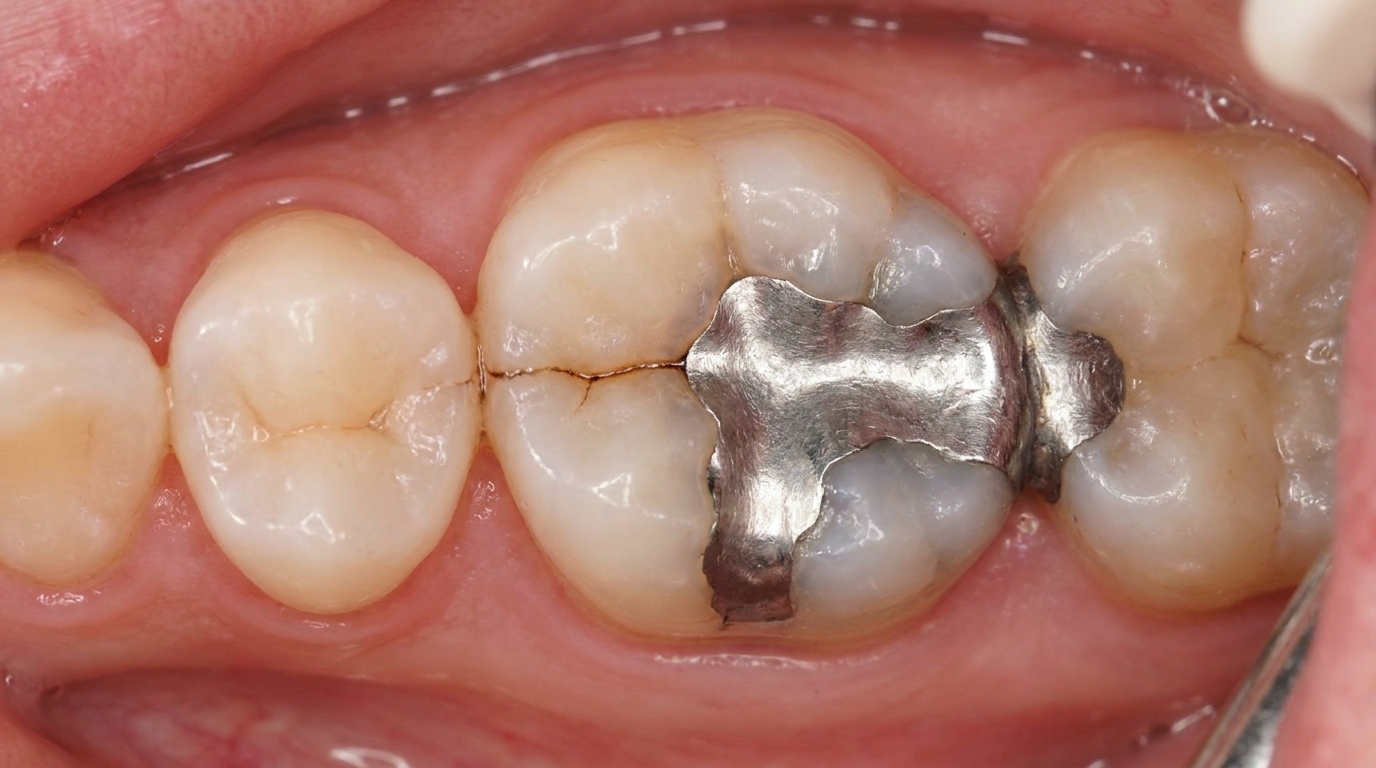
Composite (dental fillings) restorations for chipped or pitted areas.
Veneers for moderate to severe fluorosis where simpler measures are not enough, typically a more invasive option saved for more demanding cases.
Crowns for the most severely pitted teeth.
Long-term review to monitor any new dental issues and to maintain the aesthetic outcome.
A patient-centred approach matters here too. People with fluorosis often grew up worrying about how their teeth looked, and the conversation should respect both the cosmetic concern and the fact that these teeth are usually structurally healthy. Calm, unhurried discussion of conservative options before any invasive treatment is itself part of effective care, values that sit at the heart of our clinical philosophy.
What's the long-term outlook?
The outlook is excellent. Most cases of dental fluorosis remain stable and asymptomatic for life. Mild fluoride-associated opacities often fade slightly over time due to ordinary surface wear and continued surface remineralisation (mineral being put back into enamel). With conservative aesthetic management, microabrasion, resin infiltration or a small veneer where needed, most patients can be entirely comfortable with how their teeth look. The structural advantage of fluorosed enamel (greater caries resistance) is a useful long-term benefit that often goes unrecognised.
A note on this article
This article is for educational purposes only and does not constitute a clinical diagnosis. Please consult a registered dental practitioner for assessment and treatment advice.
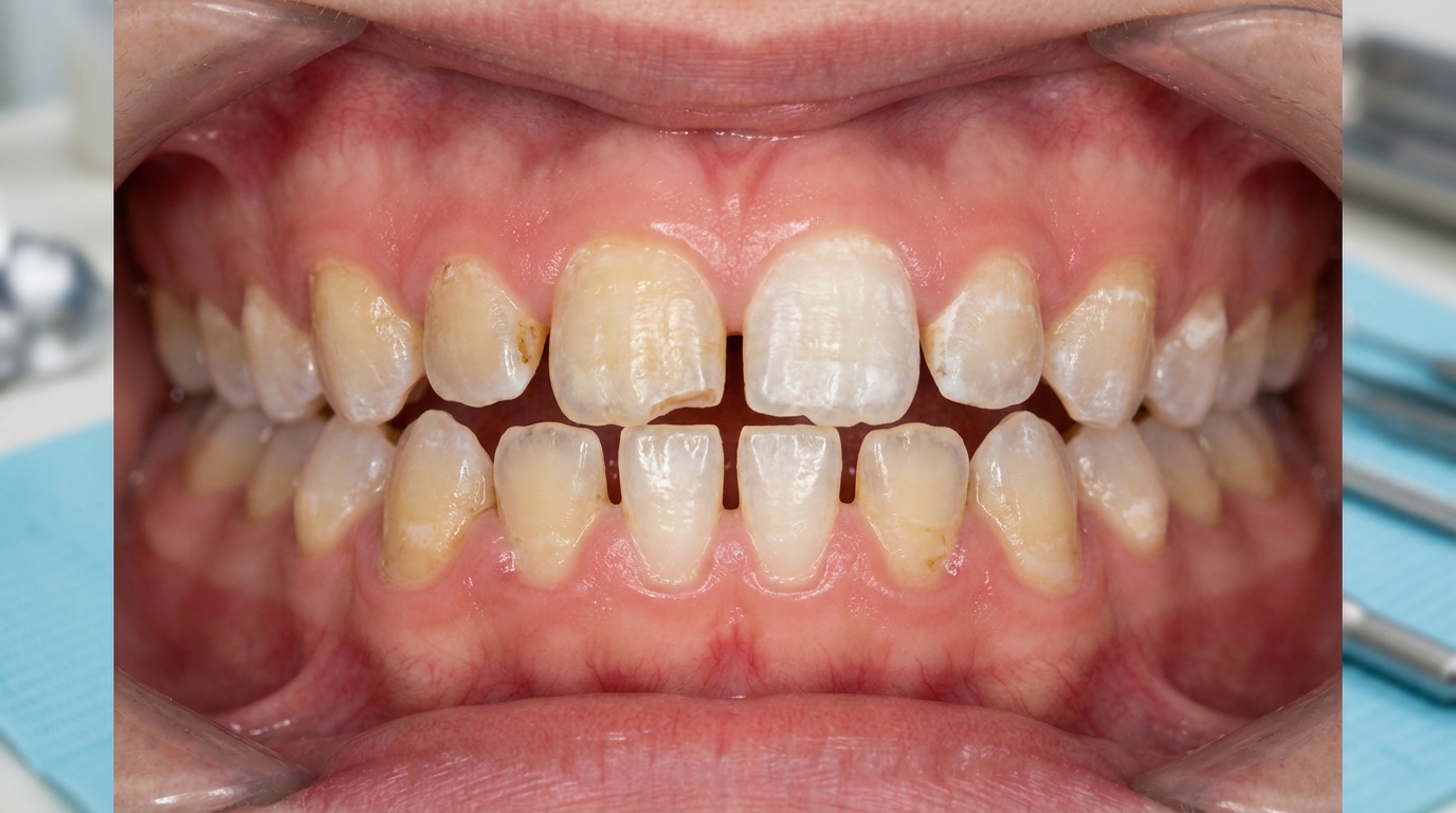
The cover image above is an AI-generated illustration based on the most common visible features of this condition described in clinical pathology references. It is not a photograph of a real case and should not be used to diagnose or rule out the condition in your own situation. If you are concerned about something you have noticed, please book an assessment with a registered dental practitioner.
References
Neville, B. W., Damm, D. D., Allen, C. M., & Chi, A. C. (2023). Oral and maxillofacial pathology (5th ed.). Elsevier. Chapter 2, Abnormalities of Teeth: Dental Fluorosis, with severity dose-dependence and water fluoridation policy, pp. 57 to 58.
Cawson, R. A., & Odell, E. W. (2017). Cawson's essentials of oral pathology and oral medicine (8th ed.). Elsevier. Chapter 2, Disorders of Development: cross-reference for fluorosis as a cause of mottled enamel.
Regezi, J. A., Sciubba, J. J., & Jordan, R. C. K. (2017). Oral pathology: Clinical pathologic correlations (7th ed.). Elsevier. Chapter on Abnormalities of Teeth: cross-reference for dental fluorosis.
Frequently asked questions
What is dental fluorosis?
Dental fluorosis is a change in the appearance of permanent tooth enamel caused by exposure to higher-than-recommended fluoride levels during tooth formation, usually between birth and 8 years of age. Mild fluorosis appears as faint white flecks or lines. Moderate fluorosis gives chalky white patches. Severe fluorosis produces brown stains, pitting and enamel breakdown.
What causes too much fluoride exposure in children?
Common sources include swallowing toothpaste (children under 6 should use a small smear), using high-fluoride toothpaste before recommended, fluoride supplements when local water is already fluoridated, infant formula made up with fluoridated water in young infants, and naturally high fluoride in some bore water. Australian drinking water fluoridation alone (0.6-1.0 ppm) is below the level that causes fluorosis.
How is mild fluorosis told apart from white patches like leukoplakia?
Fluorosis shows symmetrically on multiple teeth that formed during the same period, especially the front teeth and first molars. It has been there since the teeth erupted. Leukoplakia and other adult white patches appear in soft tissue, not on the tooth, and appear later in life. Photos and dental records help confirm fluorosis.
How is dental fluorosis treated?
Mild cases need no treatment. Moderate fluorosis can be improved with at-home or in-chair whitening, micro-abrasion (gentle removal of a thin enamel surface combined with acid), resin infiltration, composite restorations or veneers depending on severity. The aim is to even out the colour and protect the teeth long-term.