Compiled from clinical pathology references. Medically reviewed by Dr Cristian Dunker, Principal Dentist at ArtSmiles Cosmetic Dentistry.
Quick summary
Also called | Environmental enamel (the hard outer layer of the tooth) hypoplasia, Turner hypoplasia, molar-incisor hypomineralization (MIH), demarcated opacities |
How urgent? | 🟡 Worth assessing, affected teeth can be more sensitive and more prone to decay; early restoration can protect them |
Common or rare? | Very common, large studies report enamel defects in around 68% of children, and molar-incisor hypomineralization (MIH) in about 13% |
Who it affects | Children and adults whose teeth formed during a period of illness, malnutrition, trauma or other systemic stress, or whose baby teeth were injured or infected before the adult tooth came through |
Who treats it | General dentist, often in coordination with a paediatric dentist for affected children |
Based on | Neville, with cross-references in Cawson and Regezi |
What is it?
Enamel hypoplasia is the umbrella term for defects in tooth enamel formed during tooth development. The textbooks use the term broadly to describe enamel that is thinner than normal, pitted, grooved, opaque or discoloured because the cells that lay down enamel (ameloblasts), the cells that lay down enamel as a tooth is forming were disrupted during the time that particular part of the tooth was being made. Because enamel cannot remodel itself once formed, these defects are present from the moment the affected tooth comes through and remain visible for life unless the tooth is restored. Most cases are environmental (from a childhood illness or a knock to a baby tooth), with a smaller number caused by genetic conditions such as amelogenesis imperfecta.
Who tends to get it?
The textbooks describe a remarkably broad profile, since enamel formation is sensitive to many influences:
Children whose enamel formed during a period of illness, chickenpox, measles, pneumonia, gastroenteritis, otitis media, urinary tract infections, prolonged fevers.
Children with chronic medical conditions, asthma, cardiac disease, coeliac disease, hypocalcaemia, hypothyroidism, gastrointestinal malabsorption, renal disease.
Children with nutritional deficiencies, vitamin A or D deficiency, generalised malnutrition.
Premature or birth-trauma babies, including breech presentations, hypoxia, multiple births.
Children given certain medications during enamel formation, tetracyclines (now avoided in children), some chemotherapy agents, high-dose amoxicillin.
Children with chromosomal conditions, Trisomy 21 (Down syndrome).
Children whose baby teeth were knocked or developed deep decay, producing localised "Turner teeth" in the underlying adult tooth.
Children with neurological disorders, cerebral palsy, intellectual disability, sensorineural hearing defects.
Children with specific genetic syndromes affecting enamel formation.
The textbooks emphasise that mild defects are extremely common, in one study of 1,500 children in an industrialised nation, 68.4% had some form of enamel defect on permanent teeth.
What causes it?
The cause depends on the pattern:
Systemic environmental hypoplasia, produces bilateral, symmetrical defects on teeth that were forming at the same time. The site of damage corresponds to the area of the tooth being formed during the systemic event. Childhood fevers in the first 2 years of life typically affect the front teeth and first molars; events around age 4-5 affect the canines, premolars and second molars.
Turner hypoplasia, caused by trauma or infection of an overlying baby tooth that damages the developing adult tooth bud. Most commonly affects the premolars (under decayed primary molars) or upper front teeth (after a knock to the baby front teeth).
Molar-incisor hypomineralization (MIH), produces demarcated white, yellow or brown opacities on the first permanent molars, sometimes with the incisors also affected. The cause is uncertain but is linked to early childhood illness, fever, asthma or pneumonia.
Genetic causes, including amelogenesis imperfecta, tricho-dento-osseous syndrome, vitamin D-dependent rickets and others.
Local injury or infection during enamel formation can produce focal defects ("Turner teeth").
The pattern of damage and the timing of any insult often allow the dentist to estimate when the disturbance occurred during childhood.
How does it develop?
Enamel is formed by specialised cells called ameloblasts in three phases, secretion of enamel proteins, transition to mineralisation, and final maturation. When a systemic illness, nutritional deficiency, medication or local injury disrupts these cells, the enamel formed in that exact period is incomplete, abnormally mineralised, or both. Once the disruption ends, the ameloblasts recover (where possible) and continue to lay down normal enamel. The result is a permanent "snapshot" of that disruption in the tooth, visible as a pit, groove, opacity or discoloration in the part of the crown that was being formed at the time.
What might you notice?
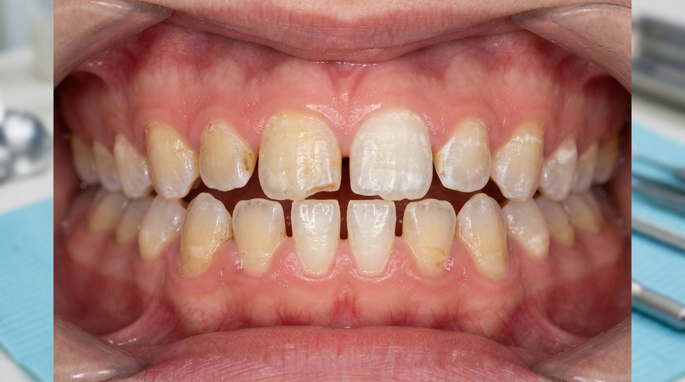
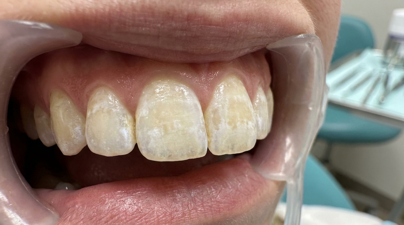
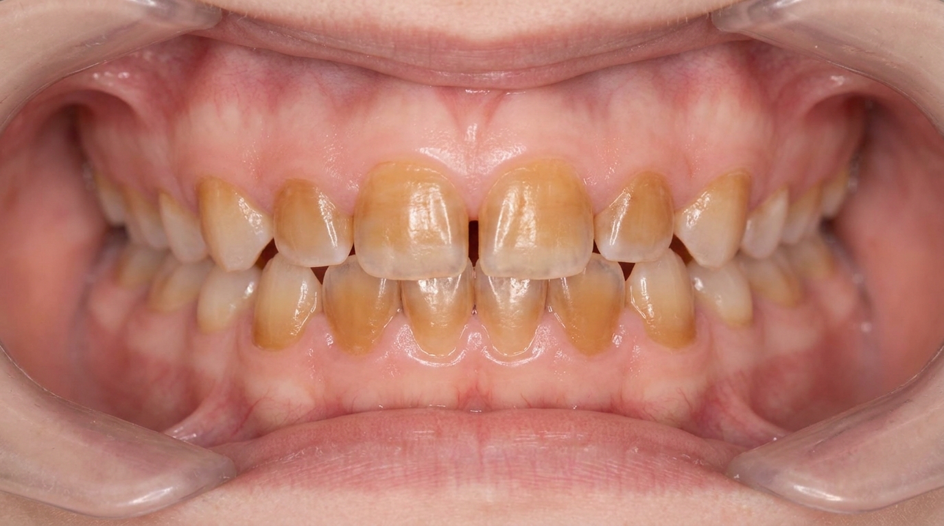
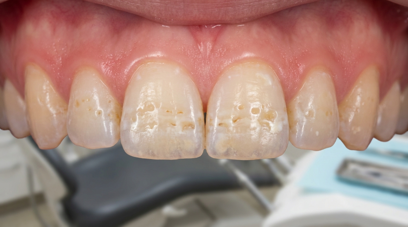
What it looks like
The pattern depends on the cause:
Horizontal rows of pits or thin enamel across symmetrical teeth, typical of a systemic illness during enamel formation.
White, yellow or brown demarcated opacities with a sharp boundary against the surrounding enamel, typical of MIH.
Severe localised hypoplasia of a single tooth (often a premolar or upper front tooth), typical of Turner hypoplasia from a damaged baby tooth.
Generalised pitting and discoloration of all teeth, suggests a generalised cause such as amelogenesis imperfecta or chronic nutritional deficiency.
Yellow or brown opacities are typically more porous and more likely to lose enamel after eruption than white opacities.
Hutchinson's incisors and mulberry molars (notched front teeth and lumpy back teeth, classic signs of congenital syphilis) in the rare case of congenital syphilis.
What it feels like
Sensitivity to cold, sweet or spicy foods, particularly common in MIH-affected molars, where the enamel allows easier passage of stimuli to the pulp.
Pain on toothbrushing in severe cases.
Difficulty achieving local anaesthetic for fillings on MIH-affected molars (well documented).
No symptoms at all in many milder cases, where the patient or parent only notices the appearance.
More frequent decay, the textbooks specifically note that areas of full-thickness enamel hypoplasia are more than twice as likely to develop caries (tooth decay).
What an X-ray might show
X-rays of severely affected teeth may show:
Reduced enamel thickness.
Irregular dentine (the softer layer beneath the enamel) surface beneath an affected tooth.
Pulp chamber narrowing if reactionary dentine (extra dentine the pulp lays down to protect itself) has formed.
Periapical changes in associated baby teeth that caused Turner hypoplasia.
What happens at the dentist?
Enamel hypoplasia is most often picked up at a routine dental check-up and clean at ArtSmiles, often when an adult tooth has just come through. The dentist will typically:
Examine the teeth carefully under good light, noting the pattern, distribution and severity of any defects.
Take a careful history about childhood illnesses, fevers, medications, accidents involving the baby teeth, and family history of similar issues.
Compare patterns, bilateral and symmetrical pattern points to a systemic cause; localised to one tooth points to a local cause; generalised pattern across all teeth raises the possibility of a hereditary condition.
Discuss restoration options appropriate to the age of the patient and the severity of the defect.
Refer to a paediatric dentist or a specialist paediatric medical team when defects suggest an underlying systemic issue.
Plan long-term care with attention to caries prevention, sensitivity management, and aesthetic restoration as the patient grows.
Is this serious?
🟡 Mild enamel hypoplasia is benign and primarily an aesthetic concern. More extensive cases, particularly MIH and severe Turner hypoplasia, produce sensitive teeth, a much higher risk of decay, difficulty with local anaesthesia, and often a need for early and ongoing restorative care. With timely intervention, even severely affected teeth can be saved and made comfortable, but late-managed cases sometimes require crowns, root canal treatment or extraction with orthodontic management of the resulting space.
Could it be something else?
Several conditions can produce similar enamel changes. The textbooks list these as the main differentials:
Dental fluorosis, diffuse mottling and pitting from excessive fluoride during development; usually symmetrical and bilateral.
Amelogenesis imperfecta, hereditary defect of enamel formation, typically affecting all teeth in both dentitions and showing in family members.
Dental caries, bacterial decay, with brown or black soft cavities rather than the smooth pits of hypoplasia.
Tetracycline staining, yellow, grey or brown bands across teeth that received tetracycline during development.
Post-eruptive tooth wear, particularly in cases where the apparent "thin enamel" is actually wear from attrition or erosion.
How is it treated?
Treatment depends on the size of the defect, the patient's age and the importance of the affected tooth.
At-home measures and habits:
Brush twice a day with fluoride toothpaste, gently to avoid further wear.
Floss daily, affected teeth often have unusual contours where plaque can accumulate.
Reduce frequency of acidic foods and drinks, affected enamel is more vulnerable.
Use a desensitising toothpaste if affected teeth are sensitive.
Avoid heavy bleaching products without dental advice, they can worsen sensitivity.
Encourage children to attend regular paediatric dental check-ups so defects are caught early and protected.
Professional steps your dentist may consider:
Topical fluoride applications, varnishes or gels, for affected teeth, particularly newly erupted molars.
Casein phosphopeptide-amorphous calcium phosphate (CPP-ACP) to remineralise mild defects and reduce sensitivity.
Resin infiltration to mask white-spot opacities without invasive drilling.
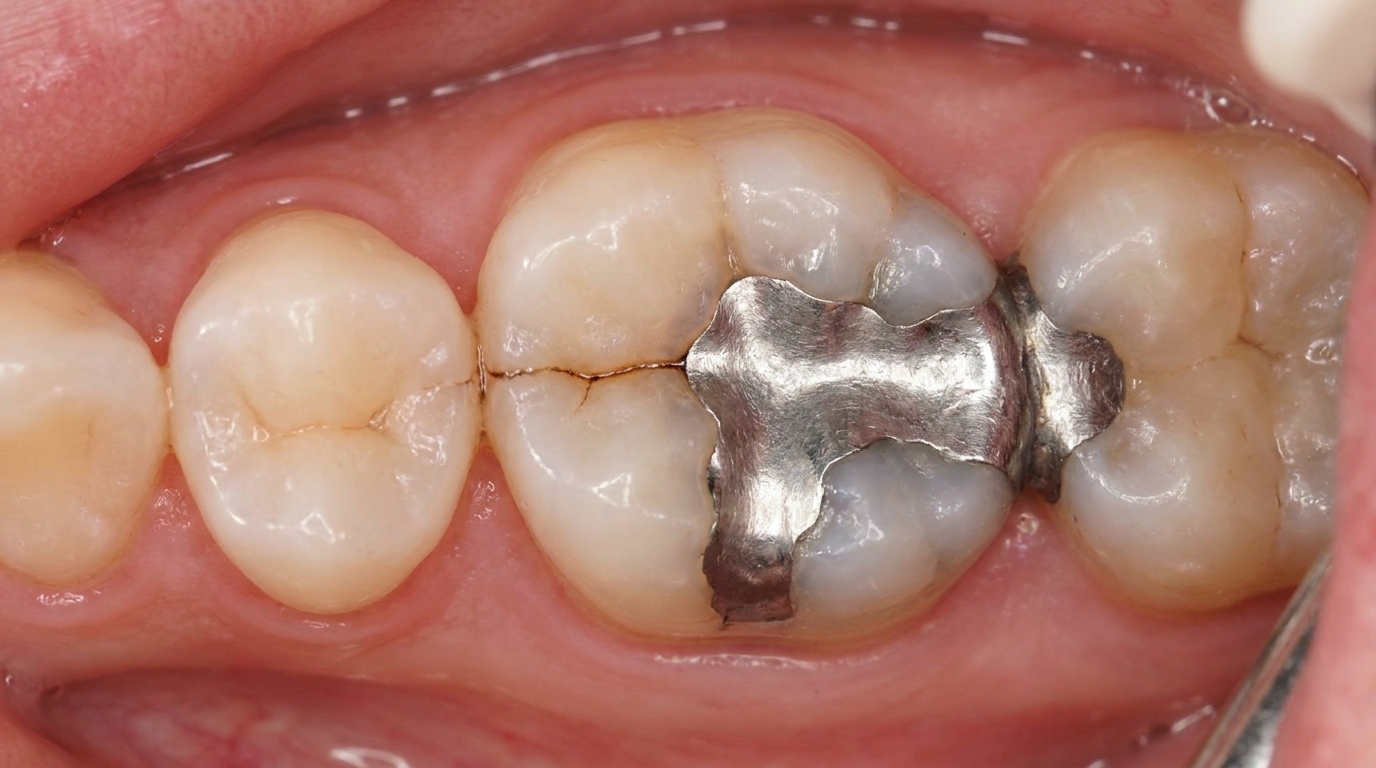
Composite (dental fillings) or resin veneers for moderate aesthetic concerns.
Preformed metal crowns for severely affected first permanent molars in children, replaced later with permanent crowns once growth is complete.
Onlays or full crowns for adults with extensive damage.
Extraction with orthodontic space management in selected severe MIH cases, particularly when the affected molar would require a lifetime of restorations.
Long-term follow-up, since affected teeth tend to need more dental care over a lifetime than unaffected ones.
A patient-centred approach is particularly important when treating children. Calm, age-appropriate explanation, generous use of topical anaesthetic before any injection, and a treatment plan that grows with the child are all part of effective care, values that sit at the heart of our clinical philosophy.
What's the long-term outlook?
The outlook depends on the severity and on whether affected teeth are caught early. Mild defects need only monitoring. Moderate defects can usually be restored with composite or resin techniques and remain comfortable for many years. Severely affected teeth, particularly MIH-affected first molars, often need staged restoration through childhood and adolescence, with full crowns once growth is complete. With consistent dental care, even severely affected teeth can usually be saved, and the long-term outlook for general dental and oral health is good.
A note on this article
This article is for educational purposes only and does not constitute a clinical diagnosis. Please consult a registered dental practitioner for assessment and treatment advice.
The cover image above is an AI-generated illustration based on the most common visible features of this condition described in clinical pathology references. It is not a photograph of a real case and should not be used to diagnose or rule out the condition in your own situation. If you are concerned about something you have noticed, please book an assessment with a registered dental practitioner.
References
Neville, B. W., Damm, D. D., Allen, C. M., & Chi, A. C. (2023). Oral and maxillofacial pathology (5th ed.). Elsevier. Chapter 2, Abnormalities of Teeth: Environmental Effects on Tooth Structure Development, including Enamel Hypoplasia, Diffuse Opacities, Demarcated Opacities, Box 2.1 Factors Associated With Enamel Defects, Turner Hypoplasia and Molar-Incisor Hypomineralization, pp. 51-55.
Cawson, R. A., & Odell, E. W. (2017). Cawson's essentials of oral pathology and oral medicine (8th ed.). Elsevier. Chapter 2, Disorders of Development: cross-reference for environmental and hereditary enamel defects.
Regezi, J. A., Sciubba, J. J., & Jordan, R. C. K. (2017). Oral pathology: Clinical pathologic correlations (7th ed.). Elsevier. Chapter 16, Abnormalities of Teeth: cross-reference for environmental enamel hypoplasia and Turner teeth.
Frequently asked questions
What is enamel hypoplasia?
Enamel hypoplasia is a permanent defect in tooth enamel that occurred during tooth formation. It can appear as thinning, pitting, grooves, or chalky white-to-brown patches on the affected teeth. Unlike adult-onset conditions, the defect is fixed in the enamel from the time the tooth formed and does not progress.
What causes enamel hypoplasia?
Causes include childhood febrile illnesses, premature birth and low birthweight, vitamin D and calcium deficiency, malnutrition, coeliac disease, infections in nearby baby teeth (leading to the so-called Turner tooth in the adult tooth above), trauma to a baby tooth, jaundice in newborns, certain medications (tetracyclines in young children), and chemotherapy in childhood. Most cases have an identifiable timing that matches early childhood.
How is it told apart from fluorosis and amelogenesis imperfecta?
Fluorosis is usually symmetrical, affects several teeth from the same period and has a typical mottled appearance. Amelogenesis imperfecta affects all teeth in both dentitions and runs in families. Single-tooth or localised enamel hypoplasia almost always has a single-event cause (illness, infection, trauma) and the timing fits the affected tooth's formation period.
How is enamel hypoplasia treated?
Minor defects need no treatment beyond maintaining good oral hygiene and topical fluoride. Larger defects on visible teeth can be improved with whitening, micro-abrasion, resin infiltration, composite restorations or veneers. Defects on back teeth that compromise structure are usually restored with composite, onlays or full-coverage crowns to prevent further breakdown.