Compiled from clinical pathology references. Medically reviewed by Dr Cristian Dunker , Principal Dentist, ArtSmiles Cosmetic Dentistry.
Quick summary
Also called | Hereditary enamel hypoplasia, hereditary enamel defect |
How urgent? | 🟡 Worth careful long-term planning, affected teeth tend to wear, chip and stain easily, and benefit from early protection |
Common or rare? | Rare, prevalence ranges from about 1 in 700 in some populations to 1 in 14,000 in others |
Who it affects | People who inherit the relevant gene change, usually with affected family members; both deciduous and permanent teeth involved |
Who treats it | General dentist working with a paediatric dentist, prosthodontist or restorative specialist; long-term staged care |
Based on | Neville, with cross-references in Cawson and Regezi |
What is it?
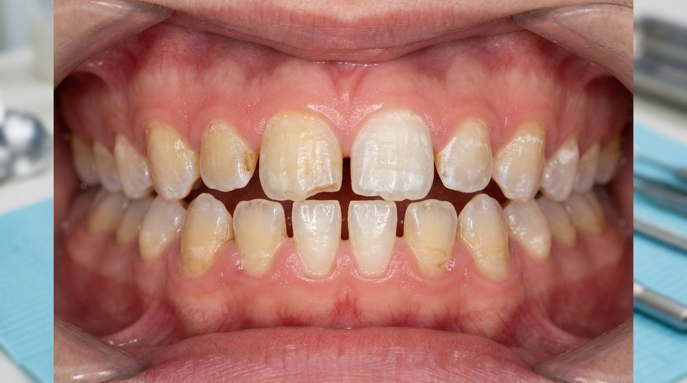
Amelogenesis imperfecta (AI) is a group of inherited conditions in which the enamel, the hard outer covering of the teeth, is not formed properly. The textbooks describe it as a defect of enamel formation that is not associated with any systemic disease or syndrome, although it can occasionally be part of broader genetic conditions. Affected teeth may be thin, pitted, mottled (patchy uneven colour), yellow or brown, fragile and prone to wear and decay. Because the underlying problem is in the genes that control enamel development, both the baby and the adult teeth are typically affected, and other family members often have similar features. With careful long-term restorative care, most affected people can have a healthy, comfortable and aesthetically pleasing dentition.
Who tends to get it?
The textbooks describe a fairly distinctive profile:
People with an inherited gene change affecting one of the proteins or processes involved in enamel formation. Common genes include FAM83H, FAM20A, ENAM and AMELX.
Both sexes affected, with X-linked patterns producing some difference between males and females (more on this below).
Often a family history, children with affected parents commonly inherit the trait, although new mutations also occur.
All teeth in both dentitions affected, the deciduous (baby) teeth and the permanent teeth show similar features.
Variable severity within affected families, even among siblings.
Prevalence varies geographically: about 1 in 700 in Sweden, 1 in 14,000 in the United States, with intermediate figures elsewhere.
What causes it?
The textbooks describe amelogenesis imperfecta as a group of single-gene disorders affecting enamel formation. Inheritance can be:
Autosomal dominant, one copy of the affected gene from one parent is enough.
Autosomal recessive, both parents must be carriers, with a 1-in-4 risk per pregnancy.
X-linked dominant or recessive, the gene sits on the X chromosome, with characteristic differences between males and females (males are affected uniformly; females show vertical stripes of normal and abnormal enamel due to random X-chromosome inactivation in different cells).
The affected genes encode proteins involved in enamel formation (AMELX, ENAM, AMBN), enzymes that process the enamel matrix (MMP20, KLK4), proteins involved in cell adhesion (LAMA3, LAMB3, COL17A1), and others involved in mineral transport, pH sensing and gene regulation. In a study of more than 270 families, just four genes (FAM83H, FAM20A, ENAM and AMELX) accounted for over 60% of cases.
There is no environmental cause, no contagious risk, and no association with diet, fluoride or local infection.
How does it develop?
Enamel forms in three main stages, secretion of an organic protein matrix, transition to mineralisation, and final maturation, all carried out by specialised cells called ameloblasts. In amelogenesis imperfecta, a gene change disrupts one of these stages, producing one of three main patterns:
Hypoplastic AI, the enamel matrix is not laid down in the right amount, so the resulting enamel is thinner than normal but generally hard and properly mineralised within its limited bulk.
Hypomaturation AI, the matrix is laid down at normal thickness but is not properly mineralised during the maturation stage. The enamel is normal in shape but soft, opaque and prone to wear.
Hypocalcified AI, the matrix is laid down but mineralisation barely begins. The enamel is so under-mineralised that it appears "cheesy" and is rapidly lost from most of the tooth, often leaving only a residual band near the gum line.
A fourth combined "hypomaturation-hypoplastic with taurodontism" group shows features of multiple types together. The exact pattern depends on which gene is affected and how.
What might you notice?
What it looks like
The textbooks describe a wide variety of appearances depending on the type:
Generalised pitted hypoplastic, pinpoint-to-pinhead pits scattered across teeth, often arranged in rows on the buccal (cheek) surfaces, with normal-thickness enamel between the pits.
Localised hypoplastic, horizontal rows or linear depressions of hypoplastic enamel, often in the middle third of the buccal surface of teeth.
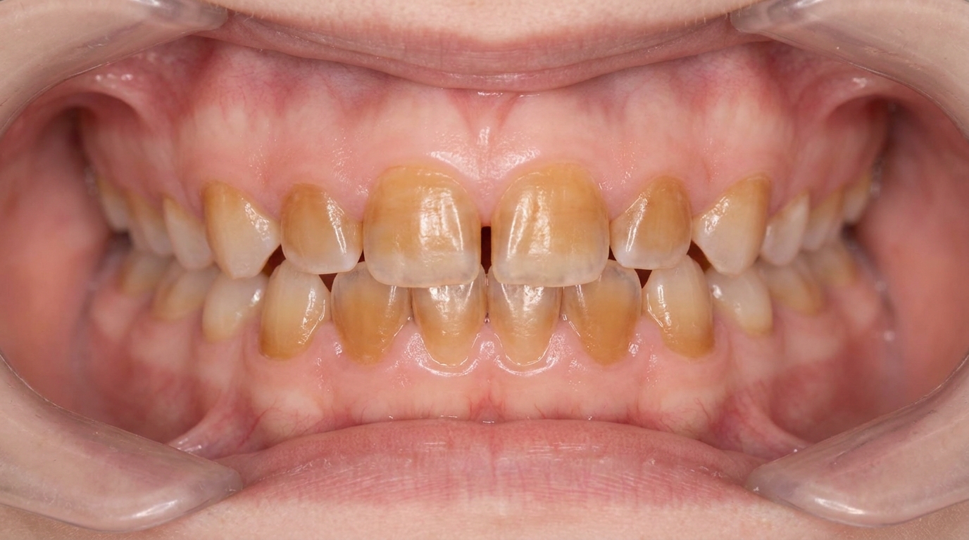
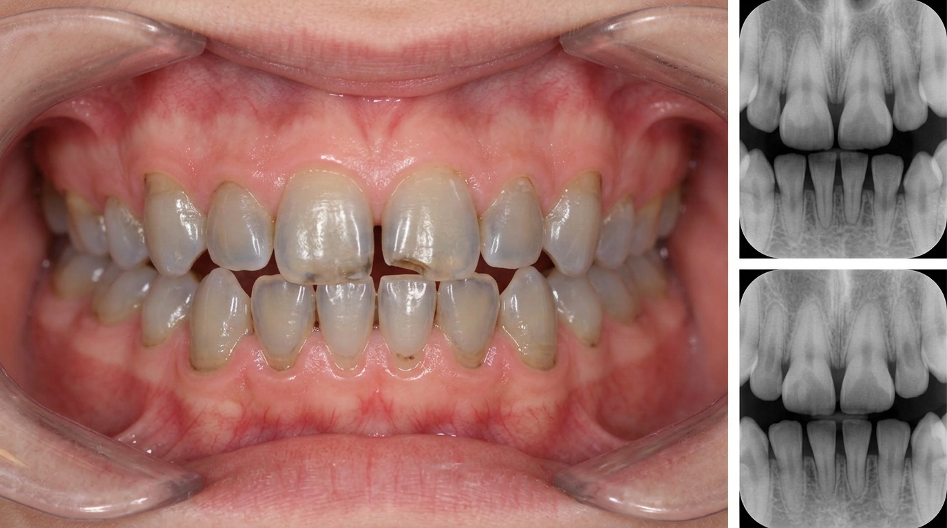
Generalised thin hypoplastic ("smooth"), small, hard, glossy yellow teeth with open contact points between adjacent teeth and often an anterior open bite (a gap between the upper and lower front teeth when biting down).
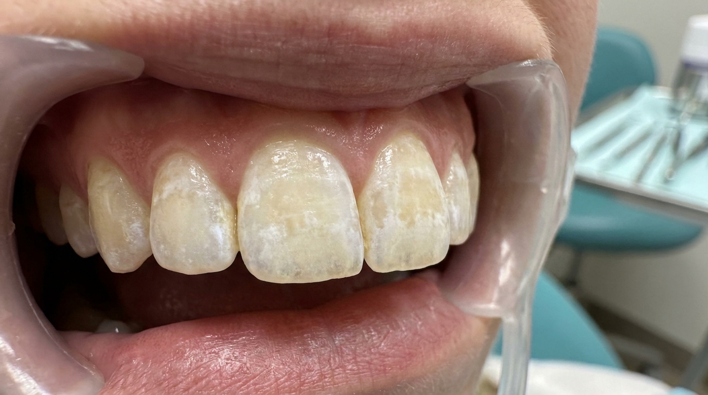
Hypomaturation, teeth of normal shape but opaque, mottled, soft enamel that wears quickly and discolours over time. A "snow-capped" pattern shows white tips on otherwise normal-coloured teeth.
Hypocalcified, soft, cheesy enamel that breaks down rapidly, often leaving teeth that look like dentine pegs with only a residual band of harder enamel near the gum line.
X-linked variants in females show vertical stripes of normal and affected enamel, reflecting random X-chromosome inactivation.
Severe attrition (gradual wear of tooth surfaces against each other) is common because the soft enamel wears quickly against the opposing teeth.
What it feels like
Amelogenesis imperfecta can produce a range of symptoms:
Sensitivity to cold, hot and sweet foods, particularly in hypomaturation and hypocalcified types.
Difficulty chewing when much of the chewing surface has worn away.
Aesthetic concern, small, yellow or mottled teeth often visible when smiling.
Open bite or difficulty meeting the teeth together in some types.
Difficulty with adequate local anaesthesia for fillings, particularly in MIH-style hypomaturation.
Increased plaque retention because of pitted, irregular surfaces.
What an X-ray might show
X-rays typically show:
A thin layer of radiopaque enamel around the teeth, which may contrast normally with the dentine in hypoplastic types or barely show in hypocalcified types.
Open contact points between adjacent teeth.
Anterior open bite or other malocclusion (when the teeth do not meet together properly).
Unerupted teeth showing resorption in some severe forms.
Taurodontism (enlarged pulp chambers in molars) in combined types.
What happens at the dentist?
Amelogenesis imperfecta is most often picked up early in childhood when the deciduous teeth come through with abnormal enamel. A dentist at ArtSmiles, typically as part of a dental check-up and clean, will commonly:
Examine all teeth carefully and document the pattern with photographs and X-rays.
Take a careful family history, since amelogenesis imperfecta usually shows in close relatives.
Distinguish from environmental enamel hypoplasia and dental fluorosis, both of which can look similar but typically have a different pattern and history.
Refer to a paediatric dentist or restorative specialist for staged long-term care.
Discuss genetic counselling for affected families who are planning further children.
Plan early protective measures, sealants, fluoride applications, preformed crowns on first molars, to slow wear and prevent decay.
Coordinate aesthetic and functional restoration as the patient grows.
Is this serious?
🟡 Amelogenesis imperfecta is not life-threatening, but without dental care it can lead to severe tooth wear, sensitivity, decay and loss of teeth. It often has a significant social and emotional impact, particularly during adolescence. With timely and well-coordinated dental care, however, most patients achieve a healthy, comfortable and good-looking dentition for life.
If you have noticed unusual enamel on your child's teeth from when they first came through, particularly if other family members have had similar issues, it is worth booking an assessment so the diagnosis can be confirmed early and a long-term care plan put in place.
Could it be something else?
Several conditions can produce similar enamel changes. The textbooks list these as the main differentials:
Environmental enamel hypoplasia, from childhood illness, trauma to baby teeth, or other systemic events. Usually affects only the teeth forming during the disturbance.
Dental fluorosis, bilateral lusterless white opacities; tied to fluoride exposure rather than family history.
Dentinogenesis imperfecta, hereditary defect of dentine rather than enamel, with translucent grey-blue teeth.
Dentine dysplasia, a related hereditary defect with abnormal dentine and short roots.
Molar-incisor hypomineralization (MIH), environmental rather than hereditary; affects only first permanent molars and possibly incisors.
Tetracycline staining, yellow, grey or brown bands from medication during enamel formation.
The combination of all teeth affected, both dentitions involved, and a positive family history is the strongest clue toward amelogenesis imperfecta.
How is it treated?
Treatment is usually long-term and staged through childhood, adolescence and into adulthood.
At-home measures and habits:
Brush twice a day with fluoride toothpaste, gently to avoid further wear.
Use a soft-bristled toothbrush and consider a desensitising toothpaste.
Avoid acidic drinks that further weaken vulnerable enamel.
Eat a balanced diet with attention to plaque-friendly snacking habits.
Attend regular dental check-ups, especially during childhood and adolescence when staged restoration is being planned.
Professional steps your dentist may consider:
Topical fluoride applications to remineralise and reduce sensitivity.
Fissure sealants (thin protective coatings on the grooves of back teeth) on newly erupted molars to protect deep grooves.
Preformed metal crowns for severely affected primary or permanent molars in children, as a holding measure until growth is complete.
Composite (dental fillings) for moderate aesthetic and functional restoration.
Veneers for the front teeth in adolescence and adulthood.
Full-coverage crowns for posterior teeth and for adult restoration of severely worn front teeth.
Full-mouth rehabilitation in adulthood, often staged over several visits with careful planning of bite, aesthetics and longevity.
Coordination with orthodontics when open bite or other malocclusion is significant.
Genetic counselling for affected families considering further children.
Long-term review, affected teeth need more dental care over a lifetime, and consistent maintenance is the key to a good outcome.
A patient-centred approach is particularly important. Children and adolescents with visible enamel defects can feel self-conscious, and a clear, hopeful long-term plan, explaining that the appearance can be substantially improved with staged care, is itself part of effective care, values that sit at the heart of our clinical philosophy.
What's the long-term outlook?
The outlook depends on the type of amelogenesis imperfecta and the consistency of dental care over a lifetime. Mild forms may need only protective care and small composite restorations. Moderate forms typically need veneers, onlays (lab-made restorations that cover part of the chewing surface) and crowns through adolescence and adulthood. Severe forms (particularly hypocalcified AI) usually need full-mouth rehabilitation with crowns by early adulthood. Across all forms, with consistent dental care, most patients can have healthy, comfortable, attractive teeth for life, and the social and aesthetic impact of the condition can be largely managed. The single most important factor is engagement with regular long-term dental review.
A note on this article
This article is for educational purposes only and does not constitute a clinical diagnosis. Please consult a registered dental practitioner for assessment and treatment advice.
The cover image above is an AI-generated illustration based on the most common visible features of this condition described in clinical pathology references. It is not a photograph of a real case and should not be used to diagnose or rule out the condition in your own situation. If you are concerned about something you have noticed, please book an assessment with a registered dental practitioner.
References
Neville, B. W., Damm, D. D., Allen, C. M., & Chi, A. C. (2023). Oral and maxillofacial pathology (5th ed.). Elsevier. Chapter 2, Abnormalities of Teeth: Amelogenesis Imperfecta, with Witkop classification, modern molecular classification, and the hypoplastic, hypomaturation and hypocalcified types and treatment, pp. 98 to 103.
Cawson, R. A., & Odell, E. W. (2017). Cawson's essentials of oral pathology and oral medicine (8th ed.). Elsevier. Chapter 2, Disorders of Development: cross-reference for hereditary enamel defects.
Regezi, J. A., Sciubba, J. J., & Jordan, R. C. K. (2017). Oral pathology: Clinical pathologic correlations (7th ed.). Elsevier. Chapter on Abnormalities of Teeth: cross-reference for amelogenesis imperfecta.