Compiled from clinical pathology references. Medically reviewed by Dr Cristian Dunker , Principal Dentist, ArtSmiles Cosmetic Dentistry.
Quick summary
Also called | Localised aggressive periodontitis, generalised aggressive periodontitis, juvenile periodontitis, early-onset periodontitis, rapidly progressive periodontitis. In the 2017 World Workshop classification, these conditions are now grouped as Stage III/IV Grade C periodontitis. |
How urgent? | 🔴 See a dentist promptly, bone loss can progress three to five times faster than ordinary gum disease, and teeth can become loose well before the patient feels much pain. |
Common or rare? | Uncommon. The condition affects roughly 1 in 1,000 people overall, with a higher rate in adolescents of African or Middle Eastern descent. |
Who it affects | Children, teenagers, and young adults, typically starting around 11 to 13 years of age, though the generalised form can present anywhere from 12 to 32 years. Often runs in families. |
Who treats it | A general dentist usually identifies it first, but specialist referral to a periodontist is the standard pathway given the speed of bone loss. |
Based on | Neville, Cawson, Laskaris |
What is it?
Aggressive periodontitis is a fast-moving form of gum disease that causes the bone supporting the teeth to break down rapidly, far more rapidly than the amount of plaque on the teeth would explain. It tends to appear in young, otherwise healthy people, and often runs in families. Under the 2017 international classification, it is now described as Stage III or IV, Grade C periodontitis to capture its severity and speed.
There are two patterns dentists recognise. The localised form mostly attacks the bone around the first molars and the front incisors, often in a strikingly symmetrical way. The generalised form is more widespread and involves additional teeth.
Who tends to get it?
Aggressive periodontitis is uncommon. Cawson reports a prevalence of around 1 in 1,000, with males and females affected roughly equally. Neville notes that the localised pattern often begins between 11 and 13 years of age, and adolescents of African or Middle Eastern descent carry around a tenfold higher risk than the general population.
The generalised pattern usually affects a slightly older age band, most patients are between 12 and 32 years. A strong family history is common in both forms, with siblings and parents sometimes carrying the same susceptibility.
It is important to separate this from the early-onset gum destruction seen in certain systemic (body-wide) conditions (Down syndrome, Papillon-Lefèvre syndrome, leukaemia, neutrophil disorders, uncontrolled diabetes, HIV). Those situations have their own causes, even though the gum picture can look similar.
What causes it?
Research points to a combination of three factors working together:
A specific bacterium. Aggregatibacter actinomycetemcomitans (formerly Actinobacillus actinomycetemcomitans) is found in more than 90% of localised disease sites. Certain strains produce a powerful leukotoxin, a chemical that disables the body's first-line defensive white blood cells.
An inherited immune quirk. Many patients have a subtle defect in how their neutrophils (a type of white blood cell) move toward and bind to bacteria. The defect is often genetic, which helps explain why the condition runs in families.
Bacterial transmission within the family. The bacterium is thought to spread between close household members, which is another reason siblings and parents can be affected.
Notably, the amount of visible plaque is often minimal, especially in the localised form. That is one of the distinguishing features: the destruction looks far worse than the level of plaque would predict.
How does it develop?
Think of the bone around a tooth as the foundation under a fence post. In ordinary gum disease, plaque builds up steadily and the foundation slowly erodes, usually over years to decades. In aggressive periodontitis, two things go wrong at once. The bacterial mix shifts toward an unusually destructive species, and the immune response that should clear it does not work properly. The leukotoxin produced by A. actinomycetemcomitans knocks out neutrophils that would normally control the infection, and the gum tissue around the tooth begins to detach from the root. Bone is dissolved away (resorbed) three to five times faster than in standard adult periodontitis. Because the bacterium can also invade the gum tissue itself, mechanical cleaning alone is not always enough to clear it.
The first molars and incisors are typically affected first, partly because they are the longest-erupted permanent teeth, giving the disease the most time to act.
What might you notice?
What it looks like
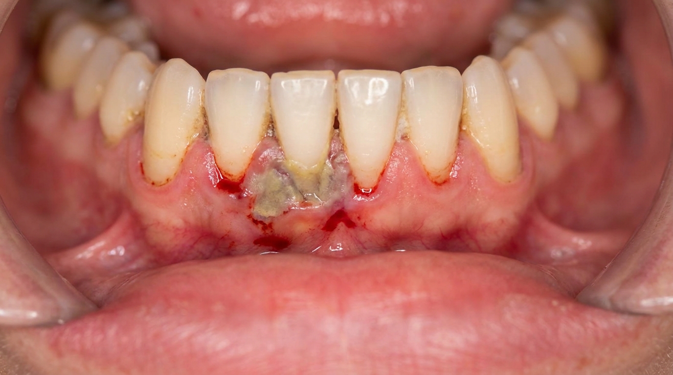
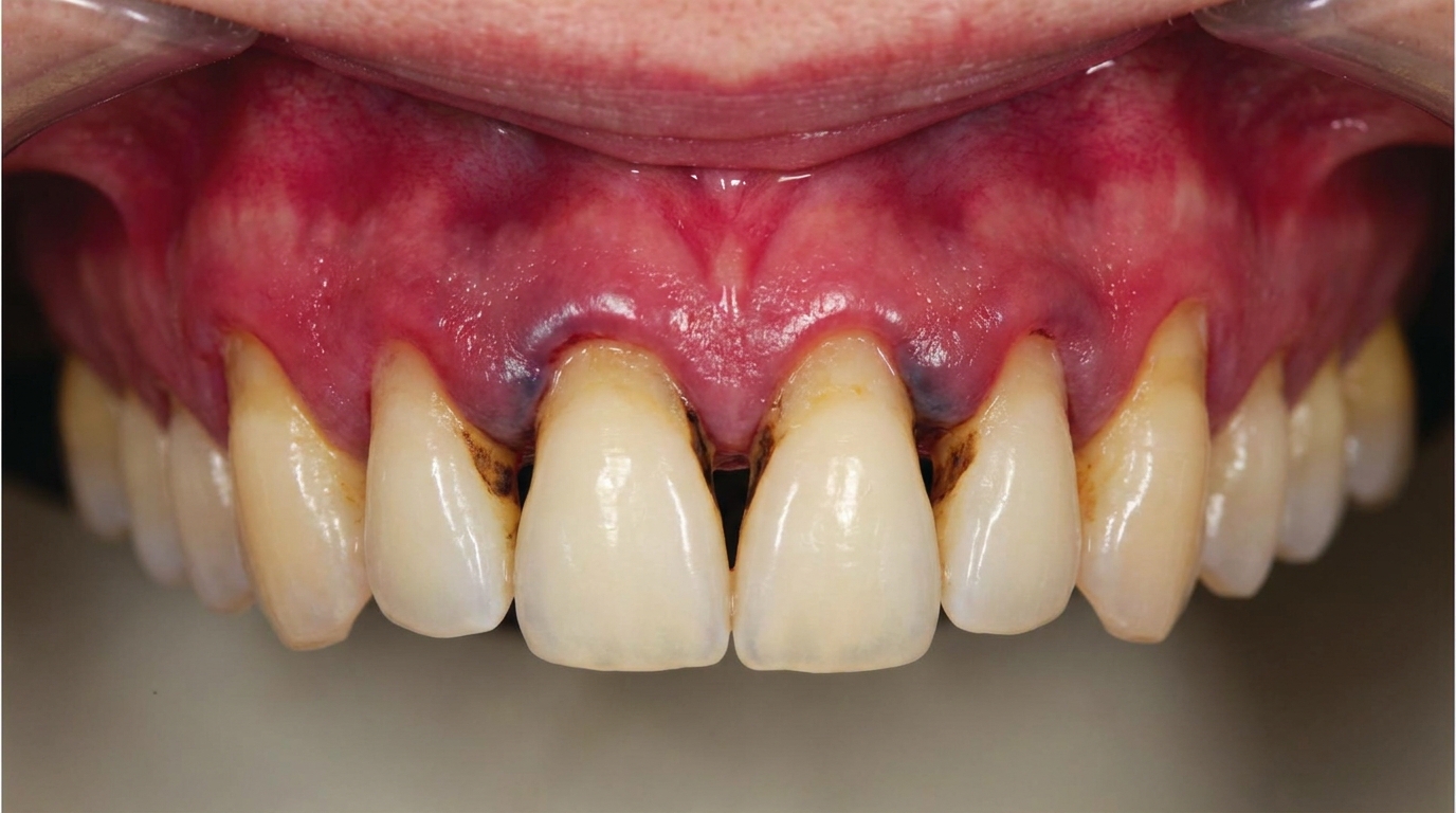
In the localised form, the gums often look surprisingly healthy on the surface, with very little plaque (the soft, sticky bacterial film on teeth) or calculus (hardened plaque, also called tartar) and only mild redness along the margin. The tip-off is usually that the front teeth begin to drift, tilt, or fan apart, sometimes with gaps opening up that were not there before. The first molars and incisors may feel loose.
In the generalised form, more teeth are involved, and the picture is closer to what most people imagine as gum disease, heavier plaque, calculus, swollen and inflamed gums, and bleeding.
What it feels like
Many people are surprised by how little discomfort there is. The condition is often painless in its early stages, which is part of why it can progress so far before being recognised. Possible signs include:
Teeth feeling loose or moving when you bite
Front teeth shifting position or developing new gaps
Bleeding from the gums when brushing or flossing (more common in the generalised form)
Bad taste or persistent bad breath
Tenderness around specific teeth, particularly first molars
What an X-ray might show
Dental X-rays are central to spotting this condition. In the localised form, the radiographs typically reveal vertical, angular bone loss around the first molars on both sides of the mouth, often producing an arc-shaped defect that runs from the back of the second premolar to the front of the second molar. The bone loss around the upper and lower incisors mirrors itself with striking symmetry, sometimes the left and right sides look almost identical. In the generalised form, bone loss is more widespread and may be horizontal, vertical, or both.
What happens at the dentist?
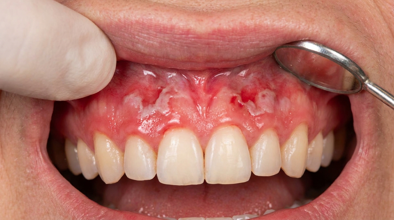
At ArtSmiles, assessment usually starts with a careful history, including any family members who lost teeth young, and a periodontal examination. Your dentist may:
Measure pocket depths around each tooth using a small probe, a pocket is the gap between the gum and the tooth. Pockets deeper than 4 mm suggest the gum has separated from the tooth.
Record clinical attachment loss at multiple sites (the total amount of gum and bone support a tooth has lost), this is considered the gold-standard way to measure how much damage has built up.
Take a full set of dental X-rays to map the pattern of bone loss. Symmetrical vertical defects around first molars and incisors are highly suggestive.
Discuss family history, since the condition often clusters in families.
Consider microbiological testing, a small bacterial sample taken from a gum pocket is sent to the lab to confirm the presence of A. actinomycetemcomitans and guide antibiotic choice.
Refer to a periodontist (gum specialist), particularly when the pattern is consistent with Stage III/IV Grade C disease.
If the picture suggests an underlying systemic cause, for example, very early childhood onset, recurrent skin infections, or unusual nail or skin findings, your dentist may also recommend medical investigations to rule out conditions such as Papillon-Lefèvre syndrome, neutrophil disorders, or undiagnosed diabetes.
Is this serious?
🔴 Yes, this is a condition that needs prompt attention. Without treatment, the localised form often continues until the affected teeth are lost, and roughly one-third of localised cases progress to a generalised pattern. The good news is that early intervention can be dramatically successful: Cawson notes that combined surgical and antibiotic treatment in the early stages can stop the disease in its tracks, and that the condition tends to slow down or 'burn out' in some patients in their twenties (a phase sometimes called post-juvenile periodontitis).
The danger is the silence of the early disease. Bone loss accumulates well before pain or obvious looseness sets in. By the time a tooth feels mobile, a significant amount of supporting bone has already been lost.
Could it be something else?
Several other conditions can produce rapid or early gum and bone breakdown. Each is distinguished from aggressive periodontitis through history, examination, X-rays, and sometimes blood tests:
Chronic periodontitis (now Stage I/II/III Grade A or B periodontitis), slow-progressing gum disease driven by plaque. How a dentist tells them apart: the rate of bone loss is much slower, the amount of plaque tends to match the destruction, and onset is usually in adulthood.
Necrotising periodontitis, destructive gum infection that follows or accompanies necrotising gingivitis. How a dentist tells them apart: presents with painful, ulcerated, 'punched-out' gum papillae, foul taste, and often systemic symptoms; commonly seen in patients who are immunocompromised (their immune system is weakened) or malnourished.
Periodontitis as a manifestation of systemic disease, including Papillon-Lefèvre syndrome, Down syndrome, leukocyte adhesion deficiency (an inherited condition where infection-fighting white blood cells cannot reach where they are needed), neutrophil disorders, leukaemia, and HIV. How a dentist tells them apart: there are non-oral signs (skin and palm-sole keratosis in Papillon-Lefèvre, recurrent infections, characteristic facial features in Down syndrome, blood abnormalities in leukaemia, immunosuppression in HIV) and the periodontal pattern is part of a wider clinical picture.
Papillon-Lefèvre syndrome, autosomal recessive disorder (a genetic pattern where both parents must carry the gene) caused by a mutation in the cathepsin C gene. How a dentist tells them apart: thick, cracked, hardened skin (called keratosis) on the palms and soles begins in early childhood, and rapidly destructive periodontitis affects both the baby teeth (deciduous teeth) and the adult teeth, often producing a 'teeth floating in soft tissue' radiographic appearance.
Down syndrome, multiple immunodeficiencies lead to early periodontal destruction. How a dentist tells them apart: the body-wide features of trisomy 21 (the genetic condition that causes Down syndrome) are present from birth.
Leukaemia, particularly acute forms can produce rapid gum breakdown. How a dentist tells them apart: additional signs include 'spontaneous' gum bleeding, gum swelling from leukaemic infiltrates, fatigue, easy bruising, and abnormal blood counts.
Drug-induced gingival overgrowth, caused by medications such as phenytoin, ciclosporin, or calcium channel blockers (e.g. nifedipine). How a dentist tells them apart: the gums are enlarged and overgrown rather than receded; bone loss is not the primary feature, and there is a clear history of the offending medication.
Hypophosphatasia, rare genetic disorder affecting bone and cementum. How a dentist tells them apart: premature loss of deciduous teeth (often the incisors) without obvious gum inflammation, plus systemic skeletal findings.
Ehlers-Danlos syndrome (type VIII), connective tissue disorder. How a dentist tells them apart: hypermobile (extra-flexible) joints (extra-flexible joints), soft and stretchy skin, easy bruising, and a defect in collagen formation (the protein that holds connective tissue together) (the protein that holds connective tissue together); teeth are commonly lost by age 30.
Langerhans cell histiocytosis (eosinophilic granuloma), produces a similar 'teeth floating in air' X-ray appearance. How a dentist tells them apart: localised tumour-like destruction of the jawbone, rather than the widespread gum pockets seen in aggressive periodontitis.
How is it treated?
Because aggressive periodontitis involves both a specific bacterium and a defect in immune function, scaling and root planing alone (a deep clean below the gum line that smooths the tooth-root surfaces) is rarely enough. Treatment is usually staged and combines several approaches.
At home, the foundation is daily plaque control:
Brushing twice daily for at least two minutes with a fluoride toothpaste; rechargeable oscillating-rotating electric toothbrushes have been shown to outperform manual brushes
Daily cleaning between the teeth (interdental cleaning), small interdental brushes are generally more effective than floss, except where teeth sit very tightly together
Antiseptic mouthwashes such as chlorhexidine, dilute sodium hypochlorite, essential-oil rinses, or povidone-iodine as an extra step alongside mechanical cleaning
Professional treatment may include:
Thorough scaling and root planing to disrupt the bacterial biofilm (the sticky bacterial layer) beneath the gum line
Combination antibiotic therapy, the combination of high-dose amoxicillin and metronidazole has been shown to be most effective in controlling A. actinomycetemcomitans, especially when started immediately after scaling. Tetracycline (often doxycycline) is another option, typically given for 2-3 weeks.
Periodontal surgery in deeper pockets, to gain access for cleaning and to recontour bone where necessary
Guided tissue regeneration with bone grafts or membrane materials for selected bony defects
Specialist (periodontist) co-management, which is the standard pathway for this category of disease
Frequent professional maintenance, typically a recall every three months, sometimes with repeat microbiological sampling
Extraction of teeth with hopeless prognosis, followed by replacement teeth (such as bridges, dentures or implants) once the disease is stabilised
What's the long-term outlook?
The outlook depends heavily on how early the condition is caught. When recognised in its early stages and treated with the right combination of mechanical cleaning and targeted antibiotics, the response can be dramatic, and Cawson notes the disease may slow down or burn out in some patients during their twenties. Long-term studies show that periodontal stability can be maintained when rigorous home care and regular professional review are kept up.
When recognition is delayed, the picture is harder. Teeth that have lost most of their bone support may not be saveable, and replacement teeth such as bridges, dentures, or implants once the disease is under control becomes part of the plan. Implants in patients with a history of high-stage, high-grade periodontitis carry an elevated risk of peri-implant disease (gum and bone infection around an implant) (gum and bone infection around an implant), so close lifelong follow-up is essential.
Because family clustering is so common, siblings and children of an affected patient should also be screened, early identification in a relative who has not yet developed obvious symptoms is one of the most useful interventions of all.
A note on this article
This article is for educational purposes only and does not constitute a clinical diagnosis. Please consult a registered dental practitioner for assessment and treatment advice.
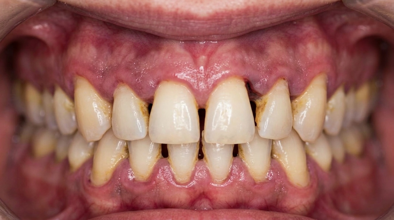
The cover image above is an AI-generated illustration based on the most common visible features of this condition described in clinical pathology references. It is not a photograph of a real case and should not be used to diagnose or rule out the condition in your own situation. If you are concerned about something you have noticed, please book an assessment with a registered dental practitioner.
References
Neville, B. W., Damm, D. D., Allen, C. M., & Chi, A. C. (2023). Oral and maxillofacial pathology (5th ed.). Elsevier. Chapter 4, Periodontal Diseases, pp. 160-167.
Cawson, R. A., & Odell, E. W. (2017). Cawson's essentials of oral pathology and oral medicine (8th ed.). Elsevier. Chapter 5, Gingivitis and Periodontitis, pp. 88-91.
Laskaris, G. (2003). Color atlas of oral diseases (3rd ed.). Thieme. Chapter 11, Periodontal Diseases, pp. 86-87.
Frequently asked questions
What is aggressive periodontitis?
Aggressive periodontitis is a form of gum disease that causes rapid loss of the bone and ligament supporting the teeth, usually in younger people (often teenagers and young adults) who appear otherwise healthy and have only modest plaque deposits. In current classifications it sits within Stage III-IV, Grade C periodontitis.
How is aggressive periodontitis different from regular gum disease?
It progresses much faster than typical chronic periodontitis, often affects specific patterns of teeth (such as first molars and incisors in the localised form), and tends to cluster in families. The amount of bone loss is striking given how little plaque is present.
Can aggressive periodontitis be treated?
Yes. Treatment combines thorough deep cleaning (scaling and root planing), often a course of antibiotics (commonly amoxicillin plus metronidazole), correction of risk factors such as smoking, and a strict supportive maintenance program. Severe cases may also need periodontal surgery, splinting or implant rehabilitation in advanced bone loss.
Will my children get aggressive periodontitis too?
There is a genetic component, so first-degree relatives have a higher risk and benefit from screening, particularly in adolescence. Early diagnosis dramatically improves the long-term outlook. A family dental review with full charting and X-rays is recommended for siblings and children of affected patients.