Compiled from clinical pathology references. Medically reviewed by Dr Cristian Dunker, Principal Dentist, ArtSmiles Cosmetic Dentistry.
When gum disease moves quickly, gum tissue ulcerating, the bone around the teeth being lost in days rather than years, and the patient feeling generally unwell, what is going on is rarely ordinary periodontitis. It is more likely necrotising ulcerative periodontitis (NUP), a severe form of gum disease that mostly affects people with reduced immunity and that needs urgent care.
This article from the team at ArtSmiles, reviewed by Dr Cristian Dunker, explains what NUP is, why it happens, and how it is treated.
Quick summary
At a glance | Detail |
|---|---|
Also called | NUP; necrotising periodontal disease; in older texts, "trench mouth periodontitis" |
How urgent? | 🔴 Urgent, the gum and supporting bone can be destroyed in days; book the next available appointment |
Common or rare? | Uncommon overall, but well recognised in people with reduced immunity |
Who it affects | Patients with HIV/AIDS, severe malnutrition, uncontrolled diabetes, on chemotherapy, or under extreme stress and exhaustion |
Who treats it | General dentist or periodontist for urgent management; coordination with the medical team for the underlying condition |
Based on | Neville, Cawson, Regezi |
What is it?
NUP is the periodontitis stage of the necrotising periodontal diseases, a small group of acute, rapidly progressive gum infections. The earlier stage is necrotising ulcerative gingivitis (NUG) (also called ANUG, acute necrotising ulcerative gingivitis), in which the destruction is limited to the gum margin. When the same destructive process extends into the periodontal ligament (the fibres that hold the tooth in its socket) and underlying bone, the condition becomes NUP.
Key features include:
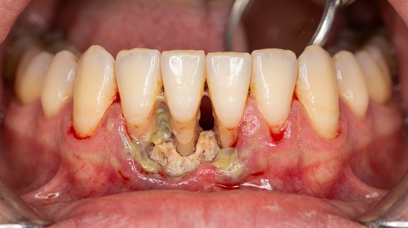
Painful, ulcerated, "punched-out" interdental papillae (the small triangles of gum between two teeth) with a grey-yellow slough on top.
Spontaneous bleeding from the gums, even at rest.
Rapid loss of attachment and bone in days to weeks.
Severe halitosis (persistent bad breath) with a characteristic foul, metallic odour.
Tooth mobility as bone support is lost.
Sometimes exposure of bone (sequestrum), a piece of dead bone separating from healthy bone, in advanced cases.
Systemic features, fever, lymph node swelling, feeling unwell, especially in immunocompromised patients (patients whose immune system is weakened by disease or medication).
Who tends to get it?
NUP is most often seen in:
Patients with HIV/AIDS, especially with low CD4 counts. It is one of the recognised oral signs of advanced HIV.
Patients on chemotherapy or other immunosuppressive therapy.
Patients with severe malnutrition, particularly children in low-resource settings.
Patients with uncontrolled diabetes.
Patients with severe stress and exhaustion, historically called "trench mouth" in soldiers in the First World War.
Heavy smokers with poor hygiene, particularly when combined with the above.
Otherwise healthy adults with adequate nutrition and reasonable hygiene very rarely develop NUP.
What causes it?
NUP is caused by an overgrowth of a particular bacterial mix, including Fusobacterium, Treponema, Selenomonas, Prevotella intermedia and other anaerobes (bacteria that thrive without oxygen), interacting with a weakened or overwhelmed immune response. Contributing factors include:
Reduced immunity, however caused.
Poor oral hygiene allowing the relevant bacteria to flourish.
Smoking, which reduces local blood flow and immune response.
Stress and exhaustion, which lower immune function.
Pre-existing periodontitis providing a reservoir of bacteria.
How does it develop?
NUP is rapid:
Bacteria accumulate at the gum margin in a person whose immune defence is reduced.
The interdental papillae ulcerate and start to break down (the NUG stage).
Without treatment, the infection extends into the periodontal ligament and bone.
Tissue is destroyed in days, with severe pain and bleeding.
In advanced cases, large areas of bone may be exposed, and the destruction can extend into the surrounding tissues (a related condition called noma in malnourished children).
What might you notice?
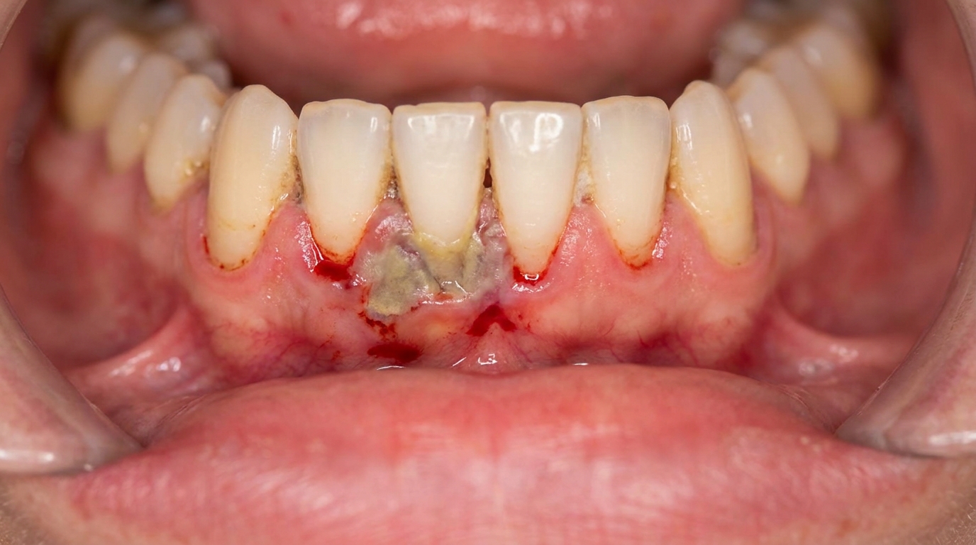
What it looks like
Ulcerated, grey-coated gum margins between the teeth, with deep cratering where the papillae used to be. The gum surface looks raw and may show exposed bone in advanced cases. Spontaneous bleeding is often visible.
What it feels like
Sudden severe gum pain that started over hours to a few days.
Spontaneous bleeding, gums that bleed without prompting.
Ulcers along the gum line with a grey film.
Very bad breath that persists despite brushing.
Loose teeth.
A bad metallic taste.
Fever and feeling unwell in many cases.
Difficulty eating because of pain.
What an X-ray might show
Bone loss between the teeth that has appeared since the last X-ray, sometimes within weeks. The pattern is often patchy and uneven, with crater-like defects in the interdental bone. Severe cases may show sequestrum (a piece of dead bone) starting to separate.
What happens at the dentist?
When NUP is suspected at ArtSmiles, the visit usually involves:
A thorough history including immune status, recent illness, medication, smoking and stress.
A careful examination with attention to ulceration, bleeding, mobility and sequestrum.
Photography and X-rays to document baseline severity.
A medical referral when there are signs of underlying immunocompromise (HIV testing, blood tests).
Immediate care, see treatment below.
A follow-up plan with frequent reviews until the disease is controlled.
Is this serious?
🔴 NUP is serious, both in itself, because of the rapid bone loss and tooth loss it can cause, and as a marker for an underlying medical problem. In severely immunocompromised patients, the disease can progress to noma, a much more aggressive condition with risk to life.
Could it be something else?
Conditions to consider include:
Acute periodontal abscess, localised, deep gum pocket with pus.
Severe ordinary chronic periodontitis, without the rapid course or systemic signs.
Primary herpetic gingivostomatitis, small clear vesicles before ulcers, mostly in children.
Recurrent aphthous stomatitis, discrete round ulcers without gum ulceration around teeth.
Leukaemia of the gum, pale, swollen, bleeding gums, with abnormal blood counts.
Drug-induced ulceration, usually less destructive of the bone.
Noma, a severe extension of NUP into the soft tissues of the face, mostly in malnourished children.
How is it treated?
Treatment combines local and systemic measures:
Gentle debridement (carefully scraping clean the affected gum margin), often with topical anaesthesia.
Antimicrobial mouthwash, usually 0.12 to 0.2% chlorhexidine twice daily.
Antibiotics, metronidazole is the mainstay, with amoxicillin sometimes added in severe cases or significant systemic involvement.
Pain relief, paracetamol, with care over NSAID use in immunocompromised patients.
Hydration and nutrition support.
Smoking cessation and stress reduction where possible.
Treatment of underlying medical conditions, HIV antiretroviral therapy, diabetes control, nutritional rehabilitation.
Long-term periodontal care once the acute phase has settled, careful cleaning, home care and recall visits.
What's the long-term outlook?
The acute disease usually settles within one to two weeks of starting treatment. The bone loss that occurred during the acute phase, however, does not return, affected teeth may need to be reshaped, splinted or, in some cases, extracted. Long-term outcomes depend heavily on the underlying medical condition and on consistent ongoing oral care.
If you have noticed sudden severe gum pain, ulceration and bleeding, particularly if you have any condition that affects your immune system, please book an urgent visit. Early treatment can prevent serious damage.
A note on this article
This article is for educational purposes only and does not constitute a clinical diagnosis. Please consult a registered dental practitioner for assessment and treatment advice.
The cover image above is an AI-generated illustration based on the most common visible features of this condition described in clinical pathology references. It is not a photograph of a real case and should not be used to diagnose or rule out the condition in your own situation. If you are concerned about something you have noticed, please book an assessment with a registered dental practitioner.
References
Neville, B. W., Damm, D. D., Allen, C. M., & Chi, A. C. (2023). Oral and maxillofacial pathology (5th ed.). Elsevier. Chapter 4, Periodontal Disease: Necrotising Periodontal Diseases.
Cawson, R. A., & Odell, E. W. (2017). Cawson's essentials of oral pathology and oral medicine (8th ed.). Elsevier. Chapter 5, Periodontal Disease.
Regezi, J. A., Sciubba, J. J., & Jordan, R. C. K. (2017). Oral pathology: Clinical pathologic correlations (7th ed.). Elsevier. Chapter 7, Periodontal Disease.
Frequently asked questions
What is necrotising ulcerative periodontitis (NUP)?
NUP is the more severe cousin of ANUG (necrotising ulcerative gingivitis). It involves the same painful, ulcerated, bleeding gums but the destruction now extends beyond the gum into the underlying bone and ligament holding the teeth. It is usually a sign of significant immune compromise.
Who is at risk of NUP?
NUP is strongly associated with severe immune suppression: HIV infection (particularly with low CD4 counts), advanced cancer, malnutrition, immunosuppressive medication and severe systemic illness. Heavy smoking and very poor oral hygiene compound the risk.
How is NUP treated?
Treatment is similar to ANUG but more involved. It includes urgent gentle debridement of the necrotic tissue, chlorhexidine rinses, pain control, and usually a course of metronidazole. The underlying immune problem must be investigated and managed, and intensive periodontal therapy follows once the acute episode is controlled.
What's the long-term outlook for NUP?
If diagnosed and treated early and the underlying cause is addressed, the outlook can be reasonable, but the gum and bone destruction does not fully reverse. Without treatment, NUP can progress rapidly to noma in severely immunocompromised patients. Lifelong supportive periodontal care is the norm.