Compiled from clinical pathology references. Medically reviewed by Dr Cristian Dunker, Principal Dentist at ArtSmiles Cosmetic Dentistry.
Quick summary
Also called | Irritation fibroma, focal fibrous hyperplasia, fibrous nodule, fibrous epulis (when on the gum) |
How urgent? | 🟢 Not urgent, benign and slow-growing; treated mainly to confirm the diagnosis and remove the lump |
Common or rare? | The most common "tumour-like" swelling in the mouth |
Who it affects | Most common in adults aged 40-60, with women slightly more often affected than men in biopsied series; sites of repeated biting or denture rubbing are particularly affected |
Who treats it | General dentist, a simple surgical excision in the dental chair under local anaesthetic is usually all that is needed |
Based on | Cawson, Neville, with cross-references in Regezi |
What is it?
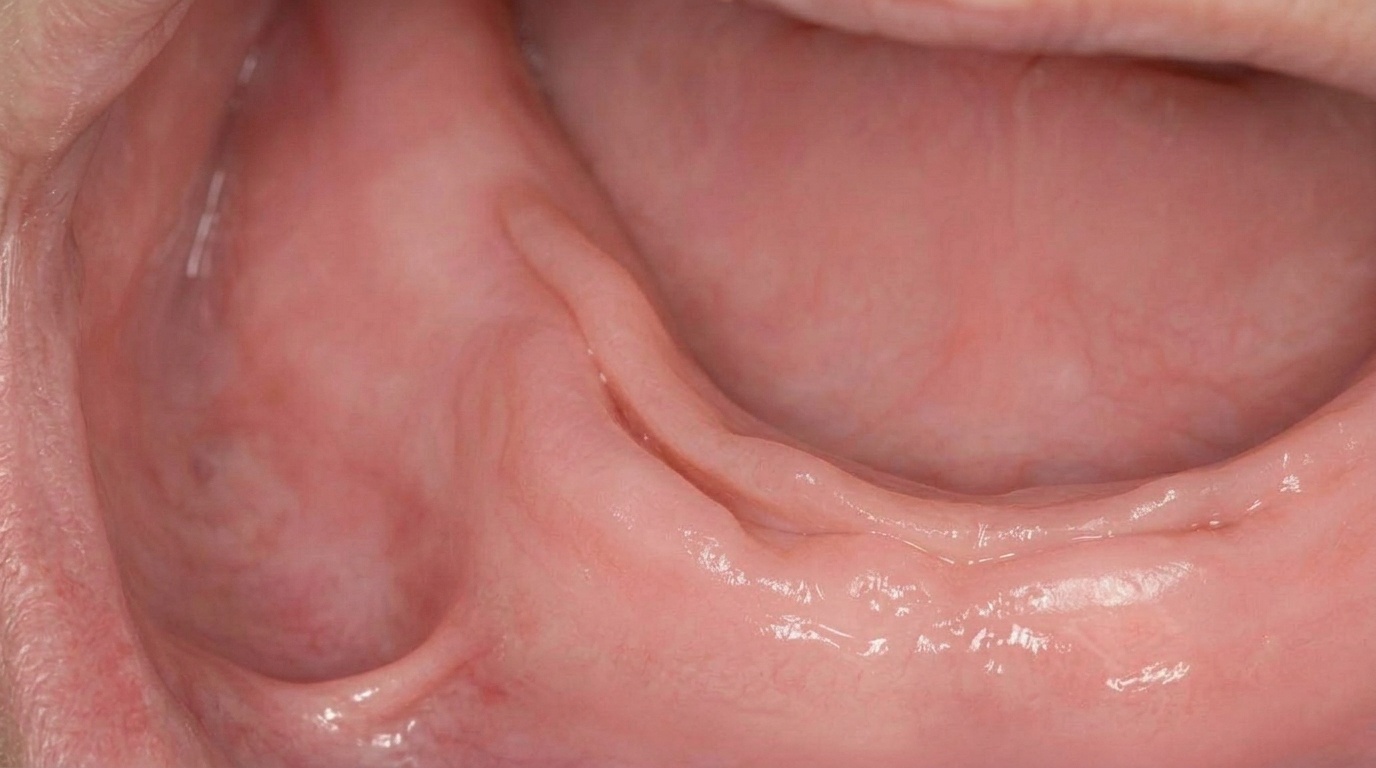
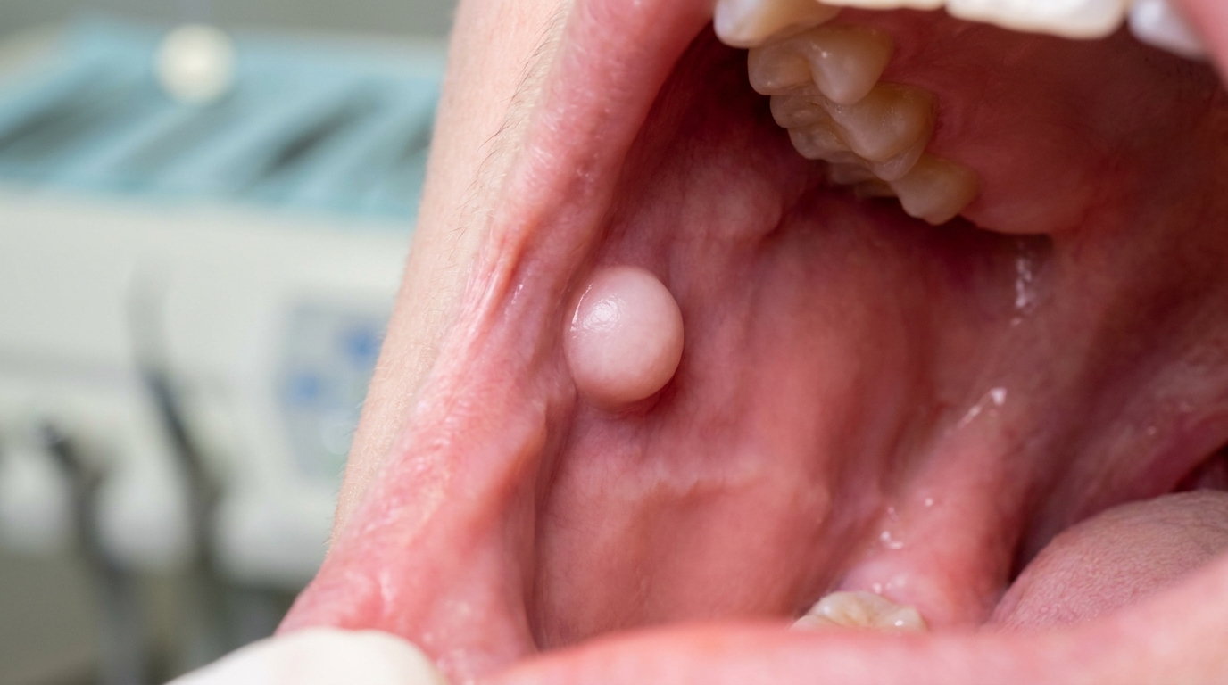
A traumatic fibroma is a small, smooth, painless lump in the mouth, made up of dense fibrous tissue covered by normal mucosa. The textbooks describe it as the most common "tumour" of the oral cavity, although they also note it is not really a true tumour, it is a hyperplastic response to chronic local irritation, such as biting the cheek, rubbing against a sharp tooth or denture, or repeated minor trauma. When the lump is on the gum, it is often called a fibrous epulis (the word "epulis" simply means "on the gum"); when it is elsewhere in the mouth, it is called a fibroma, irritation fibroma or focal fibrous hyperplasia. Whatever it is called, it is benign, slow-growing and easily managed.
Who tends to get it?
The textbooks describe a fairly recognisable pattern:
Adults of all ages, with the peak in the fourth to sixth decades (40s-60s).
Slightly more common in women than men in biopsied series, with a male-to-female ratio close to 1:2.
Common at sites of chronic biting or rubbing, the inside of the cheek along the bite line, the lateral border of the tongue, the inside of the lower lip and the gums.
Particularly common in denture wearers, where the lump may form at the edge of the denture (a related condition called epulis fissuratum when the lump fits along the denture flange).
Children sometimes have small frenal tags, a related fibrous overgrowth on the maxillary labial frenum that is harmless and rarely needs treatment.
What causes it?
The textbooks describe the cause as a reactive hyperplasia rather than a neoplasm:
Chronic biting of the cheek, the most common single cause along the bite line.
Chronic rubbing or pressure, for example, from a sharp tooth edge, a rough filling, a poorly fitting denture, or an orthodontic appliance.
Repeated minor injury that triggers the body's repair response. Over time, fibrous tissue accumulates as the area is repeatedly remodelled.
Past pyogenic granuloma, the textbooks specifically note that many fibrous epulides on the gum represent the "matured" form of a pyogenic granuloma after the initial inflammation has settled.
There is no link to viruses, infections, smoking, alcohol, or systemic disease. Traumatic fibromas are not contagious and do not run in families.
How does it develop?
A small area of mucosa that is repeatedly bumped, bitten or rubbed develops a microscopic injury. The body responds by laying down fibrous tissue to support and repair the area. With ongoing irritation, this fibrous tissue gradually accumulates into a slightly raised area, which over time becomes a firm nodule. Microscopically, the lump is made up of a nodular mass of fibrous connective tissue covered by stratified squamous epithelium, often with thickened keratin from continued surface trauma. The fibrous tissue is not encapsulated and merges gradually with the surrounding tissue. There is no dysplasia, no neoplastic feature, and the lesion does not progress to cancer.
What might you notice?
What it looks like
The classic appearance is well described:
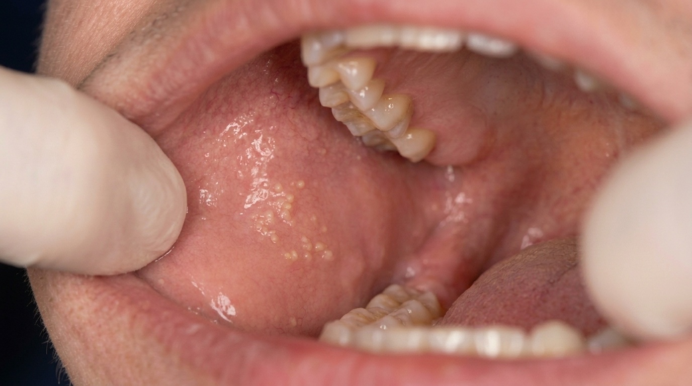
A smooth, dome-shaped or pedunculated nodule with a normal-looking pink mucosal cover.
Less than 1.5 cm in diameter in most cases, although larger lumps occur.
Most commonly on the inside of the cheek along the bite line, where the upper and lower teeth meet.
Other common sites: the lateral border of the tongue, the inside of the lower lip, and the gums (where it is called a fibrous epulis).
The surface may appear white if it has been chronically traumatised and developed extra keratin.
In darker-skinned patients, the surface may show gray-brown pigmentation.
The lump usually looks the same colour as the surrounding mucosa and has a uniform texture.
What it feels like
Most traumatic fibromas are completely painless. When symptoms occur they may include:
A firm but soft texture when pressed.
Mild discomfort if the lump is repeatedly bitten or caught between teeth.
An ulcer or rough surface if the lump itself is being chronically traumatised.
No bleeding in most cases, bleeding suggests something else, such as a pyogenic granuloma.
What an X-ray might show
Traumatic fibromas are confined to soft tissue, so X-rays do not show the lump itself. An X-ray may be relevant only if the lump is on the gum and there is concern about an underlying bone problem.
What happens at the dentist?
A traumatic fibroma is most often picked up at a routine dental check-up and clean at ArtSmiles or when the patient mentions a lump they have noticed. The dentist will typically:
Examine the lump carefully, note its size, shape, surface, colour and consistency.
Look for a clear cause, a sharp tooth, a rough restoration, a denture edge, or a habit of cheek-biting.
Take a careful history of how long the lump has been present and whether it has changed.
Address the underlying cause where possible, smoothing a sharp tooth, replacing a rough restoration, or adjusting a denture.
Discuss simple excision if the lump is troublesome, persistent or unsightly, since the diagnosis is best confirmed on histopathology.
Reassure that the lesion is benign.
Is this serious?
🟢 A traumatic fibroma is benign. It is not cancer, not contagious, and does not progress to anything dangerous. The textbooks specifically note that "recurrence is extremely rare" after conservative excision and that the only reason to remove the lump is for histopathological confirmation, comfort or appearance. Importantly, the textbooks also stress that other benign or malignant tumours can mimic the clinical appearance of a fibroma, which is why excised tissue should always be sent for microscopic examination.
Could it be something else?
Several conditions can produce a similar small nodule in the mouth. The textbooks list these as the main differentials:
Pyogenic granuloma, a fast-growing, often bleeding red lump, particularly common in pregnancy. Many "old" pyogenic granulomas mature into fibrous epulides.
Peripheral giant cell granuloma, a deep red or purple lump on the gum, often arising from the periodontal ligament; usually requires biopsy.
Peripheral ossifying fibroma, a firm gum lump that may show small calcifications on X-ray; biopsy distinguishes it from a fibrous epulis.
Mucocele, a soft, fluctuant, often bluish cyst from a damaged minor salivary gland, more common on the lower lip.
Lipoma, a soft, yellowish lump made of fat tissue, typically deeper and more compressible.
Gingival cyst of the adult, a small dome-shaped cyst, usually bluish, on the gum.
Salivary or other neoplasm, particularly when the lump is on the lip, palate or floor of the mouth and is firm or growing.
Squamous cell carcinoma, important to rule out in any persistent lump that ulcerates, bleeds or has a rough surface, particularly in older smokers or alcohol users.
How is it treated?
Treatment is straightforward and generally curative.
At-home measures and habits:
Identify any habit that may be irritating the area, cheek-biting, biting on a denture, lip-biting, and try to break it.
Maintain excellent oral hygiene so the lump and surrounding tissue stay clean.
Avoid biting on the lump while you wait for an appointment.
Professional steps your dentist may consider:
Conservative surgical excision under local anaesthetic. The lump is removed in one piece, the small wound is closed with a stitch or two, and healing usually takes a couple of weeks.
Histopathological examination of the removed tissue, essential to confirm the diagnosis and rule out the differentials above.
Address the underlying cause at the same appointment, smoothing a sharp tooth, replacing a rough filling, or adjusting a denture, so the lump does not recur.
No specific follow-up beyond routine check-ups, since recurrence is uncommon when the cause has been removed.
A patient-centred approach matters here too. People often worry that any lump in the mouth might be cancer. Calm, clear explanation of what a traumatic fibroma is, why removal and biopsy are recommended, and how quickly the area heals is itself part of effective care, values that sit at the heart of our clinical philosophy.
What's the long-term outlook?
The outlook is excellent. Once the lump has been excised and the diagnosis confirmed, recurrence is uncommon, particularly when the underlying habit or local irritant has been addressed. The mucosa heals over a couple of weeks and the area generally returns to normal. There is no need for prolonged follow-up beyond routine dental review, and there is no long-term health implication.
A note on this article
This article is for educational purposes only and does not constitute a clinical diagnosis. Please consult a registered dental practitioner for assessment and treatment advice.
The cover image above is an AI-generated illustration based on the most common visible features of this condition described in clinical pathology references. It is not a photograph of a real case and should not be used to diagnose or rule out the condition in your own situation. If you are concerned about something you have noticed, please book an assessment with a registered dental practitioner.
References
Neville, B. W., Damm, D. D., Allen, C. M., & Chi, A. C. (2023). Oral and maxillofacial pathology (5th ed.). Elsevier. Chapter 12, Soft Tissue Tumors: Fibroma (Irritation Fibroma; Traumatic Fibroma; Focal Fibrous Hyperplasia; Fibrous Nodule), the most common oral "tumour", with detailed clinical and histopathologic features, pp. 514-515.
Cawson, R. A., & Odell, E. W. (2017). Cawson's essentials of oral pathology and oral medicine (8th ed.). Elsevier. Chapter 19, Common Benign Mucosal Swellings: Fibrous Polyps, Epulides and Denture-Induced Granulomas, with key features in Box 19.1, pp. 314-315.
Regezi, J. A., Sciubba, J. J., & Jordan, R. C. K. (2017). Oral pathology: Clinical pathologic correlations (7th ed.). Elsevier. Chapter 4, Connective Tissue Lesions: cross-reference for fibrous hyperplasia and irritation fibroma.
Frequently asked questions
What causes a traumatic fibroma?
It is caused by repeated mechanical irritation of the soft tissues of the mouth, most often from cheek- or lip-biting, a sharp tooth edge, a broken filling or a rough denture flange. Over time the irritated area lays down extra collagen fibres to protect itself, gradually forming a firm pink lump.
Is a traumatic fibroma dangerous?
No. A traumatic fibroma is a benign, non-cancerous reactive overgrowth, not a tumour. It does not spread, does not turn into cancer and is not infectious. The main reasons to treat it are that it can keep getting bitten or rubbed and stays in place once the irritation is gone.
How is a traumatic fibroma treated?
A small surgical excision under local anaesthetic is the standard treatment. The lump is lifted off the lining, the wound is closed with a stitch or two, and the tissue is sent for microscopic examination to confirm the diagnosis. Identifying and removing the source of irritation (e.g. smoothing a sharp tooth edge) is just as important to prevent recurrence.
Will a traumatic fibroma come back after removal?
Recurrence is uncommon once the lump and the underlying cause of irritation are both addressed. If the same area keeps being bitten or rubbed, a new fibroma can occasionally form at the same site, so the focus is on protecting the area from further trauma.