Compiled from clinical pathology references. Medically reviewed by Dr Cristian Dunker , Principal Dentist, ArtSmiles Cosmetic Dentistry.
Quick summary
Also called | Oral lipoma, fibrolipoma (a lipoma with extra fibrous tissue), angiolipoma, spindle cell lipoma (a benign variant with spindle-shaped cells under the microscope), sialolipoma |
How urgent? | 🟡 Worth a check-up, a lipoma is benign, but a soft yellowish lump should still be examined to rule out other lesions |
Common or rare? | Uncommon in the mouth, lipomas are the most common soft-tissue tumour overall, but oral examples make up only 1 to 4% of cases |
Who it affects | Adults aged 40 and over; uncommon in children; affects men and women fairly equally |
Who treats it | General dentist, sometimes with referral to an oral surgeon for excision |
Based on | Regezi, Neville, Cawson, Laskaris |
What is it?
A lipoma is a benign (non-cancerous) lump made of fat cells. It sits just under the lining of the mouth and feels soft and squashy. Lipomas are the most common soft-tissue growth in the body, but they are uncommon inside the mouth, and when they do appear they almost always behave themselves, growing slowly, painlessly, and without spreading.
Who tends to get it?
Lipomas of the mouth and jaw are uncommon, making up only around 1 to 4 per cent of all lipomas in the body. Most people who develop one are 40 years of age or older, they are rarely seen in children. Men and women are affected in roughly equal numbers across most studies.
The buccal mucosa (the inside of the cheek) and buccal vestibule (the gutter between cheek and gum) account for almost half of all oral lipomas. Other reported sites include the tongue, lips, floor of the mouth and, less often, the gingiva. People who carry more body fat are slightly more likely to develop lipomas, although the connection is not strong.
What causes it?
The exact cause of a lipoma is not fully understood. They appear to arise as small overgrowths of fat cells that gradually form a discrete lump. Interestingly, the fat inside a lipoma behaves quite independently of the rest of the body's fat, losing weight does not shrink a lipoma, even when normal body fat is being lost.
Genetic factors are part of the picture for some people. In laboratory studies, many lipomas show consistent rearrangements involving a region of chromosome 12 (12q13,q15), which suggests that small genetic changes within the fat cells themselves may drive the lump's formation. There is no link to diet, oral hygiene or any specific lifestyle habit. Some lumps in the cheek that look like lipomas are not actually new growths at all, but a herniation (poking through) of the buccal fat pad after trauma, for example, after a blow to the cheek in a child, or after wisdom-tooth surgery in an older patient.
How does it develop?
Think of fat in the body a bit like the soft padding inside a cushion. Normally that padding is spread out evenly. In a lipoma, a small pocket of those fat cells starts to grow as its own cluster, slowly enlarging within a thin natural skin (a fibrous capsule) that keeps it neatly contained. Because the cells inside are simply mature fat cells, the lump stays soft, smooth and self-contained.
A lipoma typically grows very slowly, over months to years. Patients are often surprised to learn how long their lump has been there once they start thinking back. Several microscopic variants exist, a fibrolipoma has a tougher fibrous component mixed in, an angiolipoma contains many tiny blood vessels, and a sialolipoma develops where minor salivary gland tissue has been caught up in the fat, but they all share the same gentle, slow-growing behaviour.
What might you notice?
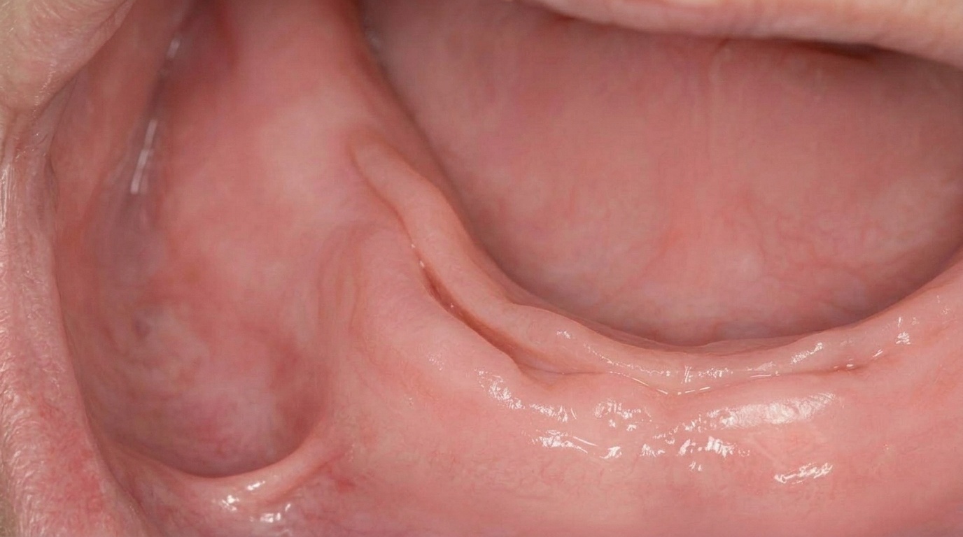
What it looks like
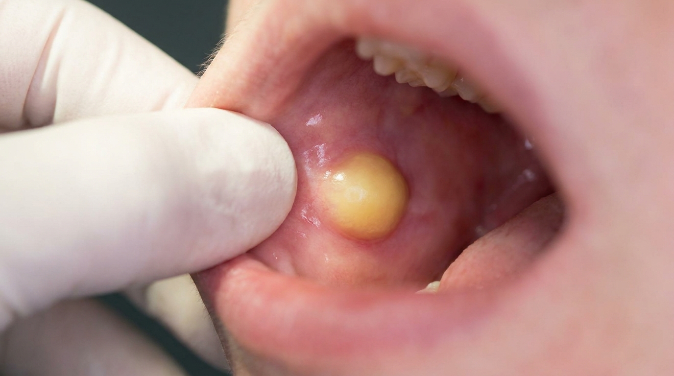
A lipoma usually appears as a soft, smooth, dome-shaped lump under the lining of the mouth. Many have a subtle yellow or yellow-white tinge, especially when they sit close to the surface, because the fat shows through the thin overlying mucosa. Small surface blood vessels are often visible running over the lump. Deeper lipomas can look pinker because the colour of the fat is masked by tissue above. Most lesions are smaller than 3 cm, although occasional examples grow larger.
Some lipomas sit on a stalk (pedunculated), while others have a broad base (sessile). The lump moves freely under the finger and is not fixed to the tissues underneath.
What it feels like
The overwhelming feature is that a lipoma does not hurt. It feels soft, sometimes so soft and squashy that it can be mistaken for a fluid-filled cyst on examination. It is not tender to touch, and it does not interfere with taste, speech or chewing unless it has grown large enough to get in the way. Some patients only become aware of theirs because they catch it between their teeth when eating.
What an X-ray might show
A lipoma is a soft-tissue lump, so dental X-rays usually show no abnormality. The role of imaging is to confirm there is no underlying bone problem that could explain the swelling.
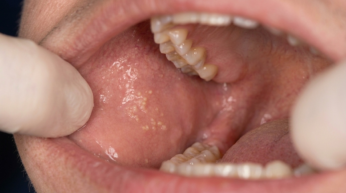
What happens at the dentist?
At ArtSmiles, your dentist will start with a careful look and gentle feel of the lump. The combination of a soft, painless, yellowish swelling in a typical site (most often the cheek) gives a strong clinical clue that the lump is a lipoma. From there, several things may happen:
A clinical record is made of the size, site, colour and feel of the lump, and how long you have noticed it.
Photographs may be taken to track any change over time.
A biopsy or excisional biopsy may be recommended. Because several other oral lumps can mimic a lipoma clinically, the only way to confirm the diagnosis with certainty is to remove the lump (or a sample of it) and have it examined under the microscope by a pathologist.
Imaging is rarely needed for a typical small lipoma in the cheek or lip. For deeper or larger lesions, your dentist may refer you for an MRI or ultrasound to map the lump before surgery.
A referral to an oral or maxillofacial surgeon may be appropriate if the lipoma is large, deep, on the tongue, or involves the floor of the mouth.
Throughout, your dentist will use plain language and explain why each step is being suggested. If anything in the appearance is unusual, for example, rapid growth, fixation to deeper tissue, ulceration or pain, further investigation will be prioritised.
Is this serious?
🟢 Usually harmless. A lipoma is benign. It does not spread, and malignant change to a fat-cell cancer (liposarcoma) from a pre-existing oral lipoma is exceptionally rare, most liposarcomas arise on their own rather than from a previous lipoma.
That said, a lump in the mouth always deserves an assessment, because soft yellow swellings can occasionally look like other lesions that do need different treatment. The reassuring news is that once a lipoma is diagnosed and removed, recurrence is uncommon and the long-term outlook is excellent.
If you have noticed a lump in your mouth that has been there for more than two weeks, it is worth booking an assessment.
Could it be something else?
Several other lumps and bumps can resemble an oral lipoma. Your dentist will work through these possibilities at the clinical examination, and a biopsy can confirm the diagnosis. The four reference textbooks list the following look-alikes:
Traumatic fibroma (irritation fibroma), a common reactive lump from cheek or lip biting that can also feel firm and submucosal. A fibroma is usually paler, firmer and develops at a known site of trauma; a lipoma is softer and often yellowish.
Mucocele, a salivary mucus-filled swelling, especially on the lower lip. A mucocele is typically translucent and bluish, fluctuates in size and may burst and refill, whereas a lipoma stays steady and yellow-tinged.
Salivary gland tumour (such as a pleomorphic adenoma), minor salivary gland tumours can present as a painless submucosal lump, particularly on the palate or upper lip. They tend to feel firmer than a lipoma, and biopsy distinguishes them.
Neurofibroma, a benign nerve-sheath lump that also presents as a smooth painless swelling. A neurofibroma feels firmer than a lipoma; multiple lesions can suggest neurofibromatosis, which prompts further work-up.
Schwannoma (neurilemmoma), another benign nerve-sheath tumour, most often on the tongue. It is firmer and more rubbery than the squashy feel of a lipoma, and microscopy is diagnostic.
Granular cell tumour, typically on the tongue, presenting as a painless smooth swelling. It tends to feel firmer and shows distinctive cells under the microscope.
Leiomyoma, a rare benign smooth-muscle tumour that can appear as a slow-growing, firm, well-defined lump, most often on the tongue. Firmness and microscopic appearance separate it from a lipoma.
Myxoma, an uncommon mesenchymal lump that, like a lipoma, is mobile and soft. The yellow hue and lobulated fatty look-and-feel of a lipoma help distinguish them clinically, with biopsy giving the final answer.
Dermoid cyst, a developmental cyst, classically in the floor of the mouth. Both can feel soft and doughy; a dermoid cyst tends to be midline and may feel more like firm putty, and imaging plus histology separates the two.
Lymphangioma, a benign lymphatic vessel malformation that can form pale, soft, translucent or nodular swellings. Sudden colour change to dark purple after bleeding into the lesion is a clue that points away from a lipoma.
Lymphoepithelial cyst, a small, yellowish nodule that can mimic the surface colour of a lipoma, most often in the floor of the mouth or on the tongue. Cysts are usually firmer and more discrete on palpation, and microscopy confirms.
Buccal fat pad herniation, not a tumour at all, but a poking-through of the cheek's natural fat pad after trauma or wisdom-tooth surgery. It can look identical to a buccal lipoma; the clinical history and surgical findings tell them apart.
Liposarcoma (and atypical lipomatous tumour), the malignant fat-cell counterpart. Genuinely rare in the mouth, but considered when a fatty lump is unusually large, deep, fixed or fast-growing. Microscopy with specific markers reliably distinguishes it from a benign lipoma.
Focal mucinosis, a small, soft mucinous nodule that can feel similar to a lipoma. Histology is needed to tell them apart.
How is it treated?
For most oral lipomas, treatment is a small surgical procedure to remove the lump.
At home, before treatment, there is nothing specific you need to do. Lipomas are not caused by oral hygiene habits, and no rinse, gel or supplement will shrink one. Your normal brushing, flossing and routine check-ups remain the best ongoing care.
At the dentist or oral surgeon, treatment may include:
Conservative local excision. This is the standard approach, performed under local anaesthetic in most cases. The lump is removed in one piece along with its thin capsule. Because lipomas are well-circumscribed and lift out cleanly, the procedure is usually quick, often half an hour or less for a small cheek lesion.
Histopathology. The removed tissue is sent to a pathology laboratory to confirm the diagnosis under the microscope. This step is important because it rules out the rare look-alikes above.
Specialist referral to an oral and maxillofacial surgeon may be appropriate where the lipoma is large, deep, on the tongue, or in the floor of the mouth, where the anatomy is more delicate.
Healing after a small intraoral excision is usually quick, with most patients back to normal eating within a few days and any sutures dissolving or being removed at a brief follow-up visit.
What's the long-term outlook?
The outlook for an oral lipoma is excellent. After conservative excision, recurrence is rare for the common subtypes. The deeper intramuscular (infiltrating) variant has a higher recurrence rate because it weaves between muscle bundles, but this form is uncommon in the mouth.
Most patients can expect a clean diagnosis from histology, complete healing of the small surgical site, and no long-term consequences. Once removed, a lipoma does not typically come back in the same place, and there is no need for ongoing imaging or surveillance for an isolated benign lesion. As always, any new lump in the mouth, wherever it appears, is worth showing to your dentist for assessment.
A note on this article
This article is for educational purposes only and does not constitute a clinical diagnosis. Please consult a registered dental practitioner for assessment and treatment advice.
The cover image above is an AI-generated illustration based on the most common visible features of this condition described in clinical pathology references. It is not a photograph of a real case and should not be used to diagnose or rule out the condition in your own situation. If you are concerned about something you have noticed, please book an assessment with a registered dental practitioner.
References
Regezi, J. A., Sciubba, J. J., & Jordan, R. C. K. (2017). Oral pathology: Clinical pathologic correlations (7th ed.). Elsevier. Chapter 7, Connective Tissue Lesions: Fat Lesions (Lipoma), pp. 182 to 183.
Neville, B. W., Damm, D. D., Allen, C. M., & Chi, A. C. (2023). Oral and maxillofacial pathology (5th ed.). Elsevier. Chapter 12, Soft Tissue Tumors: Lipoma, pp. 530 to 532.
Cawson, R. A., & Odell, E. W. (2017). Cawson's essentials of oral pathology and oral medicine (8th ed.). Elsevier. Chapter 20, Soft Tissue (Mesenchymal) Neoplasms: Lipoma and fibrolipoma, pp. 321 to 322.
Laskaris, G. (2003). Color atlas of oral diseases (3rd ed.). Thieme. Chapter 33, Benign Tumors: Lipoma, pp. 310 to 311.
Frequently asked questions
Is an oral lipoma dangerous?
Oral lipomas are benign and rarely dangerous. Malignant transformation to liposarcoma is very rare, but any lump in the mouth should be assessed by a dentist to confirm the diagnosis.
What does a lipoma in the mouth look like?
A typical oral lipoma is a soft, smooth, well-defined yellowish lump under the lining of the mouth, often on the inside of the cheek. It grows slowly and is usually painless.
Do oral lipomas need to be removed?
Most oral lipomas are removed by simple surgical excision under local anaesthetic, both to confirm the diagnosis on histopathology and to stop them being bitten or interfering with function. Recurrence after complete removal is very low.
Can oral lipomas turn into cancer?
True malignant transformation of an oral lipoma into a liposarcoma is exceptionally rare. A rapidly growing, firm or fixed lump should be checked promptly, as liposarcoma usually arises de novo rather than from a long-standing lipoma.
How are oral lipomas diagnosed?
Diagnosis starts with clinical examination of a soft, yellowish, slow-growing submucosal lump. Imaging (such as ultrasound or MRI) may be used for deeper or larger lesions, but the definitive diagnosis is made by histopathology after excisional biopsy.