Compiled from clinical pathology references. Medically reviewed by Dr Cristian Dunker , Principal Dentist, ArtSmiles Cosmetic Dentistry.
Quick summary
Also called | Gorlin cyst, calcifying cystic odontogenic tumour (CCOT), an older name still seen in some textbooks, dentinogenic ghost cell tumor (the solid form), ghost cell odontogenic carcinoma (the rare malignant form) |
How urgent? | 🟡 Worth checking, usually painless but progressive; the cyst form behaves benignly while solid and malignant variants need more involved management |
Common or rare? | Rare, most pathology services see only a handful of cases each year |
Who it affects | Most often adults in their 20s to 40s; cysts associated with odontomas (benign tumours made of disorganised tooth tissue) can appear earlier (around age 17); both jaws affected, with most cases in the front of the mouth |
Who treats it | General dentist working with an oral and maxillofacial surgeon and an oral pathologist, diagnosis depends on histopathology (examination of the removed tissue under the microscope) after surgical removal |
Based on | Cawson, Neville, with cross-references in Regezi |
What is it?
A calcifying odontogenic cyst is a rare lesion of the jawbone, first described by Gorlin and colleagues in 1962. It belongs to a small family of "ghost cell" lesions, so called because under the microscope, the lining contains pale, swollen cells that have lost their nuclei but kept the outline of the original cell. These ghost cells (large, pale cells with no nucleus that look like an outline of where a cell used to be) often calcify. Most calcifying odontogenic cysts grow as fluid-filled cysts. A small minority grow as solid masses, known as dentinogenic ghost cell tumors, and an even rarer malignant variant exists called ghost cell odontogenic carcinoma. Because the cystic, solid and malignant forms behave very differently, accurate diagnosis after surgery is important.
Who tends to get it?
The textbooks describe a fairly distinctive profile:
Mean age around 30, with most cases diagnosed between the second and fourth decades of life.
A younger subgroup, particularly when the cyst is associated with an odontoma (a developmental tooth-shaped mass), with mean age around 17.
Equal frequency in the maxilla (upper jaw) and mandible (lower jaw).
About 65% of cases occur in the incisor and canine areas, the front of the mouth.
About one-third of cases are associated with an unerupted tooth, most often a canine.
About 20% of cases occur alongside an odontoma.
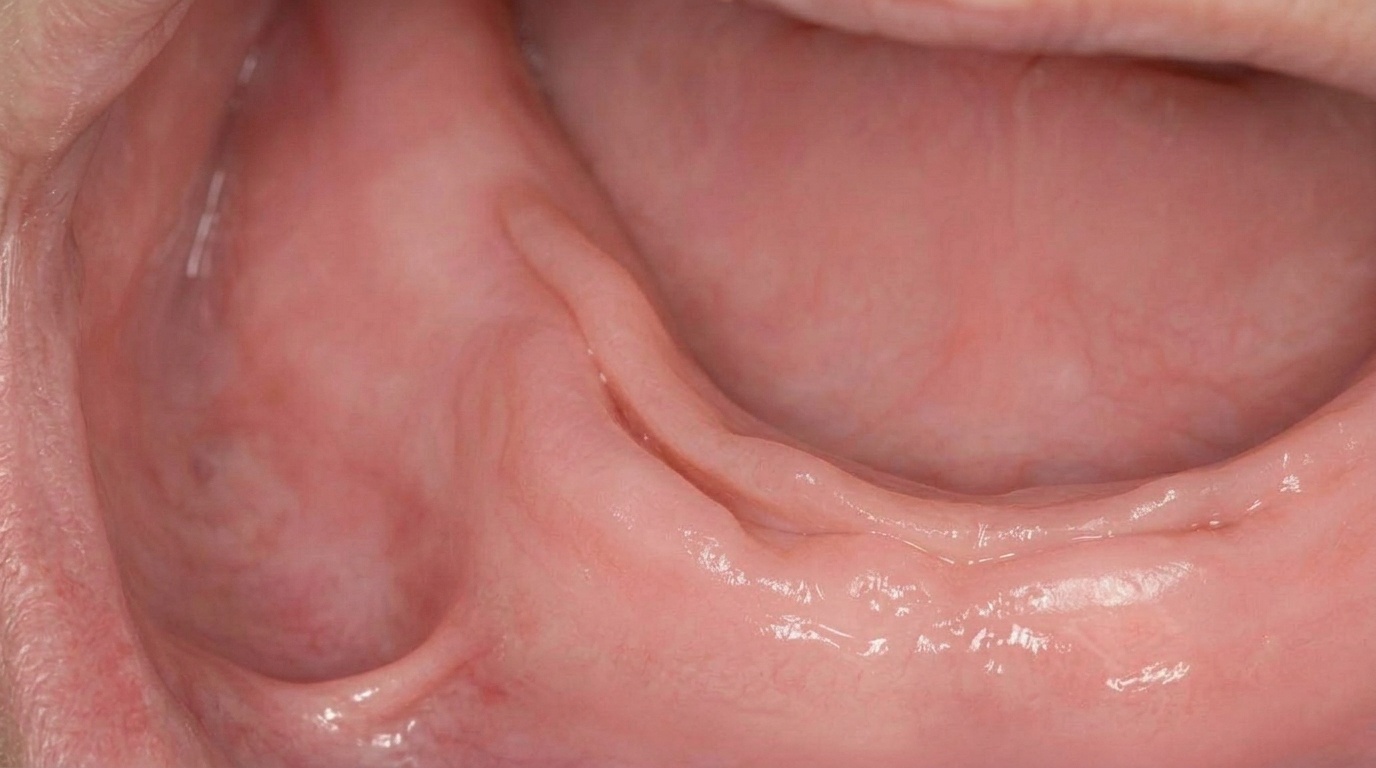
5-17% of cases are extraosseous (peripheral), appearing as a small lump on the gum rather than as an X-ray finding inside the bone. These peripheral lesions tend to occur later in life, with peak prevalence in the sixth to eighth decades.
The solid dentinogenic ghost cell tumor form is most often seen between the third and fifth decades, often in the back of the jaws.
There is no strong sex or ethnic predilection.
What causes it?
The cause is not fully understood, but the textbooks describe several relevant findings:
It develops from epithelial cells associated with tooth formation. The lining contains odontogenic epithelium, the same lineage of cells that originally formed the enamel and tooth structure.
Molecular studies have shown mutations in the CTNNB1 gene, which controls a key signalling pathway (β-catenin) involved in growth and differentiation. The same mutations are also seen in the related dentinogenic ghost cell tumor and the rare ghost cell odontogenic carcinoma.
Association with odontomas, adenomatoid odontogenic tumors and ameloblastoma (a benign but locally aggressive tumour of tooth-forming cells)s has been reported, suggesting that ghost cell lesions sit on a spectrum of odontogenic growths.
Local factors (such as inflammation or trauma) have not been clearly identified as causes; calcifying odontogenic cysts can occur in apparently healthy bone.
How does it develop?
A calcifying odontogenic cyst usually grows slowly. Within the bone, the lining produces fluid that gradually expands the cavity. The lining cells include cuboidal or columnar basal cells (resembling the ameloblasts that originally lay down enamel) and a layer of looser cells reminiscent of the stellate reticulum of a developing tooth. Some of these epithelial cells lose their nuclei and become the characteristic "ghost cells", which then accumulate and calcify within the cyst. In some cases, dentine-like material (dentinoid) forms in the wall, reflecting the cyst's odontogenic origin.
Most calcifying odontogenic cysts stay within the original bone outline and behave benignly. The solid dentinogenic ghost cell tumor form is more aggressive, it can resorb adjacent tooth roots, perforate the cortical plate (the thin outer layer of bone), or invade the maxillary sinus. The ghost cell odontogenic carcinoma form is rare but capable of local invasion and even distant spread.
What might you notice?
What it looks like
Most calcifying odontogenic cysts produce no visible change at all. When they do, they may appear as:
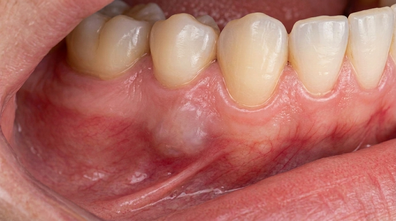
A slow, painless swelling in the jawbone, sometimes producing a slight bulge under the gum.
Facial asymmetry if the cyst has grown for many years.
A small lump on the gum in the rare peripheral form, sometimes resembling a fibroma or other gum nodule.
What it feels like
Most lesions cause no pain. When symptoms do occur, they may include:
Mild pressure or tightness in the affected area.
Loose neighbouring teeth if the cyst has resorbed adjacent roots.
Tingling or numbness if the lesion is large enough to compress a nearby nerve, particularly relevant in the lower jaw.
Pain or swelling if the cyst becomes secondarily infected.
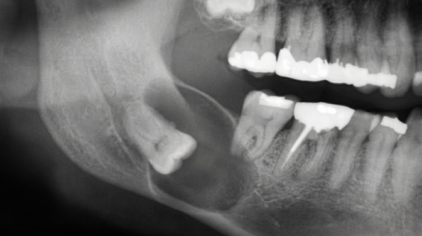
What an X-ray might show
The diagnosis is usually first suspected from imaging. Typical findings on a panoramic X-ray or a small-volume cone-beam CT (a 3D dental X-ray, often shortened to CBCT) (CBCT) scan include:
A well-circumscribed radiolucent (darker on X-ray, indicating soft tissue or fluid rather than bone) lesion, usually unilocular but occasionally multilocular.
Radiopaque flecks or toothlike densities within the radiolucency in about one-third to one-half of cases, a strong clue toward this diagnosis.
Association with an unerupted tooth, most often a canine, in about a third of cases.
Root resorption or divergence of nearby erupted teeth in larger cases.
Most cysts are 2-4 cm in greatest diameter, although lesions up to 12 cm have been reported.
The mixed radiolucent,radiopaque (lighter or white on X-ray, indicating mineralised or calcified material) appearance is one of the more distinctive imaging clues, but a definitive diagnosis still requires histopathological examination.
What happens at the dentist?
A calcifying odontogenic cyst is most often discovered incidentally on a panoramic X-ray taken for another reason, for example, before orthodontic treatment, while assessing wisdom teeth, or planning implants. A dentist at ArtSmiles, typically as part of a dental check-up and clean, will usually:
Take a careful history of any past trauma, swelling, jaw discomfort or numbness.
Examine the area for any swelling, asymmetry, or change in surrounding teeth.
Arrange additional imaging, typically a CBCT scan to map the lesion in three dimensions and to check its relation to the inferior dental nerve, the maxillary sinus, or adjacent tooth roots.
Test the vitality of nearby teeth to rule out a non-vital tooth as the source.
Refer to an oral and maxillofacial surgeon for biopsy and definitive treatment.
Coordinate with an oral pathologist for histopathological examination of the removed tissue, which is essential to confirm the diagnosis and distinguish a benign cyst from a more aggressive variant.
Is this serious?
🟡 The cystic form of a calcifying odontogenic cyst is benign and rarely recurs after careful enucleation (a minor surgical procedure where the cyst is removed cleanly from its bony cavity). The solid (dentinogenic ghost cell tumor) form is more aggressive and can recur in up to 73% of cases when treated conservatively. The malignant ghost cell odontogenic carcinoma form is rare but potentially life-threatening, with a reported 5-year survival of around 73%. Because these forms can look similar at first, an unhurried diagnostic process, including imaging, biopsy and specialist histopathology, is important. Most patients diagnosed with the cystic form, which is the most common, can expect a very good outcome with appropriate surgery.
If a routine X-ray has shown an unusual cyst-like lesion in the jaw, particularly one with mixed radiolucent and radiopaque areas, it is worth booking an assessment so the right imaging and specialist input can be arranged early.
Could it be something else?
Several other lesions can produce a similar appearance on imaging or share clinical features. The textbooks list these as the main differentials:
Dentigerous cyst, also occurs around the crown of an unerupted tooth, but is purely radiolucent and lacks ghost cells on histology.
Odontogenic keratocyst (OKC), a more aggressive cyst that grows along the bone, with a distinctive parakeratinised lining and a higher recurrence rate.
Ameloblastoma, a benign but locally aggressive odontogenic tumour with a "soap bubble" or "honeycomb" multilocular X-ray appearance.
Adenomatoid odontogenic tumor, a benign tumour, often associated with an unerupted upper canine, that can also show fine calcifications inside.
Odontoma, a developmental tooth-shaped mass that can show very dense calcifications and sometimes coexists with a calcifying odontogenic cyst.
Calcifying epithelial odontogenic tumor (Pindborg tumor), another rare odontogenic tumour with calcifications. The histopathology is distinctive but the imaging can overlap.
How is it treated?
Treatment depends on whether the lesion is the cystic form, the solid form or the rare malignant form. The textbooks describe a fairly clear hierarchy:
At-home measures and habits:
Maintain excellent oral hygiene to limit the risk of secondary infection in any cyst-like lesion.
Attend regular check-ups and X-rays while a known lesion is being monitored or after surgery.
Report any changes, new swelling, pain, numbness, or mobility of teeth, promptly.
Professional steps your dentist may consider:
Surgical enucleation with histopathological examination is the standard treatment for the cystic form. The cyst is shelled out of the bone, the cavity is cleaned, and the tissue is sent for microscopic confirmation. Recurrence after careful enucleation is uncommon.
More extensive surgery, marginal or segmental resection, for the intraosseous solid dentinogenic ghost cell tumor form, where conservative treatment alone is associated with high recurrence.
Wide excision and oncological management if the rare ghost cell odontogenic carcinoma is identified, generally coordinated through a head and neck cancer team.
Conservative excision for peripheral (gingival) lesions, which generally behave benignly with minimal chance of recurrence.
Long-term radiographic follow-up at intervals of 1, 2 and often 5 years to confirm complete healing and rule out recurrence.
A patient-centred approach is particularly important when imaging has raised the possibility of a rare lesion. Honest discussion of the diagnostic pathway, what imaging is needed, why a biopsy matters, and what the various forms mean, is itself part of effective care, values that sit at the heart of our clinical philosophy.
What's the long-term outlook?
For the most common (cystic) form, the outlook is excellent. After careful enucleation and confirmation on histopathology, recurrence is uncommon and the bone usually heals well. The solid dentinogenic ghost cell tumor form has a higher recurrence rate, particularly with conservative treatment, but with adequate surgical margins long-term control is generally good. The rare ghost cell odontogenic carcinoma form is more serious, with reported 5-year survival around 73% and a need for ongoing oncological follow-up. Across all forms, the single most important factor is accurate histopathological diagnosis after surgical removal, which is why a planned surgical biopsy and specialist input are so important from the start.
A note on this article
This article is for educational purposes only and does not constitute a clinical diagnosis. Please consult a registered dental practitioner for assessment and treatment advice.
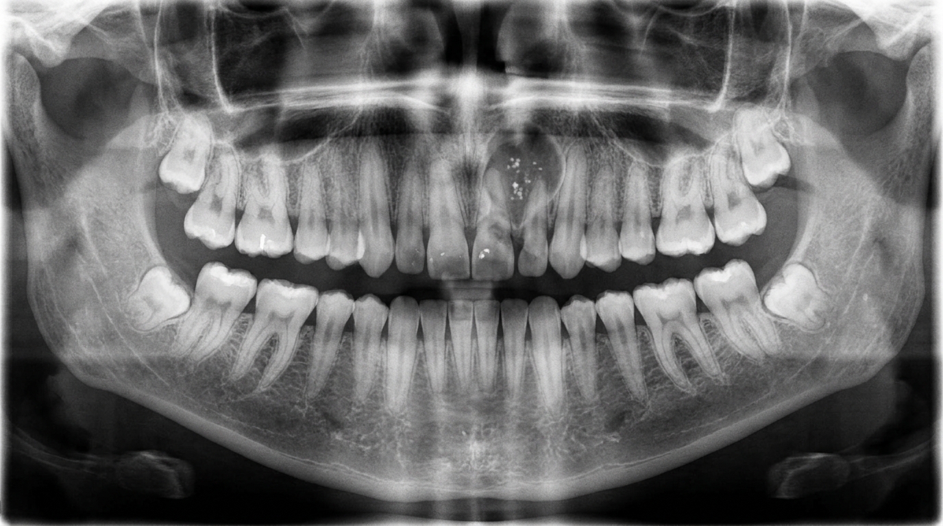
The cover image above is an AI-generated illustration based on the most common visible features of this condition described in clinical pathology references. It is not a photograph of a real case and should not be used to diagnose or rule out the condition in your own situation. If you are concerned about something you have noticed, please book an assessment with a registered dental practitioner.
References
Cawson, R. A., & Odell, E. W. (2017). Cawson's essentials of oral pathology and oral medicine (8th ed.). Elsevier. Chapter 8, Odontogenic Tumours and Tumour-like Lesions of the Jaws: Calcifying (Ghost Cell) Odontogenic Cyst, with key features in Box 8.6, pp. 143 to 144.
Neville, B. W., Damm, D. D., Allen, C. M., & Chi, A. C. (2023). Oral and maxillofacial pathology (5th ed.). Elsevier. Chapter 15, Odontogenic Cysts and Tumors: Calcifying Odontogenic Cyst (Calcifying Cystic Odontogenic Tumor; Gorlin Cyst; Dentinogenic Ghost Cell Tumor; Ghost Cell Odontogenic Carcinoma), pp. 701 to 704.
Regezi, J. A., Sciubba, J. J., & Jordan, R. C. K. (2017). Oral pathology: Clinical pathologic correlations (7th ed.). Elsevier. Chapter 10, Cysts of the Jaws and Neck: Calcifying odontogenic cyst as differential of unilocular jaw cysts.
Frequently asked questions
What is a calcifying odontogenic cyst?
A calcifying odontogenic cyst, also called Gorlin cyst, is a rare developmental cyst of the jaw containing characteristic 'ghost cells' and small areas of calcification. It is benign and usually treated by surgical removal.
How is it different from other jaw cysts?
Imaging often shows a mixed radiolucent and radiopaque appearance because of the small calcifications within the cyst. Definitive diagnosis is made by examining the removed tissue under the microscope and finding ghost cells.
Is it related to Gorlin-Goltz syndrome?
No. Despite the shared 'Gorlin' name, the calcifying odontogenic cyst (Gorlin cyst) is not the same as Gorlin-Goltz syndrome (naevoid basal cell carcinoma syndrome). The two were described by the same pathologist but are unrelated conditions.
How is it treated?
Surgical enucleation is the standard treatment. Recurrence is uncommon after complete removal. Long-term follow-up with periodic X-rays confirms the cyst has not returned.