Compiled from clinical pathology references. Medically reviewed by Dr Cristian Dunker, Principal Dentist, ArtSmiles Cosmetic Dentistry.
Quick summary
Also called | Fordyce granules, ectopic sebaceous glands of the oral mucosa |
How urgent? | 🟢 Not urgent, an entirely benign anatomical variation |
Common or rare? | Very common, present in about 80% of adults if examined carefully |
Who it affects | Both sexes, all ages and backgrounds, often more visible from puberty onwards |
Who treats it | General dentist for diagnosis and reassurance; no treatment is needed |
Based on | Neville, Cawson, with cross-references in Regezi and Laskaris |
What is it?
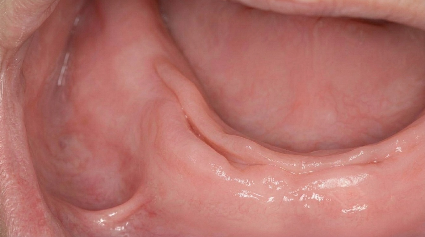
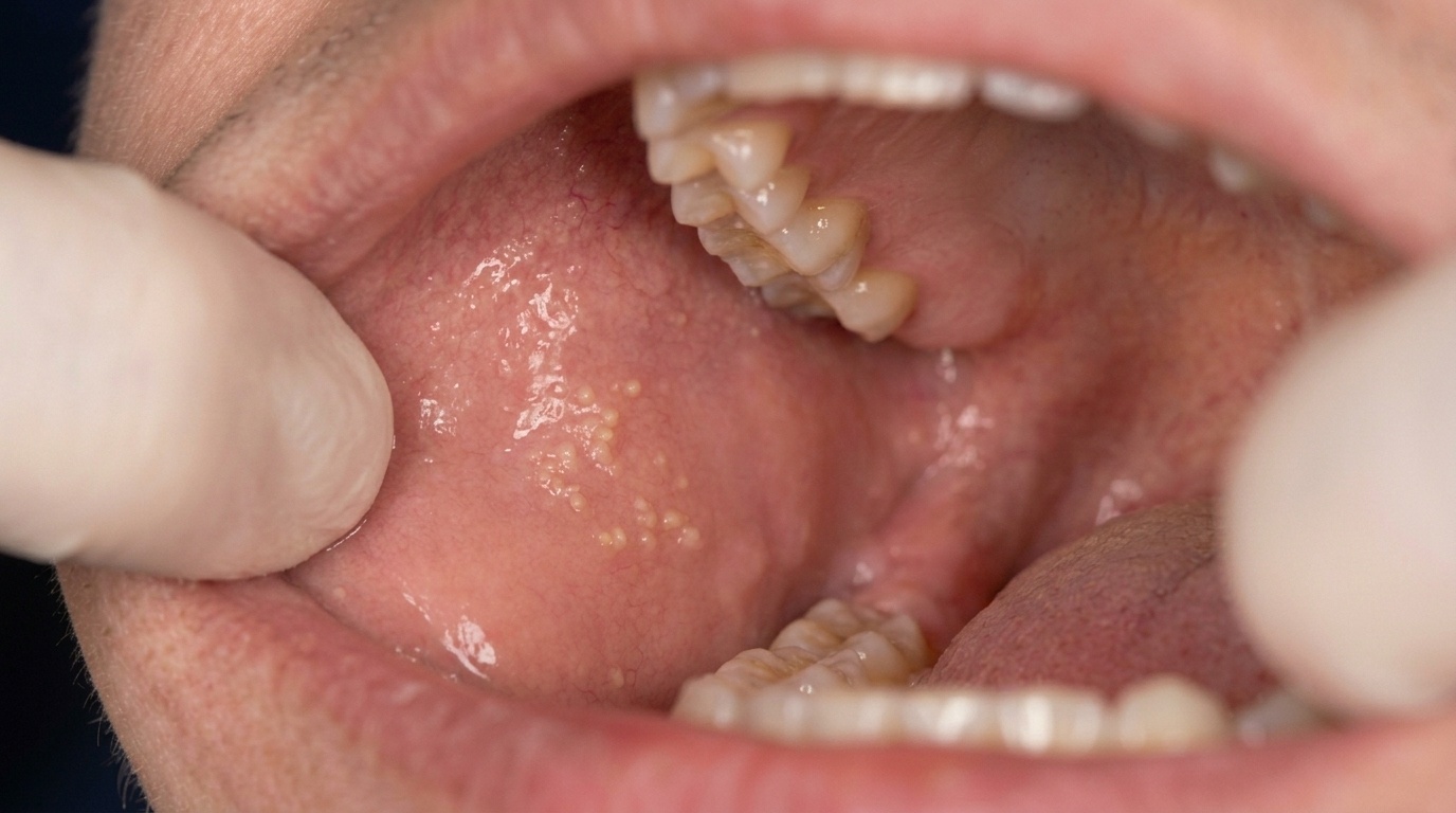
Fordyce spots (also called Fordyce granules) are tiny ectopic sebaceous glands (oil-producing glands sitting in a place where they would not normally be expected) of the mouth lining. They appear as small (1 to 3 mm) yellow-white papules (tiny soft raised spots), most often on the inside of the cheek and the vermilion (red-pink) border of the upper lip. The textbooks describe them as a harmless anatomical variant rather than a disease. They are not infectious, not cancerous, and not a sign of any underlying problem. Up to 80% of adults have at least some Fordyce spots if examined carefully.
Who tends to get it?
The textbooks describe a fairly consistent picture:
Around 80% of adults have at least some Fordyce spots if examined carefully.
Both sexes are affected, with some studies reporting a slightly higher rate in men.
All ages, with the spots typically becoming more visible from puberty onwards as the sebaceous glands respond to sex hormones.
Newborns sometimes show similar spots that can be mistaken for thrush; they then persist quietly into adulthood.
All ethnicities are affected.
Because the spots are so common, dentists often see them at routine check-ups even when the patient has not noticed them.
What causes it?
Fordyce spots come from an embryonic anomaly (a small developmental quirk during the early formation of the mouth lining). Sebaceous glands are normally found in the skin, attached to hair follicles. In Fordyce spots, the same type of gland develops in the mouth lining without an associated hair follicle. Several points worth noting:
Present from birth in most people, although not always visible at first.
More obvious after puberty because the sebaceous glands enlarge under the influence of sex hormones.
Not caused by smoking, diet, hygiene, infection or sexual activity in any meaningful way.
Not a sexually transmitted condition, despite the fact that similar spots can appear on the genitals.
How does it develop?
The course is gradual. The sebaceous glands are present from birth in the affected area, but during childhood they sit quietly and produce very little oil. From puberty onwards, hormonal stimulation enlarges the glands and they become visible as small yellow-white papules. In adulthood, the spots may slowly become more numerous or more obvious with age. They do not progress to any other condition and do not change in any harmful way.
What might you notice?
What it looks like
The classic appearance is well described:
A cluster of small yellowish-white spots on the inside of one or both cheeks.
Tiny dots on the upper lip that look like little oil droplets just below the surface.
Multiple spots in symmetrical patterns, often on both sides of the mouth.
Smooth-surfaced spots, just under the lining rather than raised above it.
More visible when the lining is stretched, for example when smiling broadly or pulling the lip away from the teeth.
What it feels like
Fordyce spots are asymptomatic (causing no symptoms). There is no pain, no itching, no bleeding and no taste change. Some people can feel the spots as slightly raised when the tongue brushes over them, but there is no discomfort. Brushing, mouthwash and other home measures do not change their appearance.
What an X-ray might show
Fordyce spots are confined to the surface lining and are not visible on dental X-rays.
What happens at the dentist?
Fordyce spots are most often noticed at a routine dental check-up and clean at ArtSmiles, either by the patient or by the dentist as part of the soft-tissue examination. The dentist will typically:
Examine the area carefully under good light, confirming the appearance: small, yellow-white, soft, scattered.
Take a brief history to confirm the spots have been there a long time and are not painful or growing.
Check the rest of the mouth to make sure nothing else is going on.
Reassure the patient in the vast majority of cases, which are diagnosed clinically without further investigation.
Photograph the area when the appearance is unusual or the patient wants to track them over time.
Discuss cosmetic options for the rare patient who is significantly bothered by visible Fordyce spots on the lip, while being honest about the limited results and recurrence rate.
Is this serious?
🟢 Fordyce spots are completely benign. They are not cancer, do not turn into cancer, are not infectious and have no association with any systemic illness. The reason they sometimes deserve attention is simply to confirm the diagnosis, since many other conditions can produce small bumps in the mouth and a careful examination provides reassurance.
If you have noticed small yellow-white spots in your mouth and would like reassurance, it is worth booking an assessment so the diagnosis can be confirmed.
Could it be something else?
A short list of conditions that can be confused with Fordyce spots:
Oral thrush (pseudomembranous candidiasis), creamy white patches that wipe off, leaving a red base, different from the firm, persistent papules of Fordyce spots.
Mucocele, a single, soft, bluish swelling most often on the lower lip.
Pyogenic granuloma, a single red, growing, easily-bleeding lump.
Lipoma, a deeper, larger, soft yellowish lump.
Mucinous cysts and small mucous retention cysts, single, soft, fluid-filled bumps.
Sebaceous hyperplasia, common on facial skin but rarely confused with intraoral Fordyce spots.
Wart (verruca vulgaris) or other HPV-related papilloma, rough, cauliflower-like surface, usually solitary.
When in doubt, a careful clinical examination, and in rare cases a small biopsy, sorts these out.
How is it treated?
The standard answer is no treatment. Fordyce spots are harmless and recurrence after any procedure is common, so most clinicians recommend leaving them alone.
At-home measures and habits:
Continue normal oral hygiene, brushing twice a day with fluoride toothpaste and flossing daily.
Avoid scrubbing or picking at the area, which can cause minor irritation without changing the spots.
Photograph the area if you want to track for any change, although Fordyce spots do not usually change in any harmful way.
Professional steps your dentist may consider:
Confirming the diagnosis by clinical examination, with a small biopsy only in rare unclear cases.
Reassuring the patient that the spots are a normal variant and need no treatment.
Discussing cosmetic options for the rare patient bothered by visible spots on the lip, including topical retinoids, CO2 or pulsed dye laser, electrodessication or micro-punch excision, while explaining the limited results and risk of scarring or recurrence.
A patient-centred approach matters here. People sometimes worry that small bumps in the mouth might be something serious, and an unhurried explanation of what Fordyce spots are and why they need no treatment is itself part of effective care, values that sit at the heart of our clinical philosophy.
What's the long-term outlook?
The long-term outlook is excellent. Fordyce spots are stable, harmless and do not require any follow-up. Once you know what they are, you can simply ignore them. Most people who once worried about the spots are reassured for life by a single explanation at the dental chair.
A note on this article
This article is for educational purposes only and does not constitute a clinical diagnosis. Please consult a registered dental practitioner for assessment and treatment advice.
The cover image above is an AI-generated illustration based on the most common visible features of this condition described in clinical pathology references. It is not a photograph of a real case and should not be used to diagnose or rule out the condition in your own situation. If you are concerned about something you have noticed, please book an assessment with a registered dental practitioner.
References
Neville, B. W., Damm, D. D., Allen, C. M., & Chi, A. C. (2023). Oral and maxillofacial pathology (5th ed.). Elsevier. Chapter 1, Developmental Defects of the Oral and Maxillofacial Region: Fordyce Granules.
Cawson, R. A., & Odell, E. W. (2017). Cawson's essentials of oral pathology and oral medicine (8th ed.). Elsevier. Chapter 2, Disorders of Development: cross-reference for Fordyce granules as an ectopic sebaceous gland variant.
Regezi, J. A., Sciubba, J. J., & Jordan, R. C. K. (2017). Oral pathology: Clinical pathologic correlations (7th ed.). Elsevier. Chapter on Cysts and Hamartomas: cross-reference for Fordyce granules.
Laskaris, G. (2006). Pocket atlas of oral diseases (2nd ed.). Thieme. Fordyce Granules entry, with clinical photographs and differential diagnosis.
Frequently asked questions
Are Fordyce spots contagious?
No. Fordyce spots are not infectious and cannot be passed to anyone else. They are simply small oil-producing glands sitting in the mouth lining where, in some people, they are visible from the surface.
Are Fordyce spots a sign of an STI?
No. Despite the fact that similar spots can appear on the genitals, Fordyce spots are a normal anatomical variant and are not sexually transmitted. They are also not caused by smoking, diet, hygiene or any specific lifestyle habit.
Do Fordyce spots need to be removed?
Usually not. They are harmless, do not turn into cancer and need no treatment. Cosmetic removal options exist for the rare patient who is significantly bothered by visible spots on the lip, but results are limited and recurrence is common, so most clinicians recommend leaving them alone.
Will Fordyce spots go away on their own?
They tend to persist throughout adult life. In some people they become slightly more obvious from puberty and then stay stable. They do not progress to any other condition and do not change in any harmful way.