Compiled from clinical pathology references. Medically reviewed by Dr Cristian Dunker , Principal Dentist, ArtSmiles Cosmetic Dentistry.
Quick summary
Also called | Pregnancy gingivitis, pregnancy tumour, pregnancy epulis (a benign red gum lump that appears during pregnancy; same lesion as a pyogenic granuloma), pregnancy granuloma, granuloma gravidarum, perimolysis (when erosion (acid-related loss of enamel from the tooth surface) is involved), ptyalism gravidarum (the medical term for excessive saliva production during pregnancy) (when excess saliva occurs) |
How urgent? | 🟡 Worth a check-up , most pregnancy-related oral changes are manageable, but bleeding gums, growing lumps, and tooth sensitivity respond best to early care |
Common or rare? | Very common , gingival inflammation affects a high proportion of pregnancies, and pregnancy-associated pyogenic granulomas occur in around 5% of pregnancies |
Who it affects | Pregnant women, most often from the first trimester onwards, with severity peaking around the seventh month |
Who treats it | General dentist, often working alongside your obstetrician or midwife |
Based on | Regezi, Neville, Cawson, and Laskaris |
What is it?
Pregnancy brings a wave of hormonal changes, and your mouth is one of the places those changes show up. The combination of rising oestrogen and progesterone, altered immune responses, dietary shifts, and morning sickness can produce a recognisable pattern of oral findings. These include exaggerated gum inflammation (pregnancy gingivitis (gum inflammation that develops or worsens during pregnancy due to hormonal changes)), localised gum lumps known as pregnancy tumours or pregnancy epulides, dental erosion from acid reflux or vomiting, occasional excess saliva, altered taste, and a heightened risk of progressing periodontal disease.
None of these conditions are unique to pregnancy. What pregnancy does is amplify how the mouth responds to plaque, acid, and minor trauma. Most signs settle after delivery once hormone levels return to baseline.
Who tends to get it?
Pregnancy-related oral changes can affect any pregnant woman, but several patterns stand out across the textbooks.
Pregnancy gingivitis is very common. Neville notes that women experience a greater susceptibility to gingivitis when exposed to the high progesterone levels of pregnancy, and Cawson reports that pre-existing gingivitis often becomes more severe in the first two months.
Pregnancy tumours (pyogenic granulomas) develop in around 5% of pregnancies, according to Regezi. Neville notes they may begin in the first trimester, and prevalence rises through the seventh month.
Dental erosion appears more often in women who experience persistent vomiting or acid reflux during pregnancy.
Severe taste alteration from prolonged vomiting in pregnancy is documented by Neville as a recognised cause of dysgeusia.
Excess salivation (ptyalism gravidarum) is uncommon but recognised, sometimes alongside hyperemesis gravidarum (severe, prolonged vomiting in pregnancy, more intense than typical morning sickness).
Women with pre-existing plaque accumulation, poor oral hygiene, or untreated periodontal disease tend to experience the most pronounced changes. Cawson and Regezi both stress that scrupulous oral hygiene can substantially reduce the severity of pregnancy-related gingival changes.
What causes it?
The driver is hormonal , but hormones rarely act alone. Across the four reference texts, the contributors include:
Rising oestrogen and progesterone, which alter the gingival response to plaque and increase blood vessel proliferation in soft tissue. Regezi describes how these hormonal shifts modify the gingival reparative response to injury, producing what was once called a pregnancy tumour.
Dental plaque and calculus, which provide the inflammatory stimulus that hormones then exaggerate , a build-up that a regular scale and clean helps keep in check.
Local irritants such as overhanging restorations, food traps, ill-fitting appliances, or rough tooth surfaces.
Repeated acid exposure from morning sickness, hyperemesis gravidarum, or pregnancy-related reflux. Neville lists pregnancy among the causes of chronic involuntary regurgitation that can lead to dental erosion (perimolysis).
Iron or folate deficiency, which Cawson notes can either trigger or exacerbate recurrent mouth ulcers during pregnancy.
Reduced or altered salivary flow in some women, which decreases the natural cleansing and buffering of the mouth.
It is important to recognise that, as Regezi puts it, significant gingival enlargement during periods of hormonal imbalance is questionable in individuals with scrupulous oral hygiene. The hormones set the stage; plaque writes the script.
How does it develop?
Think of your gum tissue as a thermostat that is normally calibrated to ignore small amounts of plaque without much fuss. During pregnancy, that thermostat becomes hypersensitive. The same plaque that previously caused mild inflammation now triggers a much larger response , more blood vessel growth, more swelling, easier bleeding. This same pattern of hormone-driven oral change is also seen later in life , for example, during menopause , though the picture differs.
When this hyper-reactive tissue meets a localised irritant , say, a food impaction site between two teeth , the response can become exaggerated enough to form a discrete lump. This lump is the pregnancy tumour, and microscopically it is a mass of small blood vessels in loose, oedematous tissue, which is why it bleeds so readily.
Meanwhile, frequent contact between tooth surfaces and stomach acid (from morning sickness) softens enamel. Saliva would normally remineralise the surface within minutes, but if vomiting is repeated, the protective window closes and enamel is gradually lost. Cawson and Neville both highlight that the buffering capacity of saliva is what determines whether erosion progresses or resolves.
After delivery, hormone levels return to baseline, the gingival hyper-response calms down, and many of these changes , including pregnancy tumours , partially or fully regress.
What might you notice?
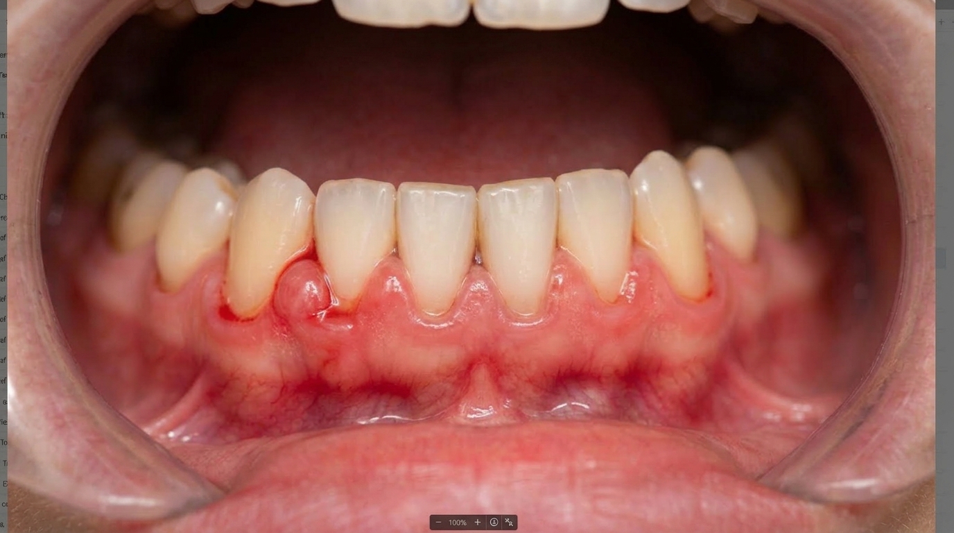
What it looks like
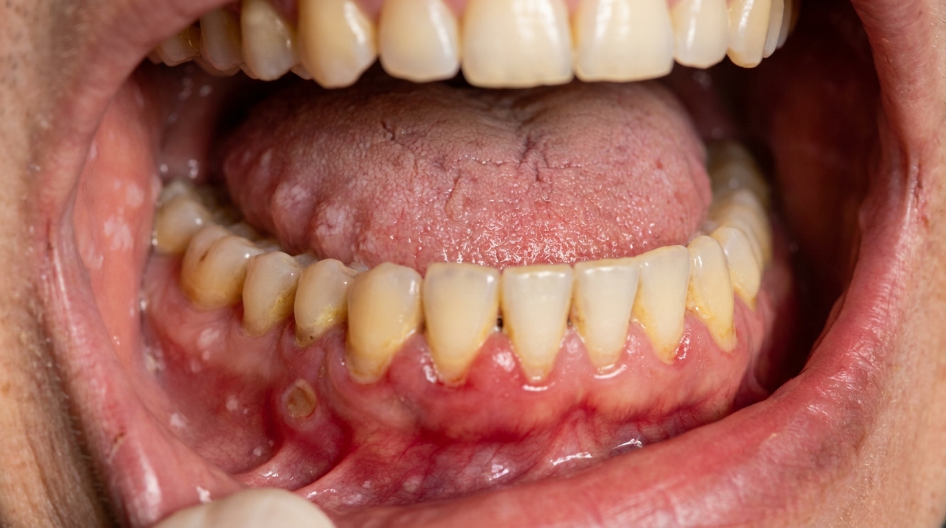
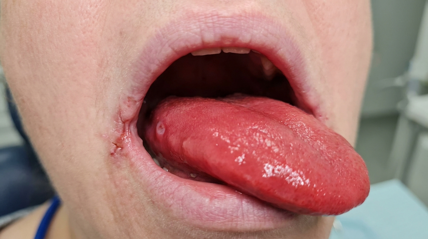
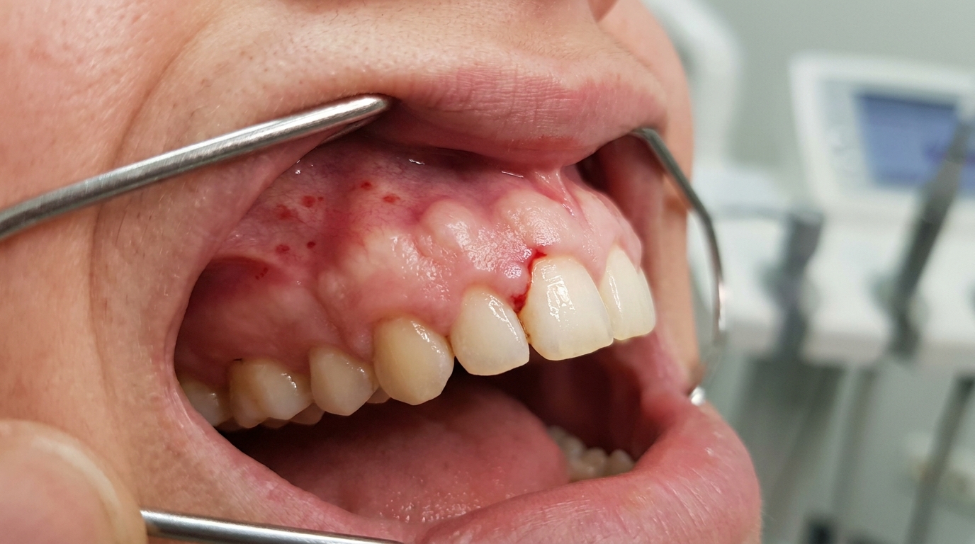
The gums often look bright red, glazed, and swollen, particularly between the teeth (the interdental papillae). Cawson describes inflammatory erythema and oedema becoming more severe in the first two months. In some areas, especially between the upper front and side teeth, a soft, dark red, sometimes purplish lump may appear and grow over weeks. This is the pregnancy tumour. Neville describes it as a smooth or lobulated mass, often pedunculated (on a stalk), with an ulcerated surface that ranges from pink to red to purple depending on its age.
If morning sickness has been a feature, the inside (palatal) surface of the upper front teeth may begin to look smoother, glassier, or thinner than before, with a yellower hue as the underlying dentine begins to show through.
What it feels like
Most pregnancy gingivitis feels like easy bleeding when brushing or flossing, sometimes with slight tenderness along the gum margin. Pregnancy tumours are typically painless but bleed alarmingly easily , Neville notes that they often bleed easily because of their extreme vascularity, and Cawson notes that they may create alarm for both the patient and clinician. Eroded tooth surfaces may feel sensitive to cold drinks, sweet foods, or acidic foods. Some women notice a metallic or altered taste, and a smaller number experience a sense of excess saliva (ptyalism), particularly when nausea is significant.
What an X-ray might show
In most pregnancy-related oral changes, X-rays add little. Cawson notes that pregnancy gingivitis shows intact crestal alveolar bone on radiographs, distinguishing it from periodontitis. Routine dental X-rays are usually deferred during pregnancy unless essential, and lead apron protection is standard when imaging is required. Cawson is explicit that only essential radiographs should be taken, the minimal radiation exposure should be given, and the patient should wear a lead apron.
What happens at the dentist?
If you are pregnant or planning a pregnancy, the team at ArtSmiles will usually take a careful history that includes how far along you are, whether you are experiencing morning sickness, your current medications, and any pre-existing gum or tooth concerns.
A gentle clinical examination focuses on:
The colour, texture, and bleeding tendency of the gums
Any localised lumps along the gum line, including their size, colour, and how easily they bleed
Signs of erosion on the inner surfaces of the upper front teeth
Plaque and calculus levels
Tooth mobility and the depth of the gingival sulcus around teeth
If a lump has developed, your dentist may monitor it during pregnancy rather than remove it, because Neville advises that treatment of pregnancy-related lesions is usually deferred unless significant functional or aesthetic problems develop, and the recurrence rate is higher when they are removed during pregnancy. Where a biopsy is ultimately needed, it can be performed under local anaesthesia.
For elective dental work , fillings, scaling, or other planned treatment , Cawson notes that the second trimester is generally the safest window. During the third trimester, some women become hypotensive when laid flat (because the enlarged uterus impedes venous return), so treatment is often performed in a more upright position. Local anaesthesia is considered desirable rather than letting a pregnant woman experience operative pain, and Cawson notes there is no evidence of risks from local anaesthesia.
Is this serious?
🟡 Worth a check-up , usually manageable, occasionally consequential.
Most pregnancy gingivitis settles with good oral hygiene and a professional clean. Pregnancy tumours, while sometimes alarming in appearance and bleeding, are benign and often shrink after delivery. Where things become more consequential is when:
Established periodontal disease progresses unchecked
Repeated vomiting causes ongoing enamel erosion
A pregnancy tumour grows large enough to interfere with eating or speaking
Iron or folate deficiency triggers recurrent mouth ulcers that affect nutrition
Left entirely unaddressed, gum inflammation can progress to attachment loss and tooth mobility, and erosion can advance to the point where teeth need restoration. None of these are emergencies, but they all respond best to early, gentle intervention.
If you've noticed any of these signs for more than two weeks, it's worth booking an assessment.
Could it be something else?
The textbooks list several conditions that can mimic the gum lumps and gum changes seen in pregnancy. Your dentist will work through these to be sure of what is actually present.
Pyogenic granuloma (non-pregnancy-related) , clinically and microscopically identical to a pregnancy tumour. The distinguishing feature is the patient context , pregnancy versus a localised irritant in a non-pregnant patient.
Peripheral giant cell granuloma , also presents as a red or red-blue gingival mass. Regezi notes it is generally clinically indistinguishable from pyogenic granuloma, although peripheral giant cell granulomas are more likely to cause bone resorption and tend to look more blue-purple than the bright red of a pregnancy tumour. Biopsy provides the definitive answer.
Peripheral ossifying fibroma , another reactive gingival lesion. Cawson and Neville both note these tend to be much lighter in colour, often pink rather than bright red, and the non-ulcerated ones can look similar to a fibroma. Biopsy distinguishes them.
Fibrous epulis (fibrous hyperplasia) , a firm, pink or red gum lump associated with poor oral hygiene, a local plaque trap, or an overhanging restoration. Cawson describes these as firm rather than soft, and not usually as deeply red as a pregnancy tumour.
Periodontal abscess , painful, red, often discharging pus, associated with a deep periodontal pocket or non-vital tooth, and usually accompanied by significant tenderness , unlike the typically painless pregnancy tumour.
Squamous papilloma , a white, spiky or cauliflower-like surface marks this out from the smooth, red surface of a pregnancy tumour.
Giant-cell epulis (in a child) , pink, red or purplish nodule anterior to the first permanent molar in a younger patient, often around the time of deciduous tooth loss or extraction.
Drug-induced gingival overgrowth , generalised firm fibrous enlargement related to phenytoin, calcium channel blockers (such as nifedipine), or ciclosporin, rather than localised reactive masses.
Leukaemic gingival infiltration , generalised swelling, often pale or purplish, with bleeding, ulceration, and other systemic signs such as pallor or bruising. Considered when the picture does not fit pregnancy alone.
Wegener's (granulomatosis with polyangiitis) , causes red-speckled 'strawberry gums' and is often accompanied by respiratory or renal symptoms.
Kaposi sarcoma, bacillary angiomatosis, or non-Hodgkin's lymphoma , Regezi lists these as less common considerations for a red gingival mass.
Metastatic cancer , rare, but Regezi notes a metastatic lesion can occasionally present as a red gingival mass. Persistent or atypical lesions are biopsied.
Postextraction granuloma (epulis granulomatosa) , a pyogenic granuloma developing in a recent extraction socket, related to bony sequestra or foreign material rather than pregnancy.
Mouth-breathing-related gingivitis , Neville notes that incomplete lip closure can produce smooth, swollen, red gingivitis of the anterior facial gingiva, mimicking pregnancy gingivitis.
Puberty gingivitis , same hormone-amplified plaque response, but in adolescents rather than pregnant women.
Diabetes-related gingivitis , Cawson notes that in poorly controlled diabetes, gingival health may deteriorate sharply, producing a similar exaggerated response to plaque.
Vitamin C deficiency (scurvy) , historically described as causing grossly swollen, congested gums.
Erosion from other causes , bulimia, hiatal hernia, frequent acidic soft drink consumption, alcoholism, or occupational acid exposure can all produce a similar pattern of dental erosion to that caused by morning sickness.
How is it treated?
Care during pregnancy is gentle, conservative, and focused on prevention.
At home, you can help by:
Brushing twice daily with a soft-bristled toothbrush and a fluoride toothpaste
Cleaning between teeth daily with floss or interdental brushes
After vomiting, rinsing with water (or a small amount of dilute bicarbonate of soda solution) and waiting around 30 minutes before brushing, so softened enamel is not abraded
Using a straw for acidic drinks where possible (and avoiding sipping them slowly)
Maintaining a balanced diet, with adequate iron and folate intake
Staying well hydrated to support saliva flow
Professional care may include:
A gentle scale and clean to remove plaque and calculus and reduce the irritant load on inflamed gums
Oral hygiene coaching tailored to areas of bleeding
Topical fluoride application to strengthen eroded enamel
Treatment of any active dental decay during the second trimester where possible
Monitoring of any pregnancy tumour, with surgical removal usually deferred unless it is causing functional or aesthetic problems. Cawson notes that a strict oral hygiene regimen can much ameliorate or abolish pregnancy gingivitis, and that pregnancy tumours generally improve after parturition. If they persist, they can be excised after the baby is born.
For pregnancy tumours that are removed during pregnancy, your dentist may discuss the higher recurrence rate so that expectations are realistic
Cawson notes that the chief precautions during dental care in pregnancy include avoiding non-essential X-rays (especially in the first trimester), avoiding intravenous sedation, keeping the patient well positioned to avoid supine hypotension, and being mindful of any pre-existing iron or folate deficiency. Drug choices are discussed with your obstetrician where needed.
What's the long-term outlook?
The outlook is generally reassuring. Cawson notes that pregnancy gingivitis improves after parturition, and Neville notes that after pregnancy and the return of normal hormone levels, some pyogenic granulomas resolve without treatment or undergo fibrous maturation and come to resemble a fibroma. Laskaris similarly notes that pregnancy granulomas may regress and sometimes disappear after delivery.
With good plaque control, most women emerge from pregnancy with their gum health restored to near baseline. Where periodontal disease was already established before pregnancy, that underlying condition will need ongoing management , pregnancy may have accelerated it, but it does not cause it on its own.
Dental erosion, unfortunately, does not reverse itself. Lost enamel is gone, but progression can be halted by addressing the underlying cause (managing reflux or hyperemesis with your obstetrician, modifying acidic intake), and any cosmetic or sensitivity issues can be addressed after delivery with conservative restorative treatment.
Any gum lump that does not resolve within a few months of delivery should be reviewed and, if it persists, biopsied , both to confirm the diagnosis and to rule out the less common conditions on the differential list.
A note on this article
This article is for educational purposes only and does not constitute a clinical diagnosis. Please consult a registered dental practitioner for assessment and treatment advice.
The cover image above is an AI-generated illustration based on the most common visible features of this condition described in clinical pathology references. It is not a photograph of a real case and should not be used to diagnose or rule out the condition in your own situation. If you are concerned about something you have noticed, please book an assessment with a registered dental practitioner.
References
Regezi, J. A., Sciubba, J. J., & Jordan, R. C. K. (2017). Oral pathology: Clinical pathologic correlations (7th ed.). Elsevier. Chapter 4 , Red-Blue Lesions (Pyogenic Granuloma, pp. 118 to 120) and Chapter 7 , Connective Tissue Lesions (Gingival Hyperplasia, pp. 164 to 166).
Neville, B. W., Damm, D. D., Allen, C. M., & Chi, A. C. (2023). Oral and maxillofacial pathology (5th ed.). Elsevier. Chapter 2 , Abnormalities of Teeth (Erosion, p. 59); Chapter 4 , Periodontal Diseases (Gingivitis, pp. 147 to 150); Chapter 11 , Salivary Gland Pathology (Sialorrhea / Ptyalism Gravidarum, p. 469); Chapter 12 , Soft Tissue Tumors (Pyogenic Granuloma, pp. 525 to 527).
Cawson, R. A., & Odell, E. W. (2017). Cawson's essentials of oral pathology and oral medicine (8th ed.). Elsevier. Chapter 5 , Gingivitis and Periodontitis (Pregnancy gingivitis, p. 80); Chapter 19 , Common Benign Mucosal Swellings (Pyogenic Granuloma and Pregnancy Epulis, pp. 316 to 319); Chapter 31 , Endocrine Disorders and Pregnancy (pp. 413 to 414).
Laskaris, G. (2003). Color atlas of oral diseases (3rd ed.). Thieme. Chapter 27 , Endocrine Diseases (Sex Hormone Disorders, pp. 252 to 253) and Chapter 35 , Tumor-like Lesions (Pregnancy Granuloma, p. 336).