Compiled from clinical pathology references. Medically reviewed by Dr Cristian Dunker , Principal Dentist, ArtSmiles Cosmetic Dentistry.
Quick summary
Also called | Eruption hematoma (when blood is in the cyst), eruption haematoma (a small collection of blood under the surface) |
How urgent? | 🟢 Not urgent, almost always harmless and resolves on its own as the tooth erupts |
Common or rare? | Common in children, frequently seen as baby teeth and first permanent teeth come through |
Who it affects | Mostly children under the age of 10, but occasionally adults whose wisdom teeth or other late teeth are erupting |
Who treats it | General dentist, most often no treatment is needed at all |
Based on | Cawson, Neville, Laskaris, with cross-references in Regezi |
What is it?
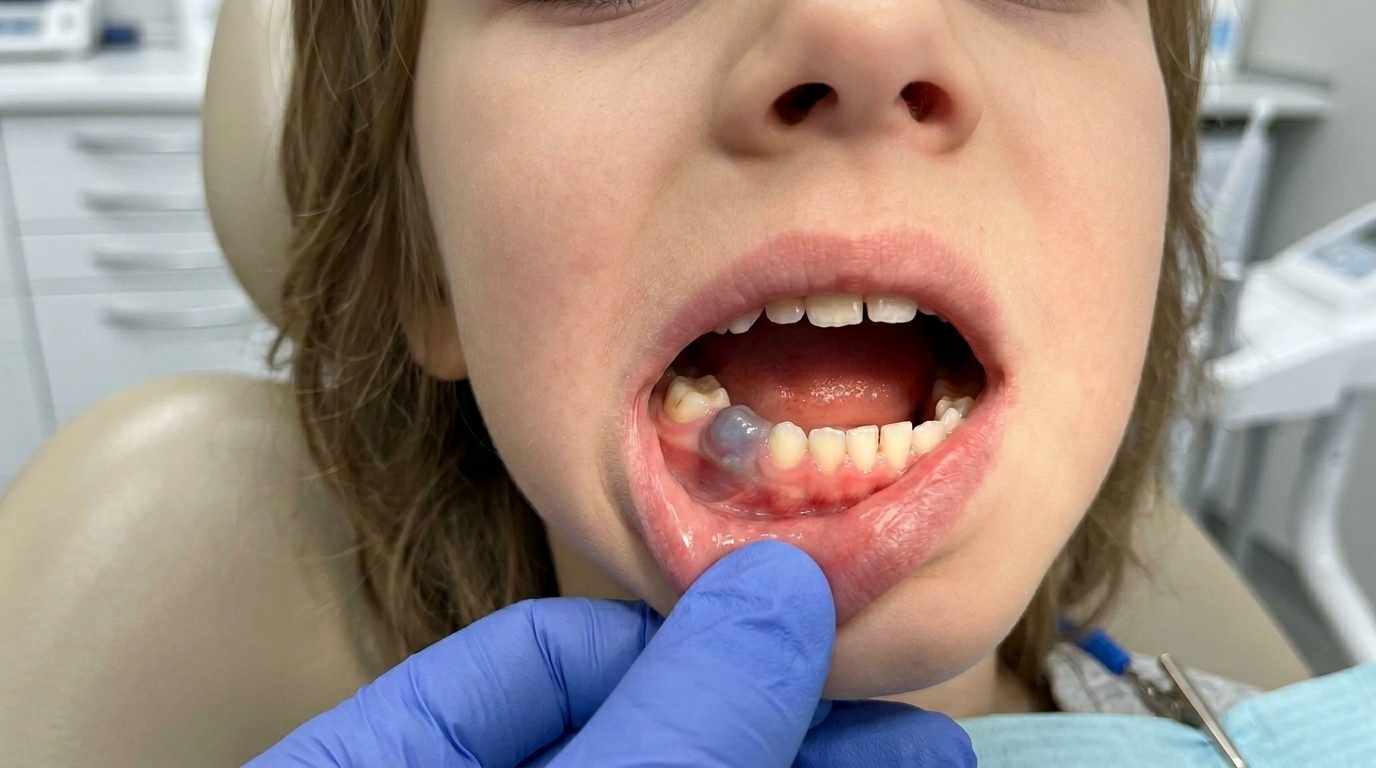
An eruption cyst is a soft, fluid-filled swelling that develops in the gum directly over a tooth that is about to come through. It is essentially the soft-tissue version of a dentigerous cyst (a fluid sac around the crown of an unerupted tooth), fluid accumulates between the crown of the erupting tooth and the gum tissue covering it. When biting or knocking on the swelling causes a small amount of bleeding into the cyst, it takes on a blue, purple or dark red colour and is then often called an eruption hematoma. Despite the dramatic colour, eruption cysts are benign, and most resolve on their own as the tooth pushes through the cyst and into the mouth.
Who tends to get it?
Eruption cysts are particularly associated with childhood:
Most often in children younger than 10, around the time baby teeth and first permanent teeth come through.
Most commonly seen over central incisors and first molars in both the baby and adult dentitions, although any erupting tooth can develop one.
Occasionally bilateral, one on each side of the mouth.
Less commonly in adults, mostly when wisdom teeth or other late teeth are erupting.
There is no strong sex or ethnic predilection. Many children develop more than one eruption cyst over the course of their dental development.
What causes it?
The textbooks describe a single, fairly simple mechanism:
As a tooth pushes its way up through the bone, the dental follicle (the soft tissue capsule around the developing crown) reaches the gum.
A fluid-filled space forms between the dental follicle and the surface of the crown, just under the gum.
The lining of this space is the reduced enamel epithelium, the thin epithelial layer that originally covered the developing crown.
If the area is traumatised (for example, by chewing on it), small blood vessels in the surrounding tissue can leak, and blood collects within the cyst, producing the classic blue or purple eruption hematoma.
There is no infection, no faulty development, and no long-term consequence.
How does it develop?
Most eruption cysts develop quietly over a few days to weeks as a tooth approaches the surface. The first thing parents typically notice is a soft, dome-shaped swelling in the gum at the spot where a tooth is expected. Some swellings stay pale and translucent until the tooth bursts through; others, particularly when a child is biting on hard food or a toy, develop the bluish hematoma appearance from minor bleeding inside.
The swelling continues to enlarge slightly until the erupting tooth's cusp finally pierces the cyst and the dome opens. The fluid drains, and the cyst essentially disappears. The tooth then continues its normal eruption.
What might you notice?
What it looks like
The classic appearance is well described:
A soft, dome-shaped swelling of the gum directly over a tooth that has not yet come through.
The swelling can be translucent, pale pink or yellowish when it contains only clear fluid.
It often becomes blue, purple or dark red when it contains blood (eruption hematoma).
The swelling is fluctuant, feels soft and a little springy, like a small water balloon.
A whitish point or a small slit may appear as the tooth begins to break through.
What it feels like
Most eruption cysts cause little or no discomfort. Some children may experience:
Mild tenderness when biting on the area.
Reluctance to eat firm foods for a few days.
Increased dribbling in toddlers.
Brief bleeding if the cyst is bumped or chewed on, particularly in older children with adult teeth coming through.
Significant pain, swelling beyond the immediate area, or fever should prompt a check, as these are not features of an uncomplicated eruption cyst.
What an X-ray might show
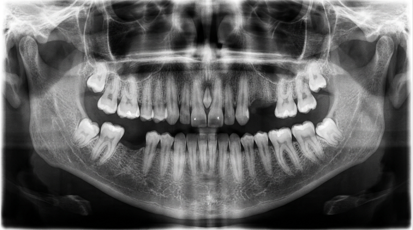
X-rays are usually not needed for an eruption cyst. The diagnosis is made on appearance and the position over an erupting tooth. If an X-ray is taken for another reason, the underlying tooth may be visible just below the gum surface, sometimes with a small radiolucent (darker on X-ray) area outlining the crown.
What happens at the dentist?
Most parents bring a child to the dentist because they have noticed a coloured swelling and are worried. A dentist at ArtSmiles, typically as part of a dental check-up and clean, will commonly:
Examine the swelling and confirm that it overlies an erupting tooth.
Press gently on the dome to confirm it is soft and fluctuant rather than firm.
Check the rest of the mouth to see whether other erupting teeth are involved or whether something else is going on.
Reassure parents and child that the appearance, while alarming, is benign and almost always resolves on its own.
Take an X-ray only if the diagnosis is unclear or if there is concern about the position of the underlying tooth.
Plan a recall in a few weeks to confirm the tooth has erupted and the swelling has settled.
Is this serious?
🟢 An eruption cyst is benign and self-limiting. It is not cancer, not contagious, and does not usually need any treatment. The dramatic colour can be alarming for parents, but reassurance and a watch-and-wait approach are almost always appropriate. The main reason to see a dentist is to confirm that what is being seen is, in fact, an eruption cyst and not one of the conditions it can mimic.
If your child has a coloured swelling that has not resolved within a few weeks, is growing rapidly, is associated with pain, or is bleeding repeatedly, it is worth booking an assessment so the appearance can be confirmed.
Could it be something else?
Several conditions can produce a blue or coloured swelling on the gum. The textbooks list these as the main differentials:
Hematoma from trauma, a similar bluish swelling, but related to a recent knock rather than an erupting tooth. Usually flat or spreading rather than dome-shaped over a tooth.
Haemangioma (a benign growth made of small blood vessels) or vascular malformation, a longer-standing reddish-blue lesion that has been present since infancy and does not resolve with eruption.
Amalgam tattoo, a flat, grey-blue area in the gum from old metal filling material; usually in older children with previous dental treatment.
Pigmented naevus, a small flat or slightly raised pigmented spot, present for some time, that does not change with eruption.
Dental abscess, a tender, often painful swelling that may have pus and is associated with a decayed or non-vital tooth, not a normally erupting one.
Oral malignant melanoma, extremely rare in children, but should be considered for any unexplained pigmented lesion that does not behave like an eruption cyst.
How is it treated?
The textbooks all agree that most eruption cysts need no treatment. The most useful steps are reassurance and follow-up.
At-home measures:
Reassure your child that the bump is harmless and will go away when the tooth comes through.
Encourage gentle brushing around the area, avoiding excessive pressure.
Stick to softer foods for a few days if the area feels tender.
Avoid letting the child chew hard or sharp objects that might bleed into the cyst.
Professional steps your dentist may consider:
No treatment, with simple monitoring as the tooth erupts, this is the most common course.
Simple unroofing of the cyst under local anaesthetic, when a tooth is not coming through after some weeks despite the cyst being clearly present. The cyst roof is gently excised, the underlying tooth is exposed, and eruption is allowed to continue.
Drainage of a tense haematoma if the swelling is unusually large or uncomfortable.
Reassessment if the appearance does not match a typical eruption cyst, with onward referral if needed.
A patient-centred approach matters here too, a young child and their parents may be quite worried by a sudden bluish swelling. Calm, clear explanation and reassurance are themselves part of effective care, values that sit at the heart of our clinical philosophy.
What's the long-term outlook?
The outlook is excellent. The textbooks describe eruption cysts as benign and self-limiting, with the tooth usually erupting normally through the cyst within days to weeks. There are no long-term consequences, and the gum overlying the new tooth heals without scarring. Children who have had an eruption cyst over one tooth may or may not develop one over a later erupting tooth, neither outcome causes any harm. Once all the teeth in the area have come through, the condition is essentially resolved.
A note on this article
This article is for educational purposes only and does not constitute a clinical diagnosis. Please consult a registered dental practitioner for assessment and treatment advice.
The cover image above is an AI-generated illustration based on the most common visible features of this condition described in clinical pathology references. It is not a photograph of a real case and should not be used to diagnose or rule out the condition in your own situation. If you are concerned about something you have noticed, please book an assessment with a registered dental practitioner.
References
Cawson, R. A., & Odell, E. W. (2017). Cawson's essentials of oral pathology and oral medicine (8th ed.). Elsevier. Chapter 7, Cysts of the Jaws: Eruption cyst with management, p. 123.
Neville, B. W., Damm, D. D., Allen, C. M., & Chi, A. C. (2023). Oral and maxillofacial pathology (5th ed.). Elsevier. Chapter 15, Odontogenic Cysts and Tumors: Eruption Cyst (Eruption Hematoma), pp. 689 to 690.
Regezi, J. A., Sciubba, J. J., & Jordan, R. C. K. (2017). Oral pathology: Clinical pathologic correlations (7th ed.). Elsevier. Chapter 10, Cysts of the Jaws and Neck: Eruption cyst as soft-tissue dentigerous cyst variant.
Laskaris, G. Pocket atlas of oral diseases. Thieme. Chapter 14, Soft-Tissue Cysts: Eruption Cyst, p. 112.
Frequently asked questions
What is an eruption cyst?
An eruption cyst is a soft, fluid-filled bluish-purple swelling on the gum directly over a tooth that is about to come through. It is essentially a small bubble of fluid in the dental sac surrounding the crown of the erupting tooth. They are most often seen in young children with their first baby teeth, or in school-aged children with their adult molars.
Is an eruption cyst painful or dangerous?
Most are painless and harmless. The bluish-purple colour can be alarming for parents, but the cyst usually bursts on its own as the tooth pushes through the gum and disappears within days to weeks. It does not damage the tooth underneath.
Does an eruption cyst need treatment?
Usually not. Most eruption cysts resolve on their own without any intervention as the tooth erupts. If the cyst becomes very large, painful or stops the tooth from coming through, a dentist may make a small painless slit (marsupialisation) to release the fluid and uncover the tooth.
How is an eruption cyst different from a normal bruised gum?
An eruption cyst is a smooth, dome-shaped, fluid-filled bubble directly over the spot where a tooth is about to come through. A normal teething gum is just red and inflamed without a clear fluid bubble. If you are unsure, a dental check-up can quickly confirm which one it is.