Compiled from clinical pathology references. Medically reviewed by Dr Cristian Dunker , Principal Dentist, ArtSmiles Cosmetic Dentistry.
Quick summary
Also called | Oral mucosal melanoma, mucosal melanoma of the mouth |
How urgent? | 🔴 Serious, although rare, oral melanoma is much more aggressive than skin melanoma; any new or changing dark patch in the mouth needs prompt assessment |
Common or rare? | Rare, accounts for less than 1% of all oral cancers and around 1-2% of all melanomas |
Who it affects | Most often adults in the fifth to seventh decades of life; slight male predilection in some series; more common in some Japanese, African and Indian populations |
Who treats it | Diagnosis often begins with a general dentist; definitive care is provided by an oral and maxillofacial surgeon, head and neck oncology team, and medical oncologist (a cancer specialist) |
Based on | Neville, Cawson, with cross-references in Regezi |
What is it?
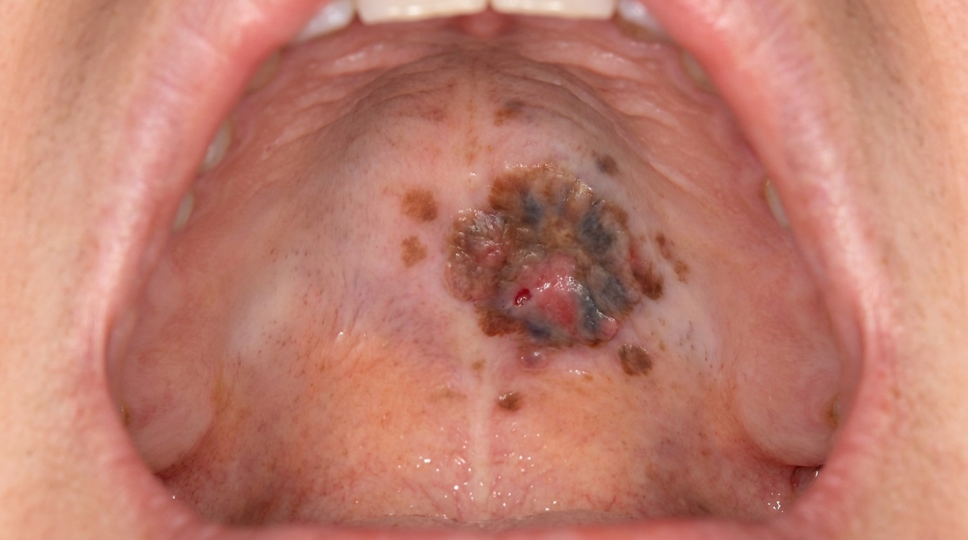
Oral malignant melanoma is a cancer that arises from the pigment-producing cells (melanocytes, the pigment-producing cells) of the lining of the mouth. The textbooks describe it as the mucosal counterpart of skin melanoma, but with two important differences. First, it is much rarer, most pigmented patches in the mouth are benign, and oral melanoma accounts for only a small percentage of mouth cancers and an even smaller percentage of all melanomas. Second, it is considerably more aggressive than its skin counterpart, with a higher rate of spread to lymph nodes and distant organs at the time of diagnosis. About 70-80% of oral melanomas occur on the hard palate (the roof of the mouth) or upper gum (maxillary alveolus), making the inside of the upper jaw a particularly important area to examine.
Who tends to get it?
The textbooks describe a fairly distinctive profile:
Most often diagnosed in the fifth to seventh decades of life (50s-70s), with a wide range across all adult ages.
Slight male predilection in many series, although some studies show no significant difference.
More common in people of Japanese, African and Indian heritage than in people of European heritage.
Hard palate or maxillary alveolus are the most common sites (70-80% of cases). Other sites include the upper lip, lower gum, buccal mucosa and tongue.
At least one in three patients has had a pigmented macule in the area for some time before the melanoma was diagnosed, sometimes for years.
What causes it?
The cause of oral melanoma is less well understood than that of skin melanoma:
Unlike skin melanoma, sun exposure does not appear to play a role, since the inside of the mouth is not exposed to ultraviolet light.
Tobacco and alcohol have been implicated as possible risk factors, but the link is weaker than for the better-known oral squamous cell carcinoma.
Pre-existing pigmented lesions, including melanotic macules and oral naevi, may be the precursor in a proportion of cases, although most oral melanomas appear to arise without an identifiable precursor.
Genetic and molecular factors are an active area of research, with some genetic mutations identified that differ from those of cutaneous melanoma.
There is no clear, consistent environmental risk factor that allows everyday prevention, which makes early recognition all the more important.
How does it develop?
Oral melanoma typically develops in stages, although these stages can be difficult to recognise clinically:
Radial (lateral) growth phase. Atypical melanocytes spread along the basal layer of the surface lining of the mouth. Clinically, this looks like a flat, dark patch with irregular borders.
Vertical growth phase. The atypical cells start to invade the underlying connective tissue. Clinically, the patch becomes raised, lumpy or ulcerated.
Lymph node (a small immune-system gland that filters tissue fluid) and distant spread. Because the mouth has a rich blood and lymphatic supply, oral melanoma tends to spread early, about a third of patients have lymph node involvement at the time of diagnosis, and about a quarter have distant metastasis (spread of cancer cells to other parts of the body).
The textbooks specifically note that the mucosal form invades blood and lymph vessels more readily than the skin form, which is one reason why outcomes are typically worse.
What might you notice?
What it looks like
The textbooks describe several classic patterns:
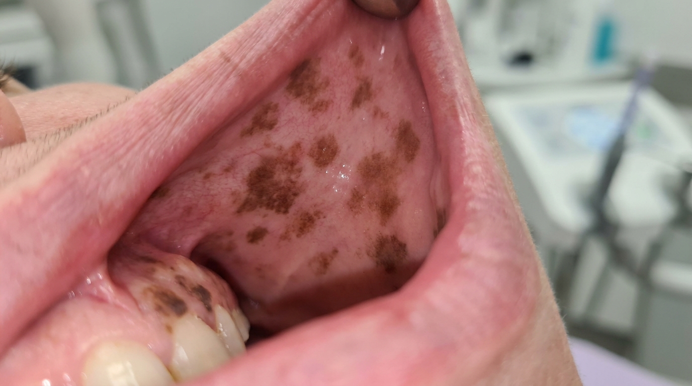
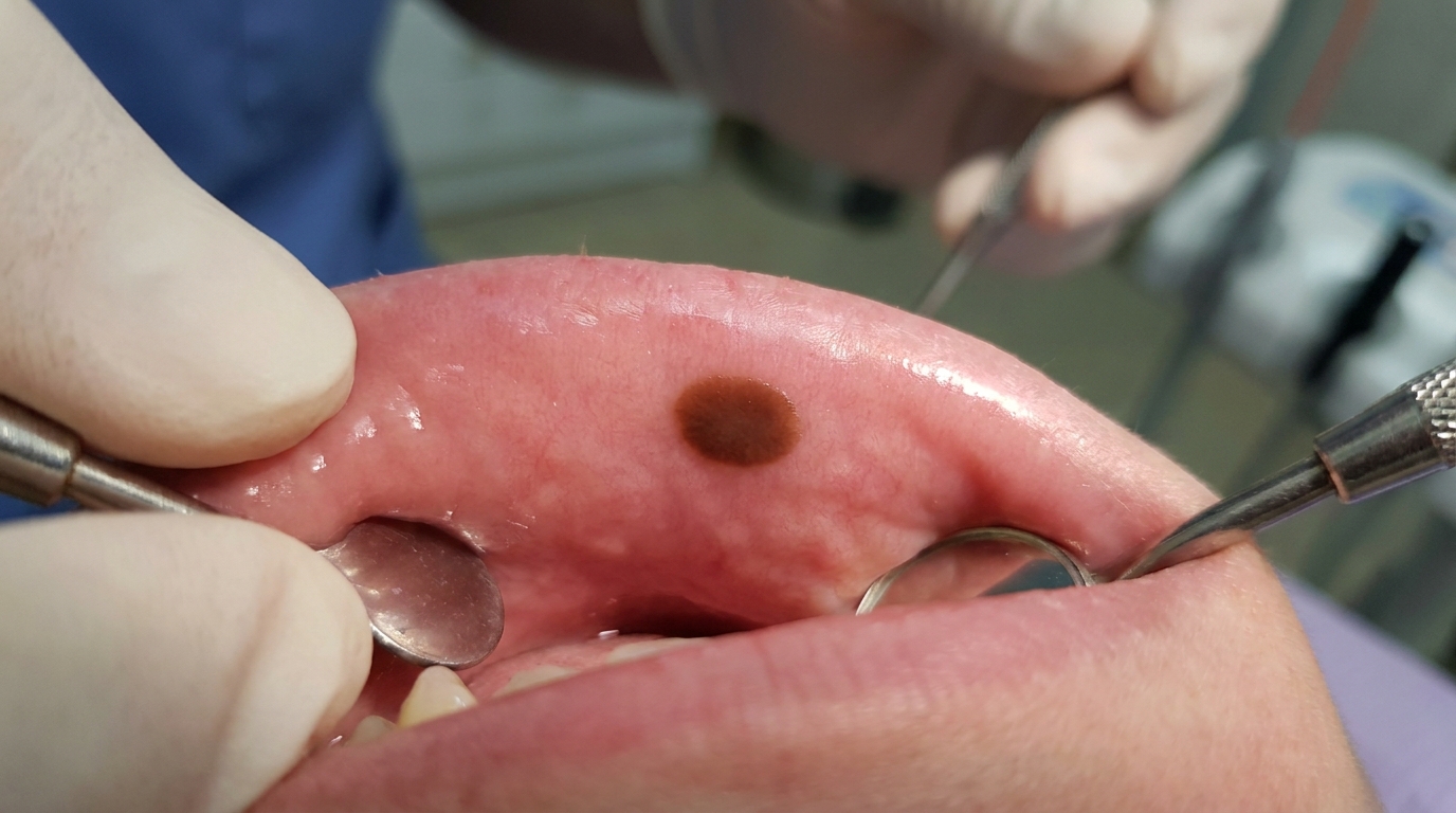
A flat, dark brown or black macule with irregular, uneven borders in the mouth.
The macule may be larger than the small physiological brown spots that some people normally have on their gums.
Mixed colours within the patch, different shades of brown, black, blue, grey or even red.
Asymmetry, one half of the lesion looks different from the other.
A raised lump or nodule developing within an existing flat patch.
Ulceration, uncommon at first but more likely as the lesion enlarges.
About 10% of oral melanomas are amelanotic, meaning they produce little or no pigment, which can make them easy to miss.
These features overlap with the well-known ABCDE warning signs of skin melanoma:
Asymmetry
Border irregularity
Colour variegation
Diameter larger than 6 mm (often)
Evolving size, shape or surface
What it feels like
Most early oral melanomas are painless. Symptoms that develop later may include:
A non-healing area that doesn't go away after a few weeks.
Discomfort or pain if the lesion ulcerates or invades nearby structures.
A loose tooth if the cancer involves the underlying bone.
Bleeding from the lesion when it is bumped or brushed.
Difficulty wearing a denture because of a new lump on the palate.
What an X-ray might show
X-rays do not show the soft-tissue lesion itself, but may show:
Irregular or "moth-eaten" bone destruction beneath the lesion when the cancer has invaded the underlying maxilla or mandible.
A widening of the maxillary sinus outline if the tumour has invaded the sinus floor.
Cervical lymph node enlargement on neck imaging.
A small-volume cone-beam CT scan, conventional CT or MRI is usually arranged once melanoma is suspected, to help map the extent before surgery.
What happens at the dentist?
Oral melanoma is sometimes picked up at a routine dental check-up and clean at ArtSmiles, particularly in patients who have a long-standing pigmented patch in the mouth that has changed. The dentist will typically:
Examine every part of the mouth carefully with good light, including under dentures and over the hard palate.
Take a careful history about how long the patch has been present and whether it has changed in size, shape, colour or surface.
Photograph the lesion and document its size as a baseline.
Distinguish the lesion from common, benign pigmented patches wherever possible.
Recommend prompt biopsy (a small tissue sample taken for laboratory examination) for any pigmented patch that is new, changing, large, raised, ulcerated or has irregular borders.
Refer to an oral and maxillofacial surgeon or head and neck oncology team without delay when melanoma is suspected.
Arrange imaging to help map the local extent and check for lymph node spread.
The textbooks specifically note that delay between first noticing a pigmented lesion and biopsy is a recurring theme in oral melanoma, partly because most pigmented patches turn out to be benign, and partly because the early lesion can look unimpressive. The practical message is that any pigmented oral lesion that is new, growing or has unusual features deserves a biopsy.
Is this serious?
🔴 Yes. Oral melanoma is rare, but it is one of the most serious cancers of the mouth. It is much more aggressive than its skin counterpart, with high rates of lymph node spread and distant metastasis at diagnosis. Five-year survival rates reported in the literature are markedly lower than for skin melanoma. The most important factor in improving outcome is early recognition and prompt treatment, which is why every persistent or changing dark patch in the mouth deserves a careful look.
If you have noticed a new dark patch in your mouth, or an existing pigmented spot that has been changing in size, shape, colour or surface, it is worth booking an assessment without delay so the right biopsy and specialist referral can be arranged.
Could it be something else?
Several conditions can produce dark patches in the mouth, and most are completely benign. The textbooks list these as the main differentials:
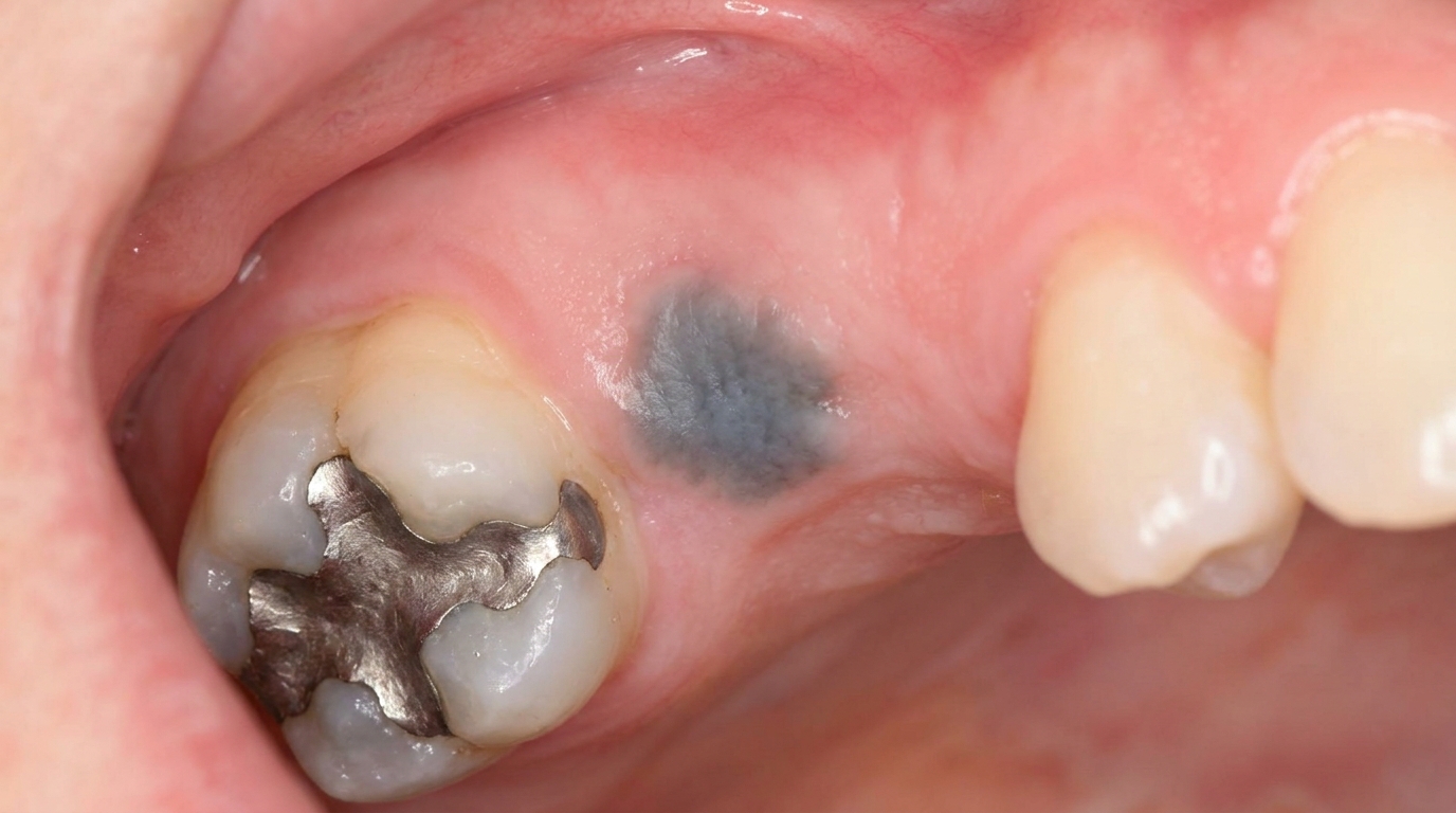
Amalgam tattoo, a stable grey or blue-black flat area near old amalgam fillings; usually shows tiny radiopaque flecks on X-ray.
Oral melanotic macule, a small, well-defined, evenly pigmented brown spot, most often on the lip or gum, with stable size and colour over time.
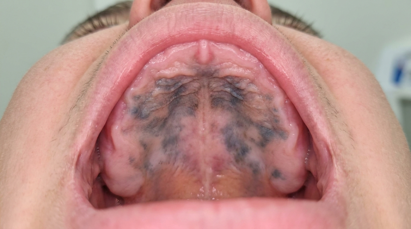
Smoker's melanosis, diffuse brown patches in heavy smokers, often on the gums and inside of the cheeks.
Physiological oral pigmentation, symmetrical, even brown pigmentation common in people with darker skin, present from a young age and stable over decades.
Drug-induced oral pigmentation, diffuse brown or grey-blue pigmentation related to medications such as minocycline, antimalarials and chemotherapy agents.
Addison's disease pigmentation, diffuse brown pigmentation as part of a systemic endocrine disease, with associated fatigue, weight loss and skin pigmentation.
Oral naevus (mole), a small, stable pigmented bump that has been present for years.
Vascular lesions, such as varices or haemangiomas, which appear bluish but are blood-filled rather than pigmented.
The combination of being new or changing, larger than a few millimetres, irregular in shape, multi-coloured, raised or ulcerated tilts the suspicion toward melanoma rather than these benign conditions.
How is it treated?
Treatment of oral melanoma is a specialist responsibility, usually within a head and neck oncology team. The textbooks describe a clear hierarchy of approaches:
Steps that begin with general dental care:
Prompt biopsy of any suspicious pigmented oral lesion, with histopathological examination and immunohistochemistry (S-100, HMB-45, SOX10, MART-1) to confirm the diagnosis.
Imaging, CT, MRI and sometimes PET-CT, to map the local extent and check for lymph node and distant spread.
Coordination with the patient's GP and the head and neck oncology team for staging (assessing how far the cancer has spread) and treatment planning.
Specialist treatment options:
Wide surgical excision with clear margins is the mainstay of treatment for resectable disease. Oral melanoma's location often makes wide margins technically challenging.
Lymph node dissection when there is clinical or imaging evidence of nodal involvement, or sentinel lymph node biopsy in selected cases.
Radiotherapy, useful for unresectable lesions, after surgery for high-risk cases, or for symptomatic palliation.
Immunotherapy (treatment that helps the body's own immune system fight cancer) with checkpoint inhibitors (such as nivolumab or pembrolizumab), which has improved outcomes in advanced melanoma over the past decade.
Targeted therapies based on molecular features of the tumour, where applicable.
Long-term, multidisciplinary follow-up, including regular oral examination, imaging and general health monitoring.
A patient-centred approach is particularly important here, where the diagnosis is frightening and the treatment journey is intense. Honest, unhurried discussion of what is being looked for, what each test is for, and what each treatment option achieves is itself part of effective care, values that sit at the heart of our clinical philosophy.
What's the long-term outlook?
The outlook for oral melanoma remains challenging, despite advances in therapy. Five-year survival rates reported in the literature have historically been around 15-30%, although outcomes have begun to improve with the introduction of immunotherapy and earlier diagnosis. Important factors include:
Tumour thickness and stage at diagnosis.
Whether lymph node or distant metastasis is present.
Whether complete surgical excision is possible.
Response to immunotherapy and other adjuvant treatments.
The most powerful single factor that patients and dentists can influence is early recognition. Every persistent or changing dark patch in the mouth deserves a careful look, most will turn out to be one of the benign differentials above, but in the rare case where it is melanoma, an early diagnosis can make a real difference to long-term outcome.
A note on this article
This article is for educational purposes only and does not constitute a clinical diagnosis. Please consult a registered dental practitioner for assessment and treatment advice.
The cover image above is an AI-generated illustration based on the most common visible features of this condition described in clinical pathology references. It is not a photograph of a real case and should not be used to diagnose or rule out the condition in your own situation. If you are concerned about something you have noticed, please book an assessment with a registered dental practitioner.
References
Neville, B. W., Damm, D. D., Allen, C. M., & Chi, A. C. (2023). Oral and maxillofacial pathology (5th ed.). Elsevier. Chapter 10, Epithelial Pathology: Mucosal Melanoma and Oral Mucosal Melanoma, with detailed clinical features, hard palate and maxillary alveolus predominance, ABCDE warning signs and treatment, pp. 435 to 441.
Cawson, R. A., & Odell, E. W. (2017). Cawson's essentials of oral pathology and oral medicine (8th ed.). Elsevier. Chapter 17, Oral Cancer: cross-reference for oral melanoma as a rare aggressive mucosal cancer.
Regezi, J. A., Sciubba, J. J., & Jordan, R. C. K. (2017). Oral pathology: Clinical pathologic correlations (7th ed.). Elsevier. Chapter on Pigmented Oral Lesions: cross-reference for oral malignant melanoma in the differential of pigmented patches.
Frequently asked questions
What is oral malignant melanoma?
Oral malignant melanoma is a rare but very aggressive cancer of the pigment-producing cells (melanocytes) in the lining of the mouth. It most often appears on the hard palate or the upper gum as a dark brown, black or sometimes red-blue patch that grows, changes shape and may bleed.
How is oral melanoma different from a harmless brown spot?
Most brown spots in the mouth are harmless (melanotic macules, amalgam tattoos, physiological pigmentation). Features that suggest melanoma include rapid growth, irregular borders, multiple colours within one lesion, bleeding or ulceration, and asymmetry. Any pigmented patch with these features needs urgent biopsy.
How is oral melanoma diagnosed and staged?
Diagnosis requires an incisional biopsy of the lesion. Imaging (CT, MRI, PET-CT) is used to look for spread to lymph nodes and distant organs. Histology confirms melanocytic origin and depth of invasion, which guide treatment and prognosis.
What is the treatment and outlook?
Treatment is wide surgical excision with clear margins, often combined with neck dissection if lymph nodes are involved. Radiotherapy and immunotherapy (checkpoint inhibitors) are used in advanced disease. Oral melanoma has a much worse prognosis than skin melanoma because it is usually diagnosed late, which is why early biopsy of any suspicious oral pigmentation is so important.