Compiled from clinical pathology references. Medically reviewed by Dr Cristian Dunker , Principal Dentist, ArtSmiles Cosmetic Dentistry.
Quick summary
Also called | Talon cusp (when on a front tooth), Leong premolar, tuberculated premolar |
How urgent? | 🟡 Worth assessing, the small extra cusp can fracture and expose the pulp (the soft nerve and blood vessel core of the tooth) before the patient is aware of any problem; early protective management can prevent this |
Common or rare? | Uncommon overall; more common in people of East Asian, Native American and Inuit descent |
Who it affects | Children and young adults, with a clear ethnic predilection; often bilateral |
Who treats it | General dentist, often working with a paediatric dentist or endodontist |
Based on | Neville, with cross-references in Cawson and Regezi |
What is it?
Dens evaginatus is a developmental tooth defect in which a small extra cusp protrudes from the surface of a tooth. The textbooks describe two main forms:
Dens evaginatus on a posterior tooth, usually a small, conical cusp projecting from the central groove of a premolar; sometimes called a Leong premolar or tuberculated premolar.
Talon cusp on an anterior tooth, a similar extra cusp on the back (palatal) surface of an upper or lower front tooth, named because it resembles an eagle's talon.
The extra cusp contains enamel, dentine and often a thin extension of the pulp. The clinical importance is that the cusp can fracture or wear away during chewing, and because the pulp extension may be very close to the surface, this can expose the pulp early, sometimes before the tooth has even fully formed. Recognising the cusp shortly after eruption and managing it carefully is the key to keeping the tooth healthy.
Who tends to get it?
The textbooks describe a fairly tight clinical group:
Strong ethnic predilection, much more common in people of East Asian (Chinese, Japanese, Korean, South-East Asian), Native American and Inuit descent.
Less common in people of European and African ancestry.
Children and young adults, the cusp is present from the moment the tooth comes through.
Both sexes affected.
Often bilateral and symmetrical, when one tooth shows the cusp, the matching tooth on the other side often does too.
Frequently associated with shovel-shaped incisors and dens invaginatus in the same individual.
What causes it?
The textbooks describe dens evaginatus as a developmental anomaly of unknown cause. Several points are useful:
Genetic predisposition appears to underlie the strong ethnic clustering.
Multifactorial inheritance is likely, with no single gene mutation identified.
No environmental factor has been clearly linked to its development, diet, illness, fluoride and trauma during pregnancy do not appear to be involved.
Often clusters with related developmental tooth shapes, shovel-shaped incisors, talon cusps (developmentals extra cusp on a front tooth that looks like an eagle's talon) and dens invaginatus frequently coexist in the same person.
How does it develop?
During tooth formation, the inner enamel epithelium normally folds smoothly to create the typical cuspal outline. In dens evaginatus, an extra outpouching of this layer produces a small additional cusp. As the tooth mineralises, the cusp acquires its own enamel covering, dentine core and often a thin extension of the dental pulp. Because the extra cusp is on the chewing surface (or palatal surface, in talon cusps), it is subjected to immediate occlusal (relating to the chewing surfaces of the teeth) forces when the tooth comes through. The cusp can fracture cleanly or wear down gradually, and either route can expose the pulp extension and lead to pulp infection, sometimes before the patient is aware of any problem at all.
What might you notice?
What it looks like
The classic appearance is well described:
A small, conical or tubercle-like cusp projecting from the central groove of a premolar's chewing surface.
A talon-shaped extra cusp on the back of a front tooth, particularly an upper lateral incisor.
Bilateral and symmetrical in many cases.
The cusp may be fractured or worn flat in older patients who have not had it protectively managed.
A deep fissure at the junction between the talon cusp and the surface of the tooth, sometimes extending down the root.
What it feels like
Most people are unaware of dens evaginatus until a dentist points it out. Symptoms when they appear may include:
A bump felt with the tongue or tooth-on-tooth contact.
A high spot in the bite when the tooth first comes through, with possible jaw discomfort.
Sharp pain if the cusp fractures and exposes the pulp.
Toothache, swelling or abscess when pulp infection develops following silent cusp fracture.
No symptoms at all when the cusp is small and the pulp extension does not reach the tip.
What an X-ray might show
X-rays often show:
A radio-opaque outline of the extra cusp sitting on the tooth's chewing surface.
A thin pulp extension running into the cusp in some cases.
Periapical changes when the pulp has become infected after cusp fracture.
A small-volume cone-beam CT (CBCT) can be useful for detailed mapping before any planned cusp reduction or pulp procedure.
What happens at the dentist?
Dens evaginatus is most often picked up at a routine dental check-up and clean at ArtSmiles when a child or young adult comes in for routine review. The dentist will typically:
Examine the chewing surfaces of premolars and the back of front teeth for any unusual cusp.
Take periapical X-rays to assess the extent of any pulp extension into the cusp.
Test the vitality of the affected tooth.
Identify any deep fissure at the base of a talon cusp, which can become a route for caries and pulp infection.
Plan staged management, typically gradual cusp reduction with desensitising agents, sometimes immediate cuspal reduction with pulp protection in larger cusps.
Refer to a paediatric dentist or endodontist when the cusp is large, has already fractured, or has caused pulp problems.
Is this serious?
🟡 The cusp itself is benign, but its tendency to fracture and expose the pulp before the tooth is fully mature makes early identification important. With careful staged management, most cusps can be safely reduced without losing pulp vitality, and the tooth can be expected to function for life.
If your child has unusual extra bumps on their teeth, it's worth booking an assessment shortly after the tooth comes through so the right protective plan can be put in place.
Could it be something else?
Several conditions can produce similar small extra projections on teeth. The textbooks list these as the main differentials:
Dens invaginatus, the opposite developmental defect, with an inward fold rather than an outward bump. The two can occur in the same tooth.
Accessory cusp (cusp of Carabelli), a small extra cusp on the inside of upper first molars, considered a normal variant rather than a defect.
Calcified deposit on a tooth, sometimes mistaken for an extra cusp; usually distinguished on close examination.
Dental caries, a soft, brown lesion rather than a hard projecting bump.
Normal cuspal anatomy, the threshold between unusual and normal can be subjective.
How is it treated?
The textbooks describe several strategies, the choice depending on cusp size, pulp extension and patient cooperation:
At-home measures and habits:
Maintain excellent oral hygiene, particularly around the base of any talon cusp, where a deep fissure can collect plaque.
Avoid biting hard objects that might suddenly fracture the cusp.
Use fluoride toothpaste to support the surrounding enamel.
Professional steps your dentist may consider:
Slow, periodic grinding of the cusp over several visits 6-8 weeks apart, with topical fluoride or stannous fluoride applied at each visit to encourage the pulp to lay down protective reactionary dentine. This is the textbook-preferred approach for mature teeth.
Single-visit cusp reduction with intentional pulpotomy, used for very large cusps or when there is significant pulp tissue inside the cusp; aims to maintain long-term vitality of the rest of the tooth.
Restoration of the resulting groove with composite resin and a small filling.
Sealing of any deep fissure at the base of a talon cusp to prevent caries access to the pulp.
Root canal treatment if pulp necrosis has already occurred, particularly important in young teeth with open apices, where regenerative endodontic (relating to root canal treatment) procedures may be needed.
Long-term follow-up X-rays to confirm the pulp has remained healthy.
A patient-centred approach matters here too. Children and young adults are not always keen on multi-visit reduction protocols, and a clear, age-appropriate explanation of why staged care matters is itself part of effective care, values that sit at the heart of our clinical philosophy.
What's the long-term outlook?
The outlook is excellent when dens evaginatus is identified and managed early. With staged cusp reduction and good oral hygiene, the affected tooth typically remains vital and functional for life. Cases caught only after pulp infection are usually still saveable with root canal treatment, although the long-term prognosis is slightly less predictable. Across all cases, regular dental review and timely action on any unusual cusp are the keys to a good long-term outcome.
A note on this article
This article is for educational purposes only and does not constitute a clinical diagnosis. Please consult a registered dental practitioner for assessment and treatment advice.
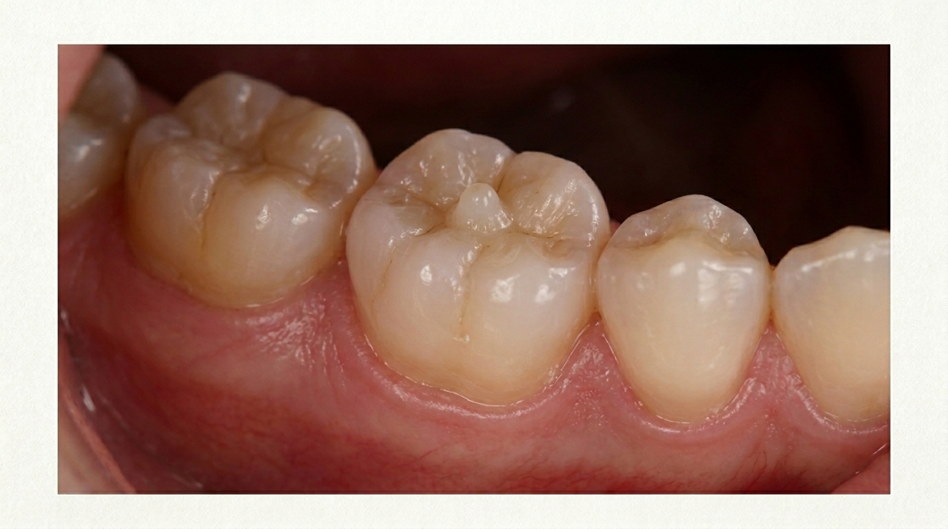
The cover image above is an AI-generated illustration based on the most common visible features of this condition described in clinical pathology references. It is not a photograph of a real case and should not be used to diagnose or rule out the condition in your own situation. If you are concerned about something you have noticed, please book an assessment with a registered dental practitioner.
References
Neville, B. W., Damm, D. D., Allen, C. M., & Chi, A. C. (2023). Oral and maxillofacial pathology (5th ed.). Elsevier. Chapter 2, Abnormalities of Teeth: Dens Evaginatus and Talon Cusp, with cuspal removal techniques and the association with shovel-shaped incisors in East Asian, Native American and Inuit populations, pp. 85 to 88.
Cawson, R. A., & Odell, E. W. (2017). Cawson's essentials of oral pathology and oral medicine (8th ed.). Elsevier. Chapter 2, Disorders of Development: cross-reference for dens evaginatus and accessory cusps.
Regezi, J. A., Sciubba, J. J., & Jordan, R. C. K. (2017). Oral pathology: Clinical pathologic correlations (7th ed.). Elsevier. Chapter on Abnormalities of Teeth: cross-reference for dens evaginatus.
Frequently asked questions
What is dens evaginatus?
Dens evaginatus is a developmental anomaly where an extra cusp-like projection (the 'tubercle') sticks out from the chewing surface of a tooth, most often a lower premolar. Inside the tubercle, the soft pulp tissue often extends close to the surface. When the tubercle wears or breaks, the pulp can quickly become inflamed or infected.
Why is dens evaginatus a problem?
Although the tooth itself looks normal apart from the small extra cusp, normal chewing wear can fracture the tubercle and expose the pulp. This can cause the pulp to die in a tooth that has never had a cavity, often in a teenager. Without early intervention, root canal treatment or extraction may be needed.
How is dens evaginatus managed?
In a young patient, the dentist may seal around the tubercle with composite resin, gradually reduce the height of the tubercle to encourage natural reparative dentine, or place a protective restoration. If the pulp is already inflamed or non-vital, root canal treatment becomes necessary. Early diagnosis at a child's check-up gives the best chance of saving the pulp.
How common is dens evaginatus?
It is uncommon overall but more frequent in people of East and South-East Asian, Indigenous American and Inuit ancestry. It usually affects lower premolars and is often bilateral. Routine dental check-ups in children and teenagers help catch it early, before the tubercle fractures.