Compiled from clinical pathology references. Medically reviewed by Dr Cristian Dunker , Principal Dentist, ArtSmiles Cosmetic Dentistry.
Quick summary
Also called | Localised argyrosis (silver staining of tissue), focal argyrosis, amalgam pigmentation |
How urgent? | 🟢 Not urgent, benign and stable; the main reason to assess is to be sure of the diagnosis |
Common or rare? | By far the most common cause of localised pigmentation in the mouth |
Who it affects | Anyone with current or past amalgam ("silver") fillings or who has had endodontic surgery (minor surgery on the tip of a tooth root); both sexes equally; usually adults |
Who treats it | General dentist for diagnosis and reassurance; treatment is rarely needed |
Based on | Neville, Cawson, with cross-references in Regezi |
What is it?
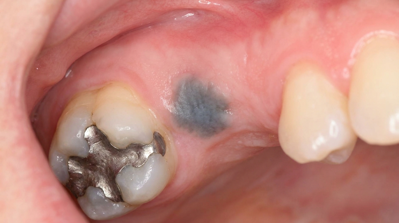
An amalgam tattoo is a flat, grey, blue or black stain in the lining of the mouth caused by tiny particles of dental amalgam, the silver-coloured filling material, becoming embedded in the soft tissue. The textbooks describe it as the most common cause of localised pigmentation in the mouth, far more common than any other type of pigmented oral lesion. Despite the alarming colour, an amalgam tattoo is harmless and does not progress to anything more serious. The reason it matters is to be sure that what looks like an amalgam tattoo is, in fact, an amalgam tattoo and not one of the rare lookalike conditions such as oral melanoma.
Who tends to get it?
Almost any adult who has had amalgam fillings is at some risk. The textbooks describe a few specific patterns:
People with old amalgam (silver) fillings, particularly those placed before tooth-coloured fillings became the routine choice.
People who have had endodontic surgery with an amalgam apicectomy retrofill (a procedure that seals the tip of a tooth root from below), fragments may be left in the surrounding bone or gum during the procedure.
People who have had old amalgam fillings replaced, small particles can fall into the gum during removal.
Both sexes equally, no significant sex predilection.
Adults, since amalgam fillings have usually been in place for years.
The likelihood is the same across all skin types and ethnic backgrounds.
What causes it?
Amalgam tattoos are caused by physical implantation of amalgam particles into the soft tissue. The textbooks describe several specific routes:
Mucosal abrasion (a small graze of the mouth lining) contaminated with amalgam dust during routine dental procedures, particularly when the rubber dam is removed and the bite is then adjusted.
Broken amalgam fragments falling into an extraction socket and being trapped as the gum heals.
Dental floss carrying tiny amalgam particles from a freshly placed filling, leaving linear stains along the gum line.
Endodontic retrofill procedures, in which amalgam is placed at the tip of a root through a small surgical opening; small fragments can be left behind in the surrounding bone or gum.
High-speed air-driven turbines can occasionally drive fine metallic particles through small breaks in the gum.
Once embedded, the particles remain in place essentially indefinitely. The body forms a small ring of fibrous tissue around larger fragments, while smaller particles disperse into the surrounding tissue and stain the supporting fibres a characteristic grey-blue.
How does it develop?
The colour comes from the amalgam itself, not from any change in the body's own cells. Amalgam is a mixture of mercury, silver, tin, copper and zinc. When tiny particles sit in the soft tissue, the silver salts bind to the connective tissue fibres, particularly the reticulin fibres (fine supporting tissue fibres) around blood vessels and nerves, and produce a fine network of grey-black staining visible at the surface as a flat patch. There is no inflammation in many cases, particularly with older, larger fragments; smaller, more dispersed particles can sometimes provoke a mild chronic inflammatory or even granulomatous (with small clusters of immune cells) response.
What might you notice?
What it looks like
The classic appearance is well described:
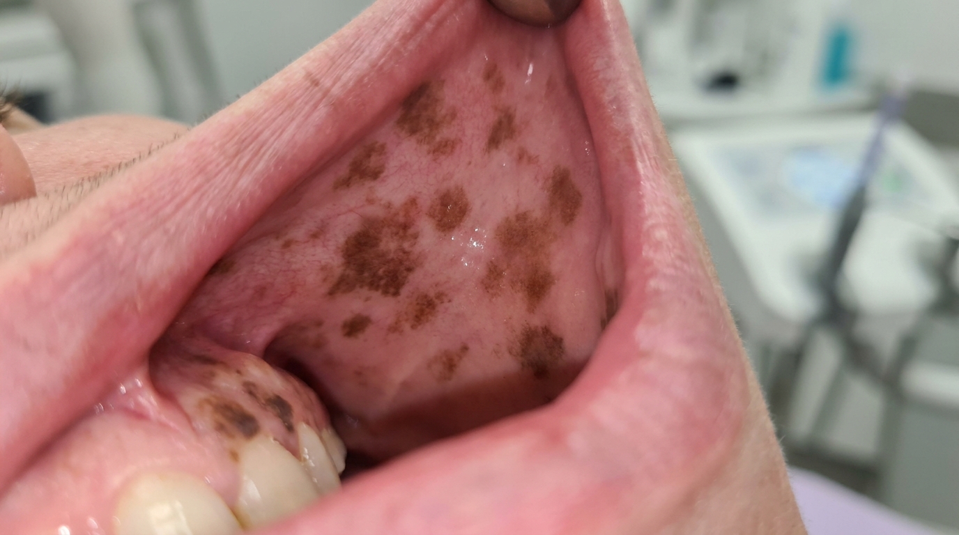
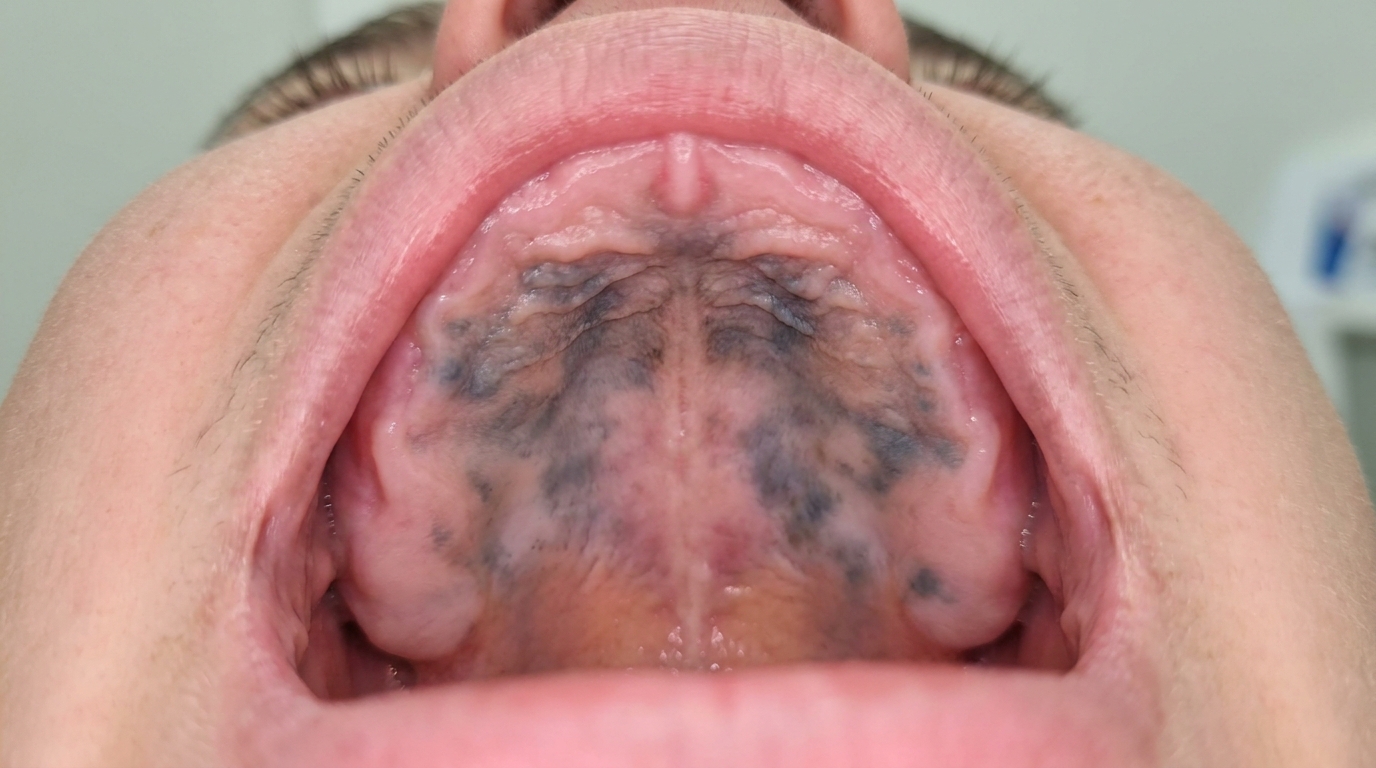
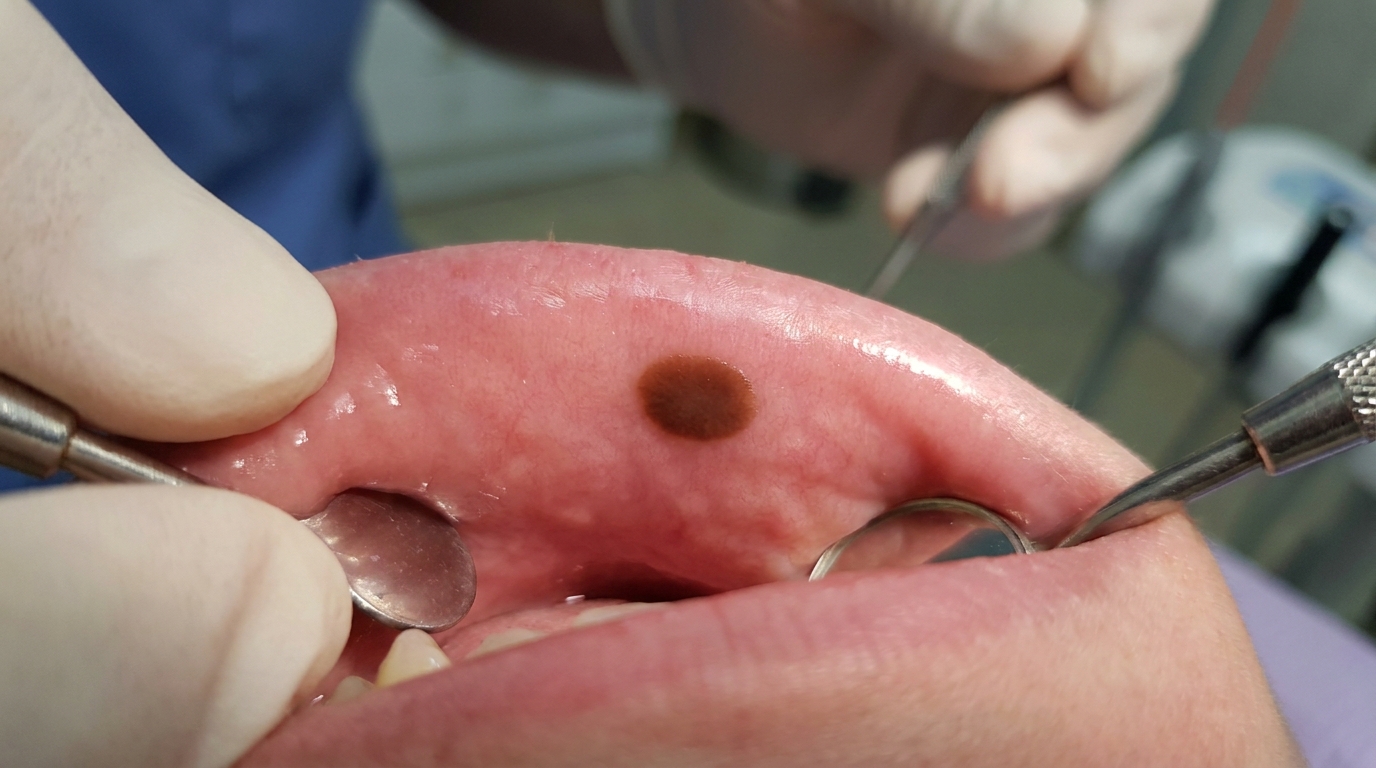
A flat, well-defined patch of grey, blue, slate or black colour.
Smooth surface, no raised area, no ulceration.
Most often on the gum, alveolar mucosa (the lining of the gum next to the cheek) or inside of the cheek, immediately adjacent to or near old amalgam fillings.
Lateral spread of the staining can occur for several months after implantation, but the patch then becomes stable.
Multiple small areas can occur, particularly when amalgam particles have been carried by floss along the gum line or where multiple amalgam-restored teeth have been treated.
What it feels like
An amalgam tattoo is asymptomatic. There is no pain, no soreness, no taste change, no bleeding and no surface roughness. The only reason it is noticed is the appearance.
What an X-ray might show
This is one of the few conditions in this article where an X-ray is genuinely useful:
A periapical X-ray (a close-up X-ray of one tooth and the bone around its root) of the area often shows tiny radiopaque (white) flecks within the soft tissue, corresponding to amalgam fragments that are large enough to be visible.
Many smaller particles are below the resolution of routine X-rays, so a normal X-ray does not rule out amalgam tattoo.
The clinical area of staining usually extends beyond the visible metallic fragments, since silver salts diffuse a short distance into surrounding tissue.
When metallic flecks are visible on X-ray, the diagnosis can usually be confirmed without biopsy.
What happens at the dentist?
An amalgam tattoo is most often picked up at a routine dental check-up and clean at ArtSmiles, or when the patient asks about a small dark patch they have noticed. The dentist will typically:
Examine the patch carefully under good light, noting its location, size, colour and edges.
Take a careful history, including past amalgam fillings, replaced fillings, extractions or endodontic surgery in the area.
Take a periapical X-ray of the area to look for the characteristic tiny radiopaque flecks.
Distinguish the patch from other pigmented lesions by location, history and clinical pattern.
Reassure the patient when the appearance and the X-ray together confirm an amalgam tattoo.
Recommend a small biopsy when no metallic fragments can be seen on X-ray and the diagnosis is not obvious clinically, the textbooks specifically suggest biopsy "to rule out the possibility of melanocytic neoplasia (a growth of the pigment-producing cells, including melanoma)" in such cases.
Is this serious?
🟢 An amalgam tattoo is benign and stable. It does not turn into cancer, does not damage surrounding tissue and does not affect general health. The reason it is worth confirming is to be sure that what looks like an amalgam tattoo is not an oral melanotic macule, an early oral malignant melanoma or one of the other lookalike conditions.
If you have noticed a grey-blue or black patch in your mouth near old fillings, particularly if it has been present for years and has not changed, it is most likely an amalgam tattoo. A check-up with an X-ray of the area can usually confirm the diagnosis without further treatment.
Could it be something else?
Several other conditions can produce a dark patch in the mouth. The textbooks list these as the main differentials:
Oral melanotic macule, a small, brown rather than grey-blue, well-defined macule, most often on the lower lip; not associated with old fillings and not visible on X-ray.
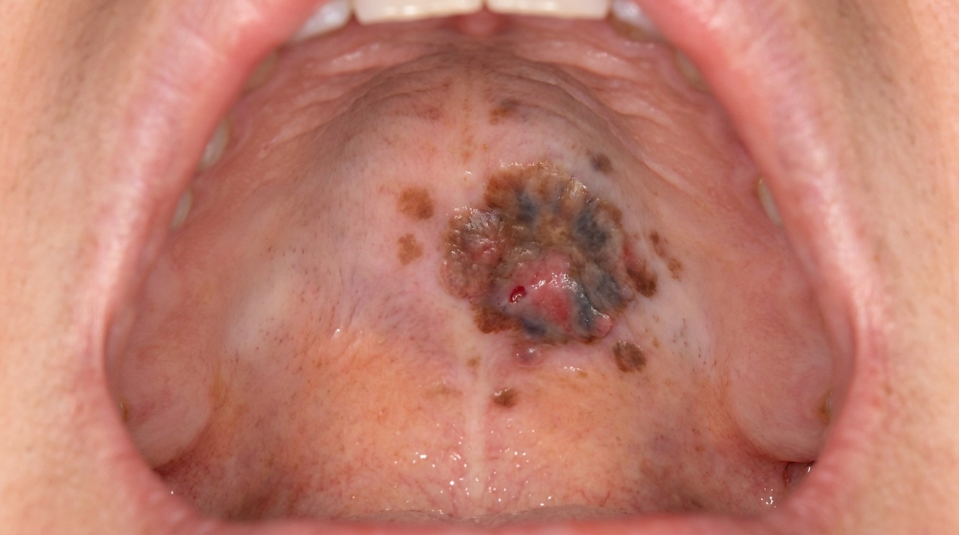
Oral malignant melanoma, a rare but serious cancer, distinguished by asymmetry, irregular borders, recent change and any raised or ulcerated component.
Oral naevus (mole), a small pigmented lesion that has been present for years; often slightly raised.
Smoker's melanosis, diffuse brown rather than focal grey-blue patches in heavy smokers.
Physiological pigmentation, symmetrical, present from childhood, not adjacent to old fillings.
Other localised exogenous pigmentations, pencil graphite tattoos (childhood injuries), intentional cosmetic tattoos, and rare metallic tattoos from broken dental burs or zirconia-titanium friction.
How is it treated?
The textbooks all agree: an amalgam tattoo generally needs no treatment.
At-home measures and habits:
Continue normal oral hygiene, brushing twice a day with fluoride toothpaste and flossing daily.
No need to avoid any food or product because of the tattoo.
Photograph the area if you want to be able to confirm later that nothing has changed.
Professional steps your dentist may consider:
Confirming the diagnosis with clinical examination and a periapical X-ray.
Reassuring the patient that the tattoo is benign and stable.
Documenting the appearance with photographs as a baseline.
Biopsy when the diagnosis is not clear clinically and no fragments are seen on X-ray.
Cosmetic removal is occasionally requested when the tattoo is in an aesthetically sensitive location such as the front gum. Options include conservative surgical excision, Q-switched ruby or alexandrite laser, and where the area is removed surgically, the use of a connective tissue graft to restore the gum colour.
Replacement of nearby amalgam fillings with tooth-coloured composite or ceramic restorations is sometimes done at the patient's request, although it does not remove the existing tattoo (which is in the soft tissue, not in the tooth).
A patient-centred approach matters here too. People sometimes worry that a "black mark" in the mouth might mean something serious. Calm, clear explanation of what an amalgam tattoo is, why it is harmless, and how easy it is to confirm with a small X-ray is itself part of effective care, values that sit at the heart of our clinical philosophy.
What's the long-term outlook?
The outlook is excellent. An amalgam tattoo is a stable, lifelong feature that does not progress to any disease. There are no long-term health implications and no need for prolonged follow-up beyond routine dental care. Many patients live for decades with an amalgam tattoo without ever realising it is there until a dentist points it out, and once explained, it is usually no longer a concern.
A note on this article
This article is for educational purposes only and does not constitute a clinical diagnosis. Please consult a registered dental practitioner for assessment and treatment advice.
The cover image above is an AI-generated illustration based on the most common visible features of this condition described in clinical pathology references. It is not a photograph of a real case and should not be used to diagnose or rule out the condition in your own situation. If you are concerned about something you have noticed, please book an assessment with a registered dental practitioner.
References
Neville, B. W., Damm, D. D., Allen, C. M., & Chi, A. C. (2023). Oral and maxillofacial pathology (5th ed.). Elsevier. Chapter 8, Physical and Chemical Injuries: Amalgam Tattoo and Other Localized Exogenous Pigmentations, with detailed clinical features and histopathology, pp. 298 to 300.
Cawson, R. A., & Odell, E. W. (2017). Cawson's essentials of oral pathology and oral medicine (8th ed.). Elsevier. Chapter 21, Melanoma and Other Pigmented Lesions: amalgam tattoo as the most common localised exogenous pigmentation in the mouth.
Regezi, J. A., Sciubba, J. J., & Jordan, R. C. K. (2017). Oral pathology: Clinical pathologic correlations (7th ed.). Elsevier. Chapter on Pigmented Lesions: cross-reference for amalgam pigmentation as a key differential of localised oral pigmentation.
Frequently asked questions
What is an amalgam tattoo?
An amalgam tattoo is a flat, grey-blue or black patch in the mouth where small particles of silver-coloured dental amalgam have been pushed into the lining during a filling, crown preparation or extraction. The particles sit harmlessly in the tissue and stain it permanently.
Is an amalgam tattoo dangerous?
No. Amalgam tattoos are completely benign and harmless. They are not allergic, not inflammatory and not a sign of mercury toxicity. The colour and pattern are mainly a cosmetic issue.
How do I know it's a tattoo and not a melanoma?
Amalgam tattoos sit near an old filling or extraction site, have stayed the same size and colour for years, and often show up on a dental x-ray as a tiny radio-opaque speck. Melanoma usually grows, changes colour, has irregular borders, and never shows up on x-ray. If there's any doubt, biopsy is the safest answer.
Can amalgam tattoos be removed?
They don't need to be removed because they are harmless. If a patient particularly wants the mark gone for cosmetic reasons (especially in the lip or front gum area), surgical excision or laser ablation can be done.