Compiled from clinical pathology references. Medically reviewed by Dr Cristian Dunker, Principal Dentist, ArtSmiles Cosmetic Dentistry.
Quick summary
Also called | Tobacco-associated melanin pigmentation, smoking-related oral pigmentation |
How urgent? | 🟢 Not urgent in itself, the pigmentation is benign and reversible; the smoking habit that causes it is the real concern |
Common or rare? | Common in regular smokers, more obvious in people with lighter mucosa |
Who it affects | Adults who smoke cigarettes, pipes or cigars; both sexes, more obvious in women in some studies |
Who treats it | General dentist for diagnosis and smoking-cessation support; no specific dental treatment is needed for the pigmentation itself |
Based on | Neville, Cawson, with cross-references in Regezi |
What is it?
Smoker's melanosis is increased melanin pigmentation (the natural brown pigment in the mouth lining) of the soft tissues of the mouth in people who smoke. The textbooks describe it as a benign, reversible response to chemicals in tobacco smoke. The lining looks brown rather than the usual pink, most often on the front gums and the inside of the lower lip. The pigmentation itself does not develop into cancer, but it is a visible marker of long-term smoking, and that habit is what matters most.
Who tends to get it?
The textbooks describe a fairly characteristic profile:
Smokers, including cigarette, pipe and cigar smokers. The amount of pigmentation tends to relate to how much and how long someone smokes.
Adults of any age, with the colour building up over months to years of regular smoking.
Both sexes, although some studies report a slightly higher rate in women, possibly because of an interaction between female hormones and melanocyte activity.
More obvious in people with lighter mucosa, where the brown stands out against the pink.
Not seen in non-smokers in a meaningful way, physiological pigmentation in non-smokers is a separate condition with a different distribution.
What causes it?
The trigger is tobacco smoke. The textbooks describe several mechanisms:
Chemicals in smoke stimulate melanocytes (the pigment-producing cells) to make more melanin.
Heat from smoke can act as a co-factor.
Repeated exposure over years allows the pigmentation to build up gradually.
Genetic susceptibility explains why some smokers develop heavy pigmentation while others develop little.
The condition is not caused by nicotine alone, switching to nicotine replacement therapy does not reproduce smoker's melanosis. It is the burning of tobacco and its by-products that drive the change.
How does it develop?
The course is gradual. With consistent smoking, melanocytes in the basal layer (the deepest, foundation layer of the lining) become more active and produce more melanin. The pigment is taken up by the surrounding cells, and over months a faint brown tinge develops, often noticed first by a dentist or dental hygienist on the front gum. Over years, the brown can darken and spread, sometimes covering large areas of the gum, cheek lining and palate. After smoking stops, the trigger is removed and the pigmentation slowly fades over months to a few years.
What might you notice?
What it looks like
The classic appearance is well described:
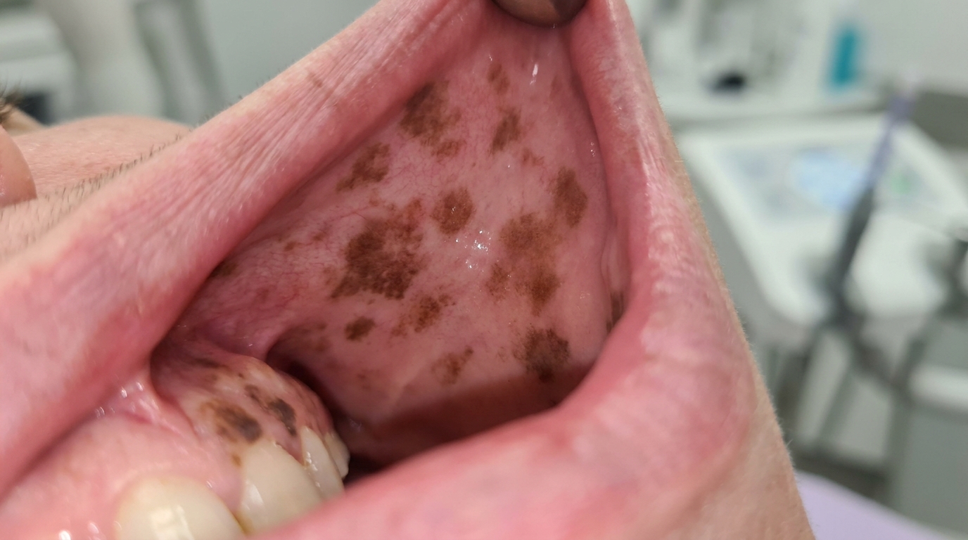
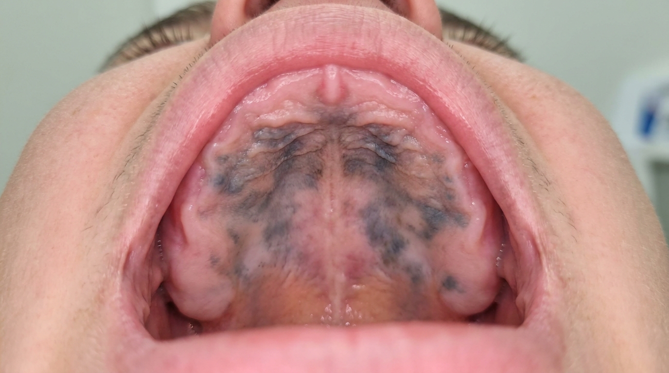
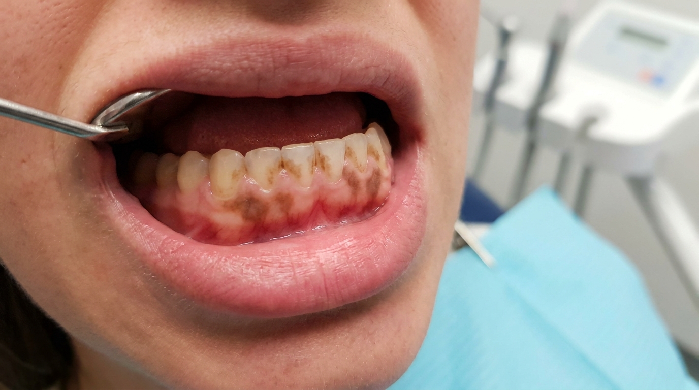
Diffuse, light to medium brown discolouration of the gums and cheek lining.
A bilateral, fairly symmetrical pattern, most often on the front lower gum just inside the lip.
Can also affect the cheek lining, the palate and the floor of the mouth.
No raised lump, no ulcer, no surface change, only colour.
What it feels like
Smoker's melanosis is asymptomatic (causing no symptoms). There is no pain, no soreness, no taste change and no bleeding. The only reason it is noticed is the appearance, and the smoking-related staining of the teeth that often goes with it.
What an X-ray might show
Smoker's melanosis is confined to the surface lining and is not visible on X-rays. X-rays may be used to assess the rest of the mouth for other smoking-related concerns, but they do not show or rule out the pigmentation itself.
What happens at the dentist?
Brown patches in the mouth are most often picked up at a routine dental check-up and clean at ArtSmiles, or when a patient asks about a colour change they have noticed. The dentist will typically:
Take a smoking history, including type of product, duration and any past attempts to quit.
Examine the area carefully under good light, noting the colour, distribution and surface texture.
Photograph the lesion as a baseline for future visits.
Distinguish from other pigmented conditions by pattern, location, history and clinical features.
Check the rest of the mouth for other findings linked to smoking, including leukoplakia, nicotinic stomatitis and any signs of early oral cancer.
Consider a small biopsy when the appearance is unusual, asymmetric, growing, raised or ulcerated, or in an unusual location.
Offer smoking-cessation support, with referral to your GP, Quitline or pharmacy if you would like help quitting.
Is this serious?
🟢 Smoker's melanosis is not dangerous in itself. It does not turn into cancer. The reason it deserves attention is what it represents, long-term smoking is associated with much more serious mouth and general health risks, including oral cancer, gum disease, cardiovascular disease, lung disease and stroke. The colour change is, in a sense, the visible part of a much bigger picture, and a useful talking point for any patient considering quitting.
If you have noticed brown discolouration of your gums or cheek lining and you smoke, it is worth booking an assessment so the diagnosis can be confirmed and we can support you with any plans to quit.
Could it be something else?
Several other conditions can produce brown discolouration of the mouth. The textbooks list these as the main differentials:
Physiological (racial) pigmentation, symmetrical, evenly distributed, present from a young age, and unrelated to smoking.
Drug-induced oral pigmentation, from medicines such as antimalarials, minocycline, oral contraceptives or some anti-cancer drugs.
Addison's disease, pigmentation of the mouth as part of an underlying adrenal insufficiency, often with skin pigmentation and other systemic symptoms.
Peutz-Jeghers syndrome, multiple small dark spots around the lips and inside the cheek in patients with intestinal polyps.
Oral melanotic macule, a single, well-defined small brown spot.
Melanocytic naevus, a true mole inside the mouth, usually solitary and present for years.
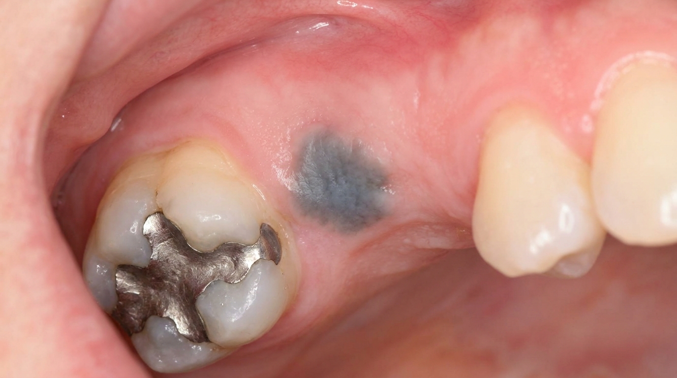
Amalgam tattoo, grey-blue rather than brown, near old metal fillings.
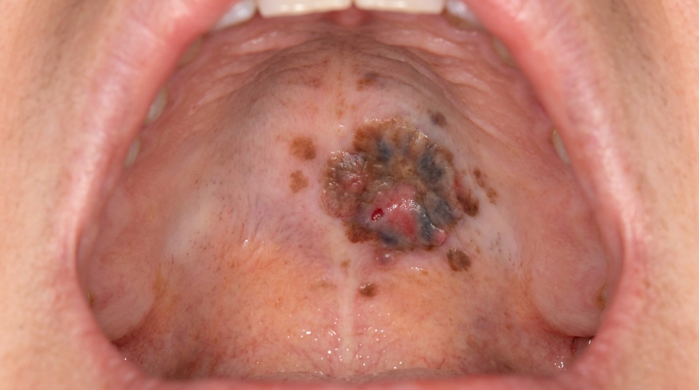
Oral malignant melanoma, a rare but serious cancer, distinguished by asymmetry, irregular borders, recent change and any raised or ulcerated component.
A careful history and examination separate most of these without needing a biopsy.
How is it treated?
Smoker's melanosis is generally not treated with any medical or surgical therapy aimed at the colour itself. Treatment is focused on the underlying habit.
At-home measures and habits:
Continue normal oral hygiene, brushing twice a day with fluoride toothpaste and flossing daily.
Photograph the area if you want to track the gradual fading of pigmentation after stopping smoking.
Plan a quit attempt with support, GPs, Quitline (13 7848) and your pharmacy can all help.
Professional steps your dentist may consider:
Confirming the diagnosis with a clinical examination and, occasionally, a small biopsy if the appearance is atypical.
Documenting the appearance with photographs as a baseline for future visits.
Smoking-cessation support, with referral to your GP or Quitline if you would like help quitting.
Checking the rest of the mouth for any smoking-related changes that deserve closer attention.
Cosmetic depigmentation procedures can be considered after smoking has stopped, but are not recommended while smoking continues because the pigmentation often returns.
A patient-centred approach matters here. Conversations about smoking can be sensitive, and a calm, non-judgemental explanation of the dental findings, what they mean, and what support is available is itself part of effective care, values that sit at the heart of our clinical philosophy.
What's the long-term outlook?
The outlook for smoker's melanosis itself is excellent. It fades when smoking stops and does not transform into anything more serious. The bigger long-term picture is shaped by the broader smoking habit, stopping smoking is one of the single most powerful steps anyone can take to improve oral and general health, regardless of age or how long they have smoked. With cessation, most patients see significant fading of pigmentation within one to three years, and a measurable reduction in their overall mouth-cancer and gum-disease risk over the same period.
A note on this article
This article is for educational purposes only and does not constitute a clinical diagnosis. Please consult a registered dental practitioner for assessment and treatment advice.
The cover image above is an AI-generated illustration based on the most common visible features of this condition described in clinical pathology references. It is not a photograph of a real case and should not be used to diagnose or rule out the condition in your own situation. If you are concerned about something you have noticed, please book an assessment with a registered dental practitioner.
References
Neville, B. W., Damm, D. D., Allen, C. M., & Chi, A. C. (2023). Oral and maxillofacial pathology (5th ed.). Elsevier. Chapter 10, Epithelial Pathology: Smoker's Melanosis, with detailed clinical features and pattern of pigmentation.
Cawson, R. A., & Odell, E. W. (2017). Cawson's essentials of oral pathology and oral medicine (8th ed.). Elsevier. Chapter 21, Melanoma and Other Pigmented Lesions: smoker's melanosis as a recognised differential of localised and diffuse oral pigmentation.
Regezi, J. A., Sciubba, J. J., & Jordan, R. C. K. (2017). Oral pathology: Clinical pathologic correlations (7th ed.). Elsevier. Chapter on Pigmented Lesions: cross-reference for smoker's melanosis as a key cause of acquired oral pigmentation.
Frequently asked questions
What is smoker's melanosis?
Smoker's melanosis is a brown to brown-grey discolouration of the mouth lining caused by tobacco smoke stimulating melanocytes to produce extra melanin. It is most common in the lower front gum and the inside of the cheek and is related to the dose and duration of smoking.
Why does smoking cause pigmentation?
Compounds in tobacco smoke (especially nicotine, benzopyrene and tar products) stimulate melanocytes to make more melanin as a protective response. The pigment is then deposited in the surface layers of the mouth lining and connective tissue.
How is it told apart from melanoma?
Smoker's melanosis is diffuse, often symmetrical, develops gradually over years of smoking, and stays stable in shape. Melanoma is single, asymmetric, grows, changes colour and may ulcerate. Any new or changing pigmented patch in a smoker should still be examined and may need biopsy.
Does the pigmentation go away if you stop smoking?
Yes, often. Most smoker's melanosis fades slowly over months to years after stopping smoking, although some pigmentation can persist. Quitting smoking is also the most important step for reducing the much more serious risks of oral cancer and gum disease.