Compiled from clinical pathology references. Medically reviewed by Dr Cristian Dunker , Principal Dentist, ArtSmiles Cosmetic Dentistry.
Quick summary
Also called | Medication-induced oral melanosis, drug-related oral discoloration |
How urgent? | 🟡 Worth identifying, the pigmentation itself is harmless, but it should be distinguished from more serious causes of oral pigmentation |
Common or rare? | Uncommon overall but well recognised in patients on certain long-term medications |
Who it affects | People taking long-term medications such as minocycline, antimalarials (medications used to prevent or treat malaria, also used for some autoimmune diseases), chemotherapy agents, oestrogen, AIDS medications, chlorhexidine mouthwash and certain psychiatric drugs |
Who treats it | General dentist for diagnosis and reassurance, in coordination with the prescribing GP or specialist |
Based on | Cawson, Neville, with cross-references in Regezi |
What is it?
Drug-induced oral pigmentation is a brown, grey, blue or black colouration of the lining of the mouth that develops as a side effect of certain medications. The textbooks describe it as a recognised but uncommon reaction in which a drug, its breakdown products, or a medication-driven increase in melanin (the natural brown pigment in skin and oral lining) production, accumulates in the oral mucosa (the soft tissue lining of the mouth). The change is usually painless and reversible, most cases gradually fade over months to years once the medication is stopped or its dose adjusted. The reason it matters is that the pigmentation can be mistaken for more serious causes of oral darkening, including oral malignant melanoma.
Who tends to get it?
The textbooks describe several recognised culprits:
Antibiotics, particularly minocycline (long-term use for acne or other chronic infections) which can produce grey-black or blue-black pigmentation in bone, gums, palate and teeth.
Antimalarials, chloroquine and hydroxychloroquine (used for malaria, lupus and rheumatoid arthritis) can produce blue-grey or brown pigmentation of the hard palate.
Chemotherapy agents, including busulfan, cyclophosphamide, doxorubicin and 5-fluorouracil, which can produce diffuse brown melanosis.
Oestrogen, including some hormonal contraceptives, can occasionally produce diffuse brown pigmentation, similar to skin changes in pregnancy ("melasma").
HIV/AIDS medications, including zidovudine (AZT), which has been associated with diffuse oral pigmentation.
Phenothiazines (an older class of antipsychotic medication), older antipsychotic medications such as chlorpromazine, with both skin and oral pigmentation possible.
Heavy metals, bismuth (in some over-the-counter stomach remedies), lead, mercury and silver salts can produce a characteristic line of pigmentation along the gum margin.
Chlorhexidine mouthwash, can produce brown surface staining of teeth and tongue, although this is more a tooth and superficial stain than true mucosal pigmentation.
People taking these medications for prolonged periods (months to years) are most at risk.
What causes it?
The textbooks describe several different mechanisms, depending on the drug:
Direct deposition of the drug or its metabolites (breakdown products of a medication) in the tissues. Minocycline and heavy metals work this way, leaving the lining itself stained.
Increased melanin production by the body's own pigment cells in response to the medication. Antimalarials and oestrogens act this way.
Drug-melanin complexes that combine drug breakdown products with melanin to form pigmented compounds.
Altered local immune response in patients on AIDS medications, which can release pigment-stimulating signals.
The exact mechanism for each drug is not always known, but the practical pattern is similar: the pigmentation tends to appear after months to years of use and gradually fades after the medication is stopped, although for drugs that deposit directly in tissue (such as minocycline), the pigmentation can persist for years.
How does it develop?
Most drug-induced oral pigmentation develops slowly and quietly. The patient usually does not notice the change until a partner, family member or dentist points it out. Microscopically, the affected mucosa shows increased melanin in the basal layer (the deepest, foundation layer of the mouth lining) or in melanin-laden macrophages in the underlying connective tissue, depending on the drug. There is no dysplasia (abnormal cell changes that can be a step toward cancer), no inflammation specific to malignancy, and no destruction of normal tissue.
What might you notice?
What it looks like
Patterns vary by drug:
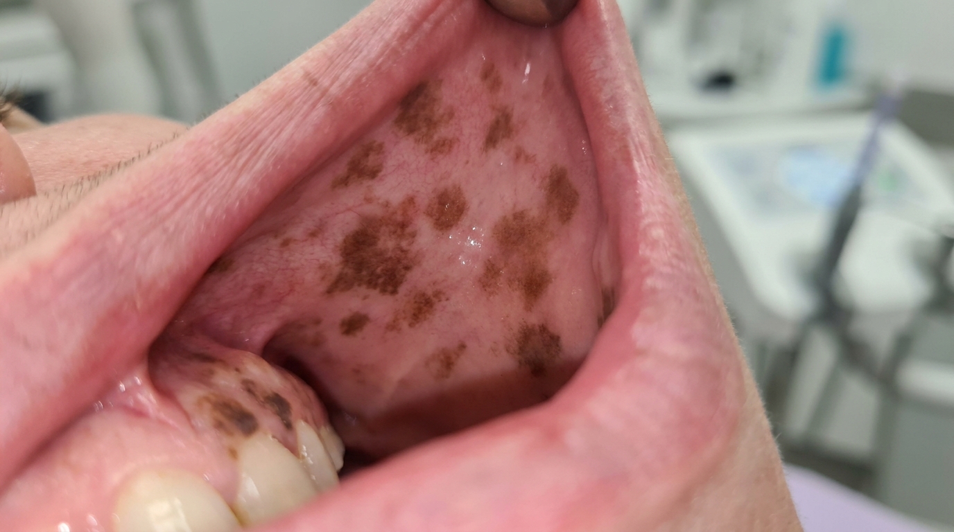
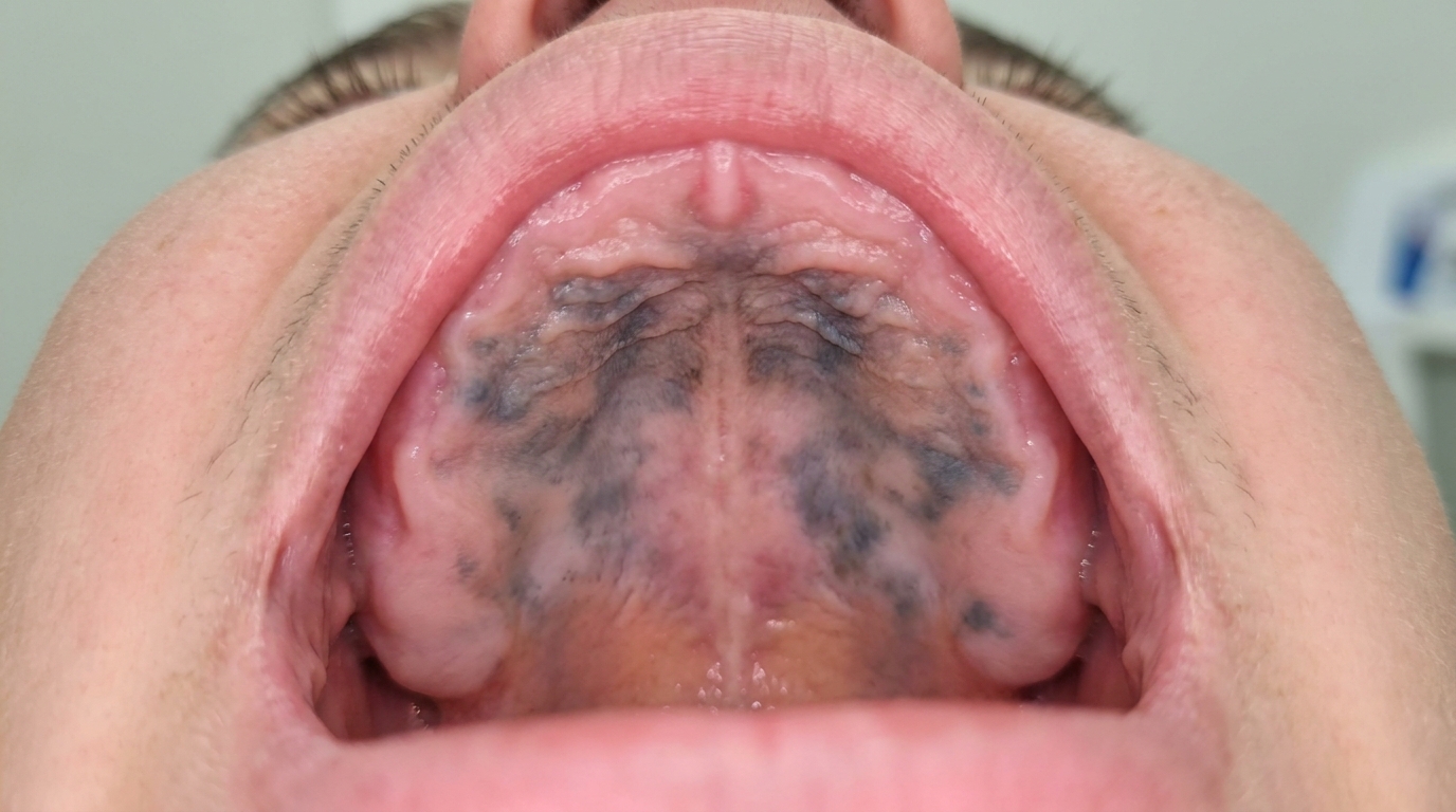
Diffuse, symmetrical brown pigmentation of the gums, inside of the cheeks and tongue, often resembling racial pigmentation but new in adulthood.
Grey or blue-grey patches on the hard palate (typical of antimalarials).
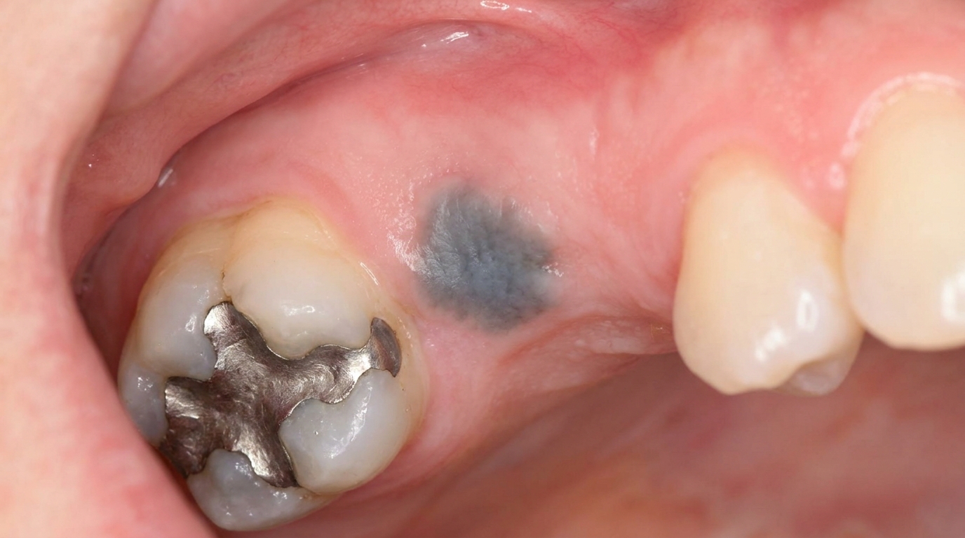
Black or blue-black pigmentation of the gums, palate, bone or teeth (typical of long-term minocycline).
A discrete pigmented line along the gum margin (heavy metal lines).
Brown surface staining of teeth and the back of the tongue (chlorhexidine mouthwash).
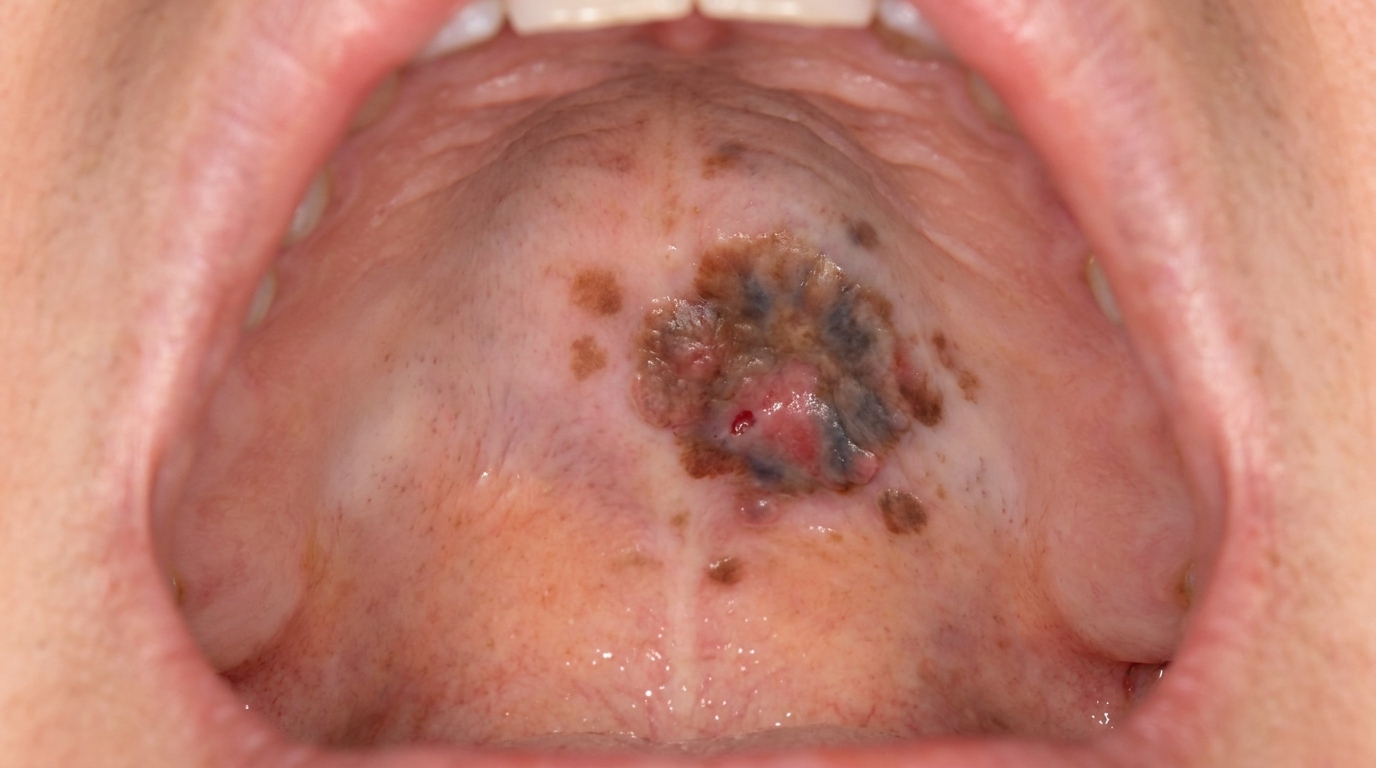
No raised, ulcerated or asymmetric features, these would prompt further investigation for melanoma.
What it feels like
Drug-induced oral pigmentation is asymptomatic. Patients do not feel pain, soreness or change in taste from the pigmentation itself. Some of the underlying medications cause other oral side effects (dry mouth, oral candidiasis (a yeast infection of the mouth), gingival overgrowth (enlarged or swollen gums)), but those are separate issues.
What an X-ray might show
Drug-induced pigmentation is confined to soft tissue and is not visible on dental X-rays. In rare cases of long-term minocycline use, the underlying jawbone may show pigmentation visible on biopsy (a small tissue sample taken for laboratory examination), but not on routine imaging.
What happens at the dentist?
Drug-induced oral pigmentation is most often picked up at a routine dental check-up and clean at ArtSmiles. The dentist will typically:
Examine the mouth carefully under good light, noting where the pigmentation is, how symmetrical it is, and whether it has changed.
Take a thorough medication history, including over-the-counter products, mouthwashes and recent prescriptions, looking for any of the recognised culprits.
Compare the appearance with physiological oral pigmentation that may have been present from childhood.
Discuss the timing, drug-induced pigmentation typically appears in adulthood after starting a medication, while physiological pigmentation has been there since childhood.
Liaise with the GP or specialist who prescribed the medication, particularly when the pigmentation is unsightly or causing concern.
Recommend biopsy only when the appearance is atypical, for example, when there is asymmetry, a focal raised area, or features that could suggest melanoma.
Is this serious?
🟡 The pigmentation itself is harmless and does not damage the tissues. The reason it matters is to be confident that it is drug-induced rather than something more serious. Once the diagnosis is made, the most important conversation is whether the medication needs to continue, can be stopped, or can be substituted, that decision rests with the prescribing doctor and depends on the underlying condition being treated.
If you are taking a long-term medication and have noticed new brown, grey or blue patches in your mouth, it is worth booking an assessment so the change can be confirmed as drug-related and any concerns ruled out.
Could it be something else?
Several other conditions can produce diffuse oral pigmentation. The textbooks list these as the main differentials:
Physiological oral pigmentation, symmetrical, present since childhood, common in people with darker skin.
Smoker's melanosis, diffuse brown patches in heavy smokers, particularly on the front gums and inside of the lips.
Addison's disease pigmentation, diffuse brown pigmentation as part of a systemic endocrine disease, with associated fatigue, weight loss and skin pigmentation.
Post-inflammatory pigmentation, brown areas following chronic inflammation, particularly in lichen planus.
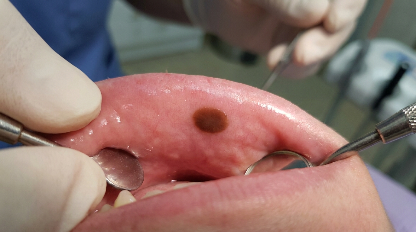
Oral melanotic macule, a single small, well-defined brown patch.
Oral malignant melanoma, a rare but serious cancer, distinguished by asymmetry, irregular borders, recent change and any raised or ulcerated component.
HIV-associated melanosis, diffuse brown pigmentation that may overlap with antiretroviral medication effects.
How is it treated?
Treatment is usually unnecessary, focusing instead on identifying the cause and reassurance.
At-home measures and habits:
Continue your prescribed medication unless your doctor advises otherwise. Do not stop a medication on your own because of mouth pigmentation, the underlying condition being treated matters more.
Maintain excellent oral hygiene with brushing twice a day and flossing daily.
Continue any rinses your dentist has recommended, but discuss whether to limit chlorhexidine if surface staining is becoming a problem.
Professional steps your dentist may consider:
Confirming the diagnosis by clinical examination and a careful drug history.
Discussing the appearance and timeline with the patient and reassuring them when the pattern is consistent with drug-induced pigmentation.
Coordinating with the prescribing GP or specialist to discuss whether the medication can be stopped, reduced or substituted, particularly if the pigmentation is causing significant cosmetic concern.
Biopsy in any atypical case, a single asymmetric or raised area, or a focal patch that does not match the rest of the pigmentation pattern.
Documentation with photographs as a baseline, particularly for monitoring patients on chronic chemotherapy or antimalarial therapy.
Cosmetic depigmentation procedures are occasionally requested for residual pigmentation after stopping the drug, although the textbooks regard these as cosmetic and not medically necessary.
A patient-centred approach matters here. Drug-induced pigmentation is often discovered while the patient is being treated for another, sometimes serious, condition. Calm, clear explanation that the pigmentation is harmless and does not require any change to important medications without medical advice is itself part of effective care, values that sit at the heart of our clinical philosophy.
What's the long-term outlook?
The outlook is excellent. For most drug-induced oral pigmentation, the colour gradually fades over months to years once the medication is stopped or its dose reduced. For drugs that deposit directly in the tissues (such as minocycline), the colour may persist for many years, but it remains harmless. The most important factor in the outcome is good communication between the patient, the dentist and the prescribing doctor, so that the right medication decisions are made for the right reasons.
A note on this article
This article is for educational purposes only and does not constitute a clinical diagnosis. Please consult a registered dental practitioner for assessment and treatment advice.
The cover image above is an AI-generated illustration based on the most common visible features of this condition described in clinical pathology references. It is not a photograph of a real case and should not be used to diagnose or rule out the condition in your own situation. If you are concerned about something you have noticed, please book an assessment with a registered dental practitioner.
References
Cawson, R. A., & Odell, E. W. (2017). Cawson's essentials of oral pathology and oral medicine (8th ed.). Elsevier. Chapter 21, Melanoma and Other Pigmented Lesions: drug-induced pigmentation including chlorhexidine and phenothiazines, pp. 327 to 328.
Neville, B. W., Damm, D. D., Allen, C. M., & Chi, A. C. (2023). Oral and maxillofacial pathology (5th ed.). Elsevier. Chapter 8, Physical and Chemical Injuries: Drug-Related Discolorations of the Oral Mucosa, including estrogen, chemotherapy agents, AIDS medications, minocycline and antimalarials, pp. 305 to 311.
Regezi, J. A., Sciubba, J. J., & Jordan, R. C. K. (2017). Oral pathology: Clinical pathologic correlations (7th ed.). Elsevier. Chapter on Pigmented Lesions: cross-reference for drug-related pigmentation as a cause of diffuse oral pigmentation.