Compiled from clinical pathology references. Medically reviewed by Dr Cristian Dunker , Principal Dentist, ArtSmiles Cosmetic Dentistry.
Quick summary
Also called | Racial pigmentation, ethnic pigmentation, physiologic pigmentation |
How urgent? | 🟢 Not urgent, physiological pigmentation is a normal variation, not a disease |
Common or rare? | Very common, particularly in people with darker skin |
Who it affects | Most often people of African, South Asian, East Asian, Pacific Islander or Mediterranean heritage; usually present from a young age and stable through adult life |
Who treats it | No treatment is needed; a general dentist can confirm the diagnosis |
Based on | Cawson, with cross-references in Neville and Regezi |
What is it?
Physiological oral pigmentation is a benign, symmetrical brown colouration of the gums and other parts of the mouth that reflects the normal amount of melanin (the natural brown pigment in skin and oral lining) in a person's mucosa (the soft tissue lining of the mouth). It is not a disease, not a sign of any underlying problem, and not a precursor to anything dangerous. The textbooks describe it as the most common form of oral pigmentation worldwide, particularly in people with darker skin. Recognising it as a normal variation is important so it isn't mistaken for one of the more serious causes of oral pigmentation.
Who tends to get it?
The textbooks describe a fairly consistent picture:
People with darker skin are more often affected. Physiological pigmentation is very common in people of African, South Asian, East Asian, Pacific Islander, Mediterranean, Middle Eastern and Latin American heritage.
Lighter-skinned people can also have small areas of physiological pigmentation, although it is less obvious.
Both sexes are affected equally.
Pigmentation usually appears during the first decade of life and remains stable thereafter.
It does not change in colour, size or distribution from year to year, which is one of its most useful diagnostic features.
What causes it?
The cause is normal melanin production by the body's pigment-producing cells:
Melanocytes (the pigment-producing cells) in the basal layer (the deepest, foundation layer of the mouth lining) of the oral lining produce melanin, just as they do in the skin.
The amount of melanin produced varies between individuals, populations and even between different parts of the same mouth.
Genetics determine the overall melanin output, which is why pigmentation patterns tend to run in families and ethnic groups.
No external factor, no infection, medication, food or habit, is needed for physiological pigmentation to develop.
It is important to note that physiological pigmentation can be made more obvious by smoking, which independently increases melanocyte activity. When a person of any background develops new or worsening pigmentation in adulthood, smoking is one of the things to consider.
How does it develop?
The pigmentation appears as the oral lining matures during early childhood. Melanocytes in the basal layer of the surface epithelium (the surface layer of the lining) release melanin into the surrounding cells, where it accumulates and gives the tissue its brown colour. Over time, the pattern stabilises and remains essentially unchanged for life. Microscopically, there is no abnormality of cell structure, no inflammation and no neoplastic (related to abnormal new tissue growth) activity, just a normal increase in melanin pigment.
What might you notice?
What it looks like
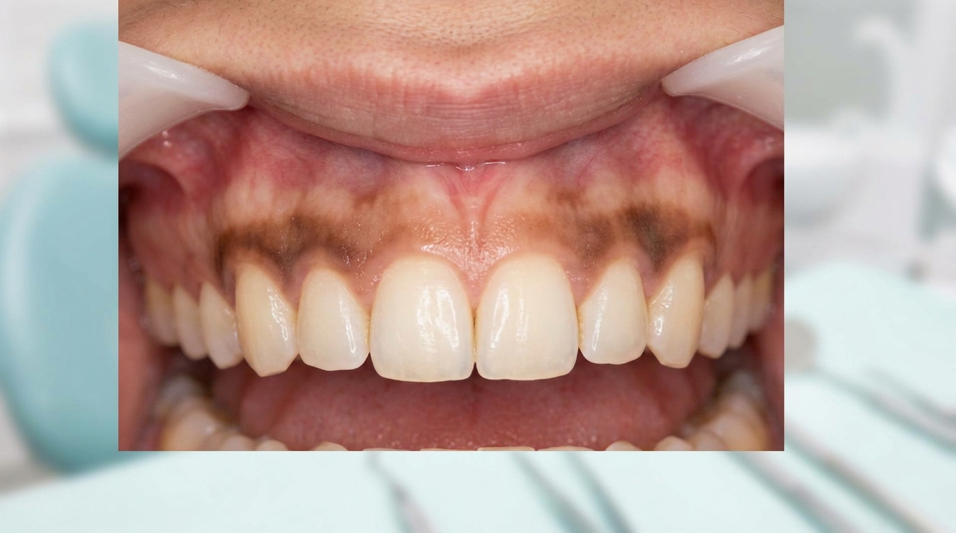
The classic appearance is well described:
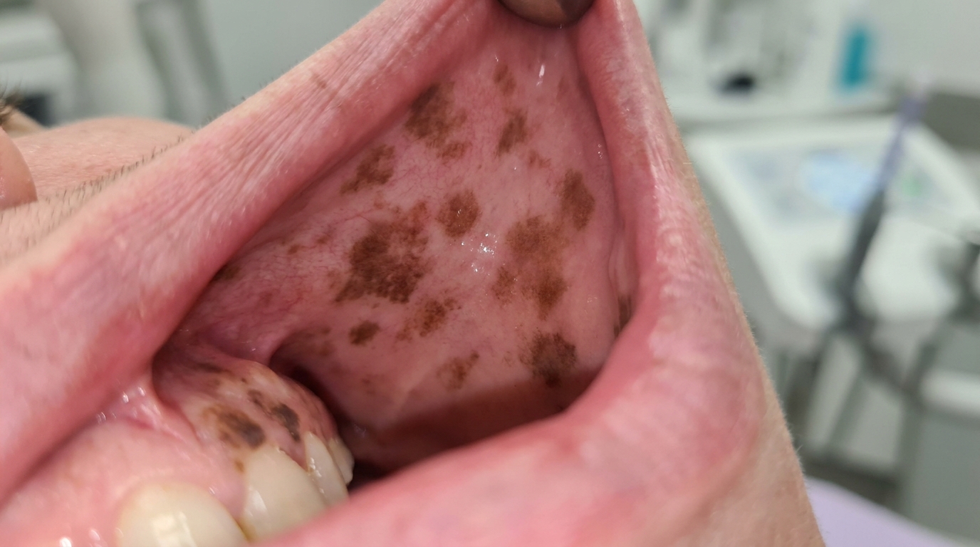
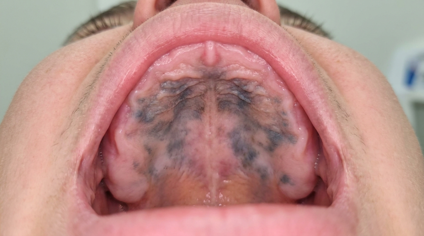
A diffuse, symmetrical brown colouration of the attached gum (the firm band of gum bound tightly to the underlying bone), particularly the band of gum just above the teeth.
The colour ranges from light tan to medium brown to a deep, almost blackish brown, depending on the individual.
Both upper and lower gums are typically involved.
The colour is even within each area, without irregular borders, mottling or focal darker spots.
Other oral surfaces can also be involved, including the inside of the cheeks, the lips, the soft palate and the under-surface of the tongue.
The pigmentation has been present for as long as the patient can remember and has not changed in adulthood.
What it feels like
Physiological oral pigmentation produces no symptoms at all:
No pain, soreness or burning.
No taste change.
No bleeding or surface irregularity.
No effect on eating, brushing or speaking.
What an X-ray might show
X-rays are not relevant to physiological pigmentation, since the change is confined to the soft tissue and does not affect the underlying bone or teeth.
What happens at the dentist?
Physiological pigmentation is most often noted at a routine dental check-up and clean at ArtSmiles, either as part of a normal examination or in response to a patient's question. The dentist will typically:
Examine the entire mouth carefully, noting where the pigmentation is, how symmetrical it is, and how stable it has been.
Take a careful history about how long the pigmentation has been present and whether it has changed in adulthood.
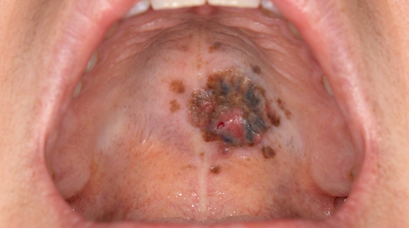
Look for any focal darker spots, raised areas or asymmetries within or beyond the area of physiological pigmentation, which would warrant further investigation.
Check for smoking habits that might be increasing pigmentation.
Reassure the patient that physiological pigmentation is a normal variation that needs no treatment.
Consider biopsy (a small tissue sample taken for laboratory examination) only when the appearance is atypical or when there is any feature that suggests a different diagnosis.
Is this serious?
🟢 No. Physiological oral pigmentation is benign and does not progress to any disease. The textbooks specifically classify it as a normal variation. The reason it is worth understanding is to make sure that it is not confused with other, more serious causes of oral pigmentation, particularly oral malignant melanoma.
If you are unsure whether the brown areas in your mouth are normal physiological pigmentation or something else, particularly if you have noticed any change in the appearance over recent months or years, it is worth booking an assessment so the diagnosis can be confirmed.
Could it be something else?
Several other conditions can produce brown or dark patches in the mouth. The textbooks list these as the main differentials:
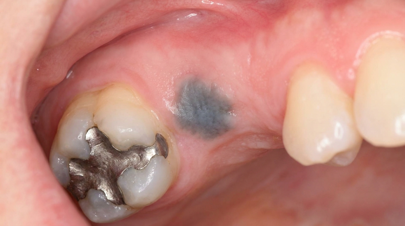
Amalgam tattoo, a localised grey or blue-black flat patch near old amalgam fillings. Tiny radiopaque flecks may be visible on an X-ray.
Oral melanotic macule, a single, small, well-defined brown patch, most often on the lower lip or gum.
Smoker's melanosis, diffuse brown patches in heavy smokers, often on the front gums and inside of the lips. Resolves over years if smoking stops.
Drug-induced oral pigmentation, diffuse brown or grey-blue pigmentation related to medications such as minocycline, antimalarials or chemotherapy agents.
Addison's disease pigmentation, diffuse brown pigmentation as part of a systemic endocrine disease, with associated fatigue, weight loss and skin pigmentation.
Post-inflammatory pigmentation, brown areas that follow chronic inflammation, especially in lichen planus, in people with darker skin.
Oral malignant melanoma, a rare but serious cancer that typically appears as an asymmetrical, irregularly bordered, growing dark patch most often on the hard palate or upper gum.
The combination of symmetry, stability over many years, and no change in adulthood is the strongest clue that an area of pigmentation is physiological rather than something else.
How is it treated?
The textbooks all agree: physiological oral pigmentation needs no treatment.
At-home measures and habits:
Continue normal oral hygiene, brushing twice a day with fluoride toothpaste and flossing daily.
No need to avoid any food, drink or product because of the pigmentation itself.
Avoid smoking, which can increase pigmentation and adds many other oral and general health risks.
Professional steps your dentist may consider:
Confirming the diagnosis by clinical examination.
Reassuring the patient that the appearance is a normal variation.
Documenting the appearance with photographs as a baseline.
Continuing routine dental check-ups, with attention to any new or focal pigmented lesion that does not match the existing physiological pattern.
Cosmetic depigmentation procedures (gingival depigmentation by laser, scalpel or chemical methods) are occasionally requested by patients on aesthetic grounds; the textbooks describe these as cosmetic rather than medically necessary, with possible recurrence over time.
A patient-centred approach matters here. Some patients with physiological pigmentation worry, sometimes for years, that the brown areas in their mouth might be something serious. Calm, clear explanation of what physiological pigmentation is, how it is recognised, and why it does not need treatment is itself part of effective care, values that sit at the heart of our clinical philosophy.
What's the long-term outlook?
The outlook is excellent. Physiological oral pigmentation is a stable, lifelong feature of the mouth that does not progress to any disease and has no long-term health implications. Routine dental review is sufficient, with attention to any new or focal pigmented lesion that develops outside the established physiological pattern. Most people who once worried about the pigmentation are reassured for life by a single explanation at the dental chair.
A note on this article
This article is for educational purposes only and does not constitute a clinical diagnosis. Please consult a registered dental practitioner for assessment and treatment advice.
The cover image above is an AI-generated illustration based on the most common visible features of this condition described in clinical pathology references. It is not a photograph of a real case and should not be used to diagnose or rule out the condition in your own situation. If you are concerned about something you have noticed, please book an assessment with a registered dental practitioner.
References
Cawson, R. A., & Odell, E. W. (2017). Cawson's essentials of oral pathology and oral medicine (8th ed.). Elsevier. Chapter 21, Melanoma and Other Pigmented Lesions: Physiological and racial pigmentation, with summary chart 21.1, pp. 327 to 328.
Neville, B. W., Damm, D. D., Allen, C. M., & Chi, A. C. (2023). Oral and maxillofacial pathology (5th ed.). Elsevier. Chapter 10, Epithelial Pathology: cross-reference for physiologic pigmentation as the most common cause of diffuse symmetrical oral pigmentation.
Regezi, J. A., Sciubba, J. J., & Jordan, R. C. K. (2017). Oral pathology: Clinical pathologic correlations (7th ed.). Elsevier. Chapter on Pigmented Lesions: cross-reference for physiologic oral pigmentation.