Compiled from clinical pathology references. Medically reviewed by Dr Cristian Dunker , Principal Dentist, ArtSmiles Cosmetic Dentistry.
Quick summary
Also called | Focal melanosis, labial melanotic macule (when on the lip) |
How urgent? | 🟢 Not urgent, benign and stable; usually treated only to confirm the diagnosis or for cosmetic reasons |
Common or rare? | The most common oral melanocytic (involving the pigment-producing cells) lesion submitted to oral pathology laboratories |
Who it affects | Adults of any age, with a mean age at diagnosis of around 42 years and roughly twice as many women as men |
Who treats it | General dentist for diagnosis and excisional biopsy (small surgery to remove the whole spot for laboratory examination) when indicated |
Based on | Neville, Cawson, with cross-references in Regezi |
What is it?
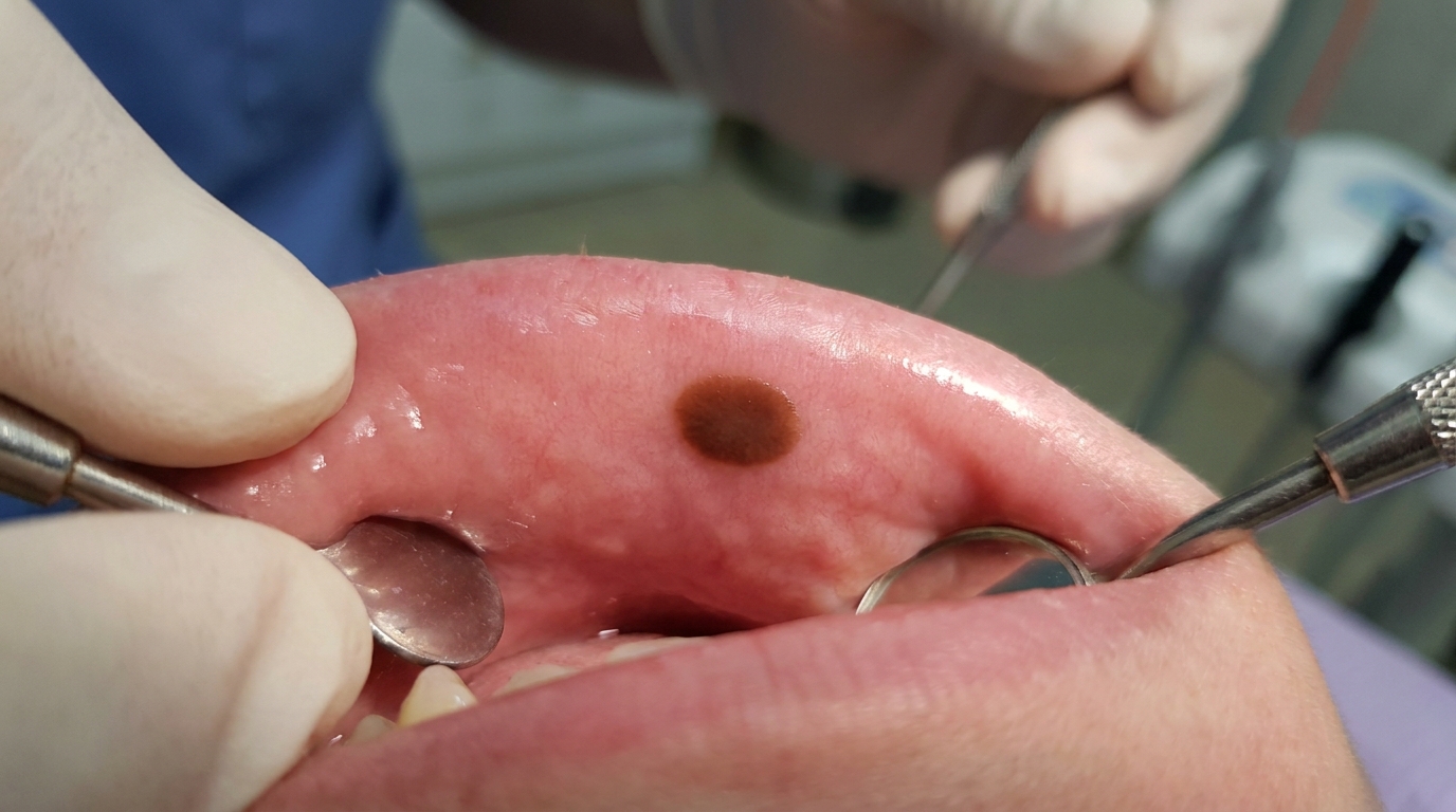
An oral melanotic macule is a small, flat, well-defined brown spot in the lining of the mouth, caused by a focal increase in melanin (the natural brown pigment in skin and oral lining) in the surface cells. The textbooks describe it as the most common oral melanocytic lesion submitted to oral pathology laboratories. Most are entirely harmless and stay the same size and colour for years. The reason a dentist will sometimes recommend removing one is to confirm the diagnosis on histopathology (microscopic examination of a tissue sample), particularly when the lesion is in a location where oral malignant melanoma is more likely (such as the hard palate or upper gum), or when the appearance has changed.
Who tends to get it?
The textbooks describe a fairly recognisable profile:
Mean age at diagnosis around 42, with a wide range across all adult ages.
Roughly twice as many women as men in biopsied series.
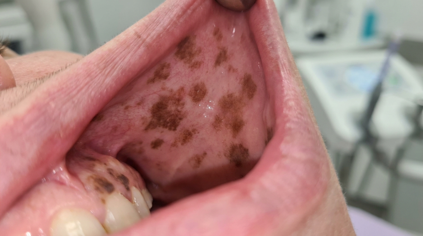
Most common location is the lower lip vermilion (about a third of cases), followed by the inside of the cheek (buccal mucosa, the soft tissue lining of the mouth), the gum, and the palate.
Usually solitary, about 17% of patients have multiple macules.
Typically less than 1 cm in greatest diameter.
Stable in size and colour once formed.
What causes it?
The exact cause is unclear, but the textbooks describe several recognised contributors:
Focal increase in melanin within the basal layer (the deepest, foundation layer of the mouth lining) of the lining, with or without an increase in the number of melanocytes (the cells that make melanin).
Possibly related to chronic minor irritation, chronic biting, friction or sun exposure (for lip lesions), although this link is not consistent.
Some cases are associated with systemic conditions, including Addison's disease, Peutz-Jeghers syndrome, McCune-Albright syndrome, and other rare syndromes (Box 10.1 in Neville).
Chronic autoimmune inflammation, particularly erosive lichen planus or pemphigoid, can produce focal pigmentation.
Sun exposure is implicated specifically for the labial melanotic macule on the lower lip vermilion.
In the great majority of cases, however, no specific underlying cause is identified.
How does it develop?
A focal increase in melanin develops in the basal layer of the oral lining. Microscopically, there is increased pigment in the basal and parabasal cells of an otherwise normal stratified squamous epithelium (the layered surface lining of the mouth). Some pigment may also be seen "free" in the underlying connective tissue or within melanophages (melanin-laden macrophages). Once the lesion is fully formed, it generally remains stable rather than continuing to enlarge, a feature that helps distinguish it from melanoma, which typically continues to change.
What might you notice?
What it looks like
The classic appearance is well described:
A single, small, flat brown patch in the mouth.
Well-demarcated, clear edges, with sharp transition to surrounding mucosa.
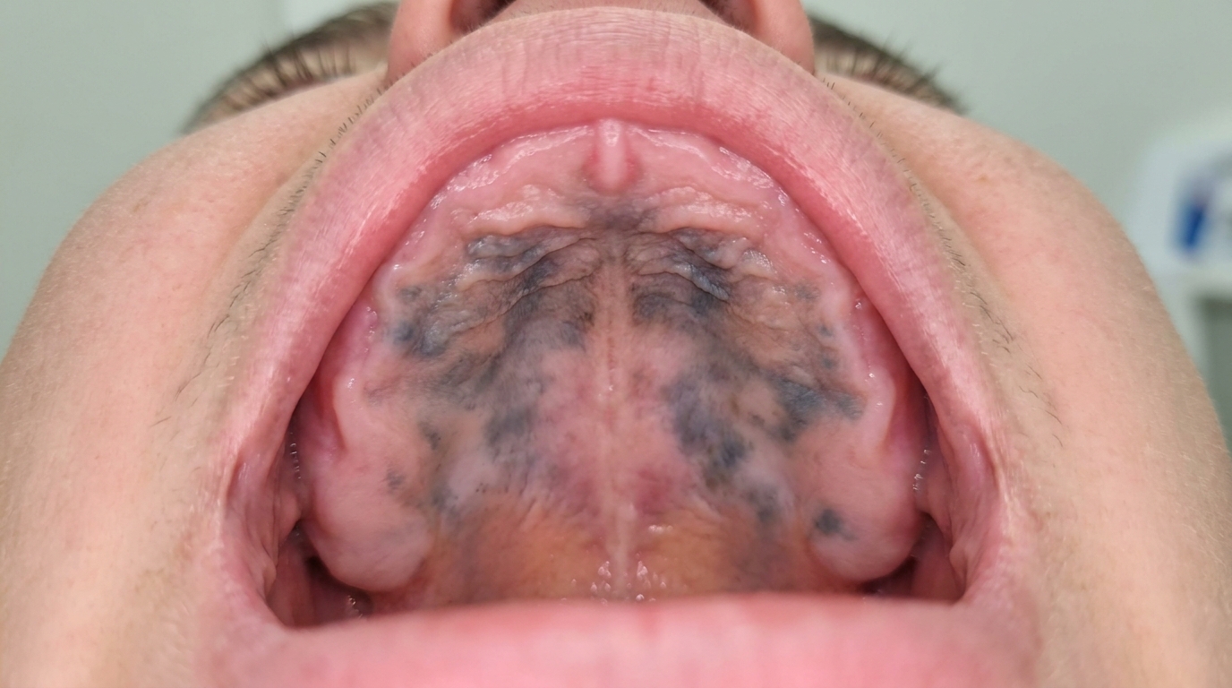
Uniformly tan to dark brown, occasionally blue or near-black.
Round or oval shape.
Less than 1 cm in greatest diameter.
Most often on the lower lip vermilion, gum, inside of the cheek or palate.
Stable for years without change in size, shape or colour.
What it feels like
An oral melanotic macule is asymptomatic (causing no symptoms). There is no pain, no roughness, no bleeding and no taste change. The lining feels normal to the tongue.
What an X-ray might show
Oral melanotic macules are confined to the surface lining and not visible on X-rays.
What happens at the dentist?
A small dark spot on the lip or in the mouth is most often picked up by the patient or by their dentist at a routine dental check-up and clean at ArtSmiles. The dentist will typically:
Examine the spot carefully under good light, noting its size, location, shape, colour and edges.
Take a careful history about how long the spot has been present and whether it has changed.
Compare with physiological pigmentation and other patterns in the rest of the mouth.
Photograph and measure the lesion as a baseline.
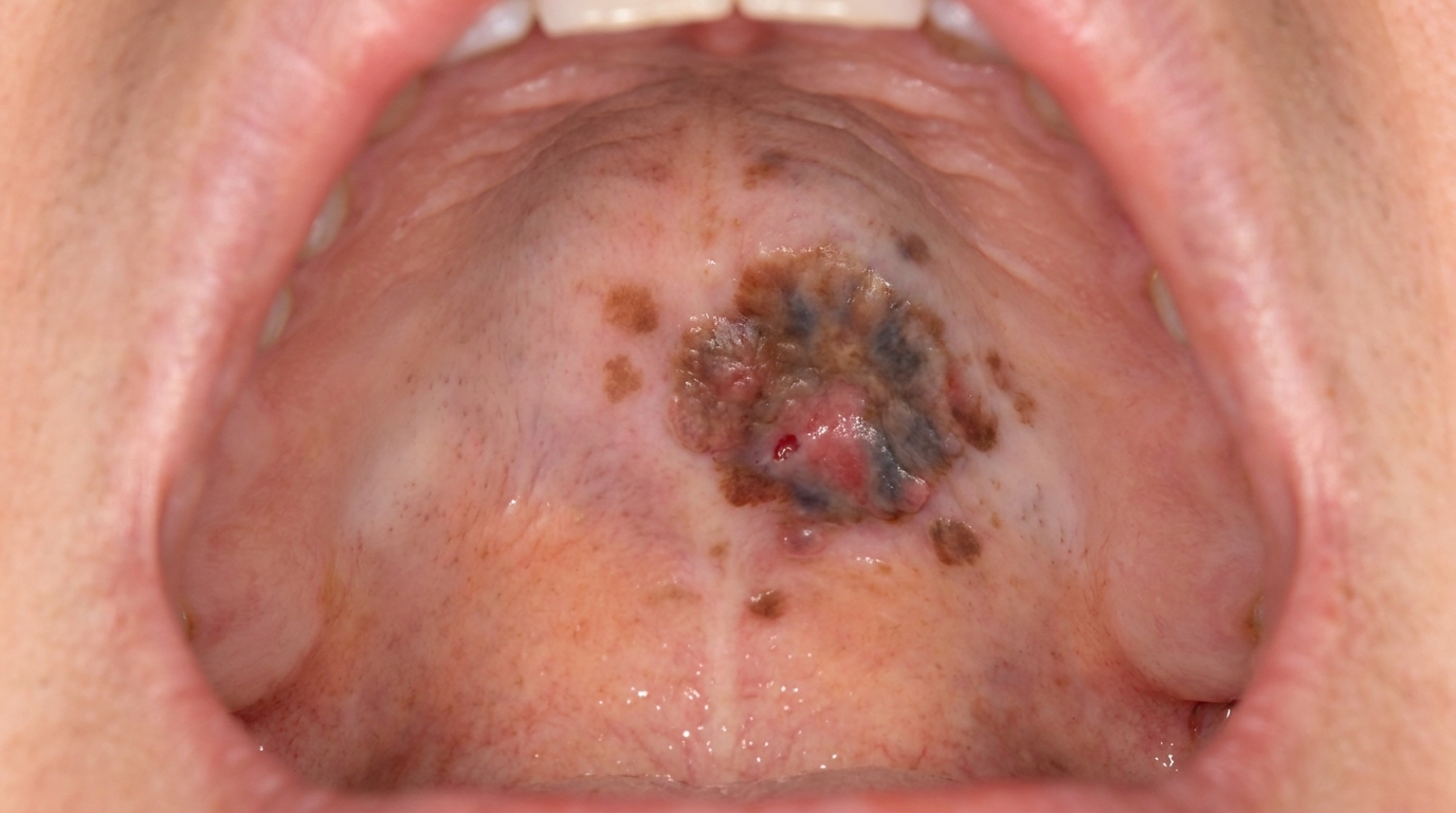
Recommend excisional biopsy when the spot is on the palate or upper gum, has been present for an unknown duration, has any irregular features (asymmetry, ragged borders, mixed colour, recent change), or is in an aesthetically important area where the patient would like it removed.
Discuss alternative treatments, laser ablation, cryosurgery (freezing treatment) or electrocautery (treatment using a fine heated tip), for cosmetic removal, while reminding the patient that these methods do not preserve tissue for microscopic examination.
The textbooks specifically advise that all oral pigmented macules of recent onset, large size, irregular pigmentation, unknown duration or recent enlargement should be biopsied to exclude early melanoma.
Is this serious?
🟢 An oral melanotic macule is benign. The textbooks specifically describe it as having no malignant potential, with only a single case ever reported of apparent malignant transformation. The reason for vigilance is not the lesion itself but the fact that early oral melanoma can look very similar in its earliest stages, and the only way to distinguish them with certainty is microscopic examination after excision.
If you have noticed a brown spot on your lip or in your mouth, particularly if it is new, growing, or has irregular features, it is worth booking an assessment so the diagnosis can be confirmed.
Could it be something else?
Several other conditions can produce a small dark spot in the mouth. The textbooks list these as the main differentials:
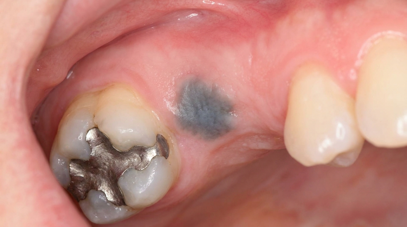
Amalgam tattoo, a grey or blue-black flat patch near old amalgam fillings, often with tiny radiopaque flecks visible on X-ray.
Oral naevus (mole), a small, often slightly raised pigmented lesion that has been present for years.
Smoker's melanosis, diffuse brown patches in heavy smokers, usually larger and more spread out than a single macule.
Oral malignant melanoma, a rare but serious cancer, distinguished by asymmetry, irregular borders, recent change, and any raised or ulcerated component.
Peutz-Jeghers syndrome, multiple small dark spots around the lips and inside the mouth, with associated intestinal polyps; usually present from childhood with a positive family history.
Lentigo simplex, a flat brown lesion of skin or vermilion that may slowly fade over years; histologically distinct.
Drug-related, post-inflammatory or systemic pigmentation, usually diffuse rather than a single small macule.
The combination of being small, well-defined, uniform in colour, and stable over months to years is the strongest clue toward a benign melanotic macule rather than a melanoma.
How is it treated?
Treatment is straightforward.
At-home measures and habits:
Continue normal oral hygiene, brushing twice a day with fluoride toothpaste and flossing daily.
No need to avoid any food or product because of the macule itself.
Use lip balm with sunscreen (SPF 30+) if the macule is on the lower lip and you spend a lot of time outdoors.
Photograph any pigmented spot you are watching, so changes can be detected reliably.
Professional steps your dentist may consider:
Excisional biopsy under local anaesthetic, preferred for lesions in higher-risk locations or with any atypical feature, since it both removes the lesion and provides tissue for histopathological diagnosis.
Histopathological examination to confirm a benign melanotic macule and rule out other diagnoses.
Cosmetic removal with laser, electrocautery or cryosurgery for purely aesthetic concerns, with the patient understanding that no tissue is preserved for microscopic examination.
No treatment with periodic review when the lesion is small, classic in appearance, in a low-risk location, and stable on long-term observation.
Documentation with photographs as a baseline for future visits, even when no treatment is performed.
A patient-centred approach matters here too. Patients often worry that a small spot in the mouth might be cancer, and an unhurried explanation of how a benign melanotic macule can be distinguished from melanoma, and why a small biopsy might be recommended for peace of mind, is itself part of effective care, values that sit at the heart of our clinical philosophy.
What's the long-term outlook?
The outlook is excellent. Once a lesion has been excised and confirmed as a benign melanotic macule on microscopy, recurrence is uncommon and there is no long-term health implication. Lesions left in place can be monitored at routine dental check-ups, with biopsy reserved for any change in size, shape, colour or surface. Most patients with oral melanotic macules are reassured for life by a single explanation and a short follow-up plan.
A note on this article
This article is for educational purposes only and does not constitute a clinical diagnosis. Please consult a registered dental practitioner for assessment and treatment advice.
The cover image above is an AI-generated illustration based on the most common visible features of this condition described in clinical pathology references. It is not a photograph of a real case and should not be used to diagnose or rule out the condition in your own situation. If you are concerned about something you have noticed, please book an assessment with a registered dental practitioner.
References
Neville, B. W., Damm, D. D., Allen, C. M., & Chi, A. C. (2023). Oral and maxillofacial pathology (5th ed.). Elsevier. Chapter 10, Epithelial Pathology: Oral Melanotic Macule (Focal Melanosis), with detailed clinical features and Box 10.1 syndromic associations, pp. 372 to 373.
Cawson, R. A., & Odell, E. W. (2017). Cawson's essentials of oral pathology and oral medicine (8th ed.). Elsevier. Chapter 21, Melanoma and Other Pigmented Lesions: Melanotic macule, with summary chart 21.1, p. 328.
Regezi, J. A., Sciubba, J. J., & Jordan, R. C. K. (2017). Oral pathology: Clinical pathologic correlations (7th ed.). Elsevier. Chapter on Pigmented Lesions: cross-reference for oral melanotic macule and focal melanosis.
Frequently asked questions
What is an oral melanotic macule?
An oral melanotic macule is a small, flat, well-defined brown to black patch in the mouth caused by harmless extra melanin in the lining. It is usually less than 1 cm across and most often appears on the lower lip, gums, cheek or palate.
Are melanotic macules dangerous?
Single, stable melanotic macules are completely benign. Multiple macules can sometimes be part of inherited syndromes (like Peutz-Jeghers syndrome) that need investigation, so it is worth mentioning if there are several or if there is a family history of polyps or unexplained pigmentation.
How is it told apart from melanoma?
A melanotic macule stays the same size, shape and colour over time. Melanoma grows, changes colour, has irregular borders, may bleed or ulcerate, and often shows multiple shades within one lesion. Any pigmented patch that grows or changes warrants prompt biopsy.
Does a melanotic macule need treatment?
No treatment is needed for typical melanotic macules. Photographs help track stability over time. Biopsy is reserved for lesions that change, look atypical, or where melanoma cannot be ruled out clinically.