Compiled from clinical pathology references. Medically reviewed by Dr Cristian Dunker, Principal Dentist, ArtSmiles Cosmetic Dentistry.
A cyst hidden inside the jawbone is the kind of thing most people only learn about because their dentist took a routine X-ray. Odontogenic keratocysts (OKCs) are a particular kind of jaw cyst, well known for growing quietly along the bone with very little outward sign, and equally well known for sometimes coming back after treatment.
This article from the team at ArtSmiles, reviewed by Dr Cristian Dunker, explains what an OKC is, why it forms, and how modern surgery and follow-up keep the long-term outlook good.
Quick summary
At a glance | Detail |
|---|---|
Also called | OKC; keratocystic odontogenic tumour (older WHO name) |
How urgent? | 🟡 Surgical referral is recommended, the cyst will continue to grow if untreated, but it is not cancer and is not an emergency |
Common or rare? | Uncommon, but the third most common odontogenic cyst overall |
Who it affects | Most often adults in their 20s to 40s, slightly more often men; can affect any age |
Who treats it | Oral and maxillofacial surgeon, with the dentist coordinating imaging and follow-up |
Based on | Neville, Cawson, Regezi, Laskaris |
What is it?
The word odontogenic means "tooth-related", these cysts arise from cell rests left behind after teeth develop. Keratocyst refers to the cyst lining, which produces a flaky protein called keratin (the same protein in skin, hair and nails). Together, an OKC is:
A developmental cyst of the jawbone.
Benign but locally aggressive.
Lined by a thin, corrugated, parakeratinised epithelium (a microscopic feature, the cyst lining shows a wavy surface and a thin layer of dead keratin, that pathologists rely on for diagnosis).
Often filled with a cheesy, keratin-rich material rather than clear fluid.
For many years OKCs were classified as a tumour (the keratocystic odontogenic tumour), because of their growth pattern; the World Health Organization later returned them to the cyst category, but the aggressive behaviour is well recognised.
Who tends to get it?
OKCs can appear at any age but are most often diagnosed in people in their 20s to 40s. They are slightly more common in men than in women. They show up on every continent and in every population.
A particular pattern to mention is Gorlin-Goltz syndrome (also called naevoid basal cell carcinoma syndrome). In this inherited condition, patients develop multiple OKCs, basal cell skin cancers from a young age, and a number of other features. Anyone diagnosed with multiple OKCs, especially in their teens or twenties, should be reviewed by a specialist for this possibility.
What causes it?
Most OKCs are sporadic, meaning they happen for no clear external reason. The underlying biology involves changes in a gene called PTCH1, which normally helps control cell growth. When PTCH1 stops working as it should, in either a single cyst (sporadic OKC) or in every cell of the body (Gorlin-Goltz syndrome), the cyst lining grows actively and the cyst expands.
Other contributing biological features include:
Active growth signals from the cyst lining itself.
An expanding cyst cavity that displaces and weakens bone.
A tendency for tiny daughter cysts (small satellite cysts that bud off from the main lining) to develop into the surrounding bone, which is one reason recurrence is more common than in other jaw cysts.
OKCs are not caused by infection, dental decay, dental trauma or anything in the diet.
How does it develop?
The classic course of an OKC is slow, quiet expansion within the bone:
Origin from cell rests. A small group of cells left over from early tooth development begins to form a tiny cyst.
Growth along the bone. Unlike many cysts, OKCs grow more along the length of the jaw than outwards, which is why the face often looks normal even when the cyst is large.
Bone thinning. As the cyst expands, the surrounding bone is gradually eroded.
Tooth displacement. Adjacent teeth may be pushed sideways, but their roots usually keep their length (they are not eaten away as much as in some other cysts).
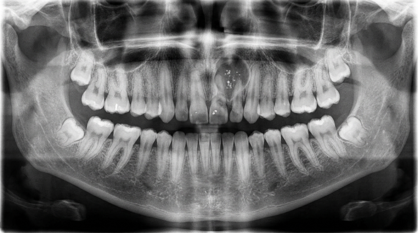
Discovery. Most OKCs are found incidentally on a panoramic dental X-ray taken for another reason. Some are only noticed when they reach a size that causes facial swelling or pain.
What might you notice?
Many patients notice nothing at all and the cyst is found on imaging. When symptoms appear, they may include:
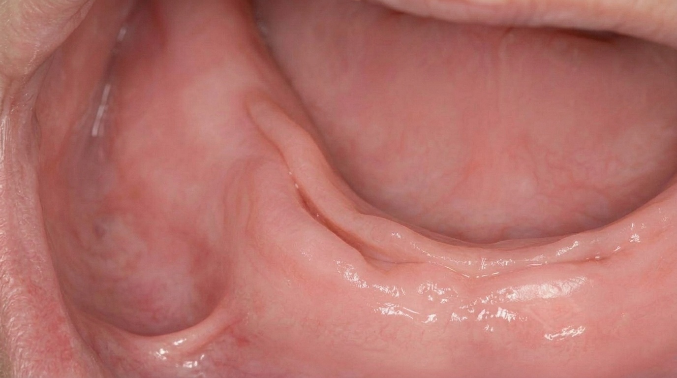
A slow swelling at the back of the lower jaw, often felt from the inside of the cheek before any visible change to the face.
A vague aching or pressure feeling in the jaw.
Numbness or tingling of the lower lip, if the cyst has grown close to the inferior alveolar nerve (the main sensory nerve of the lower jaw).
A tooth that has been pushed out of line.
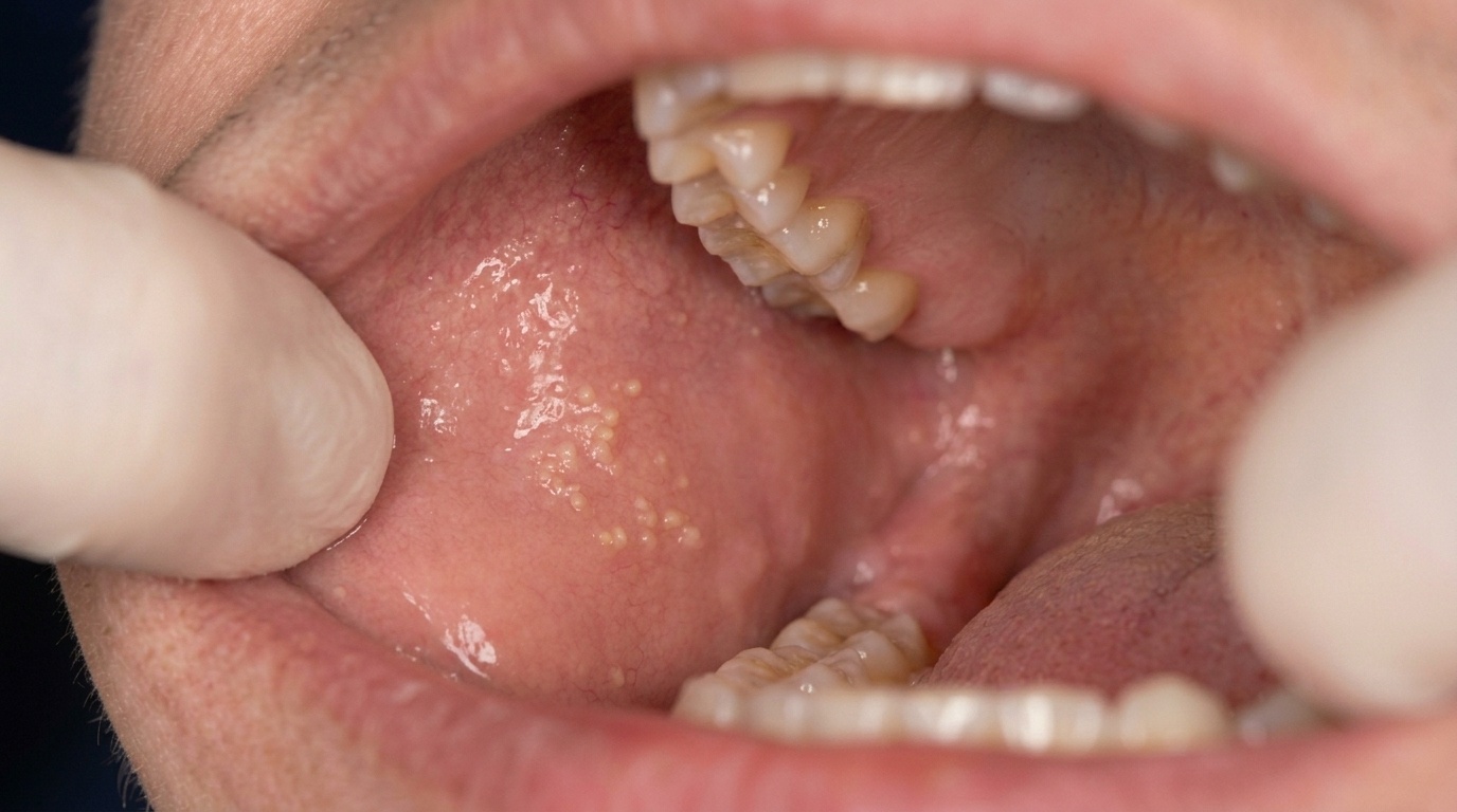
Discharge of cheesy material if the cyst becomes infected and drains into the mouth.
A pathological fracture of the jaw if the cyst is very large and the bone has been thinned over years.
The most important point is that most OKCs are silent until imaging finds them, which is one of the reasons regular dental check-ups (with appropriate X-rays at appropriate intervals) matter.
What happens at the dentist?
When a suspicious dark area is seen on a dental X-ray, the path forward usually looks like this:
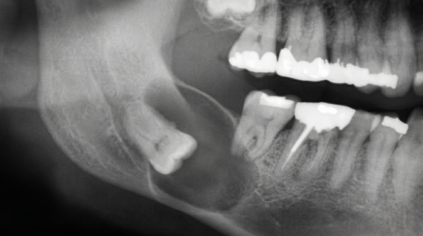
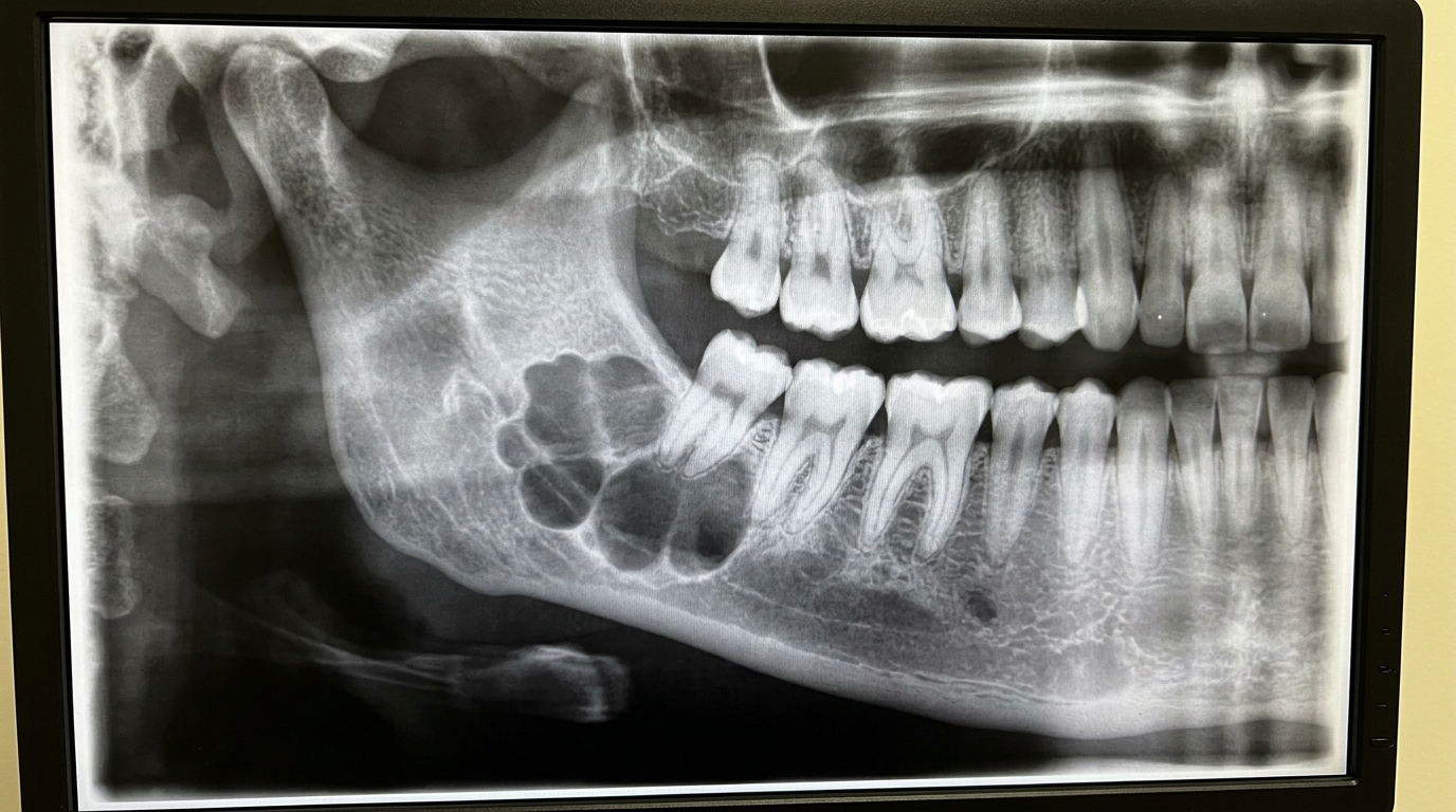
A careful look at the X-ray. Most OKCs appear as a well-defined dark area in the bone, often with a smooth, slightly scalloped edge, most commonly in the angle and ramus of the lower jaw.
Additional imaging. A panoramic X-ray gives a good overview. A cone-beam CT (CBCT, a 3D dental X-ray) shows the size and the relationship to nerves, teeth and the inferior border of the jaw.
Discussion of findings. We explain what we see, what it might be and why a specialist opinion is recommended.
Referral to an oral and maxillofacial surgeon. OKCs are managed surgically, and your treating surgeon will recommend the most appropriate technique.
Histopathology (examination of the removed tissue under the microscope) of the cyst lining, this is the only way to confirm an OKC. Several other cysts can look identical on imaging.
Long-term follow-up. Most OKCs are followed up for at least five years (and sometimes longer) with periodic X-rays to check for recurrence.
If multiple cysts are found, your surgeon may also arrange skin and genetic assessment for Gorlin-Goltz syndrome.
Is this serious?
🟡 OKCs are not cancer. They do not spread to other parts of the body. The reasons we treat them seriously are:
They keep growing. Unlike some cysts, OKCs do not stop on their own.
They thin the jawbone. Large untreated cysts weaken the jaw and can lead to fracture.
They recur. Recurrence rates after simple removal range from about 25% to 62% in published series, depending on technique.
They can become infected if they communicate with a tooth or the mouth.
They have a syndrome association, in some patients they are a sign of Gorlin-Goltz syndrome.
A small minority can transform, very rarely, into a more aggressive lesion. This is why long-term follow-up is recommended.
Could it be something else?
A dark area in the jaw on X-ray can have several explanations. Common possibilities include:
Dentigerous cyst, a cyst around the crown of an unerupted tooth.
Radicular (periapical) cyst, a cyst at the tip of a non-vital tooth root.
Ameloblastoma, a benign but locally aggressive odontogenic tumour, often multilocular.
Calcifying odontogenic cyst (Gorlin cyst, CCOT), usually contains visible calcifications.
Lateral periodontal cyst, a small developmental cyst beside a vital tooth.
Residual cyst, a periapical cyst that remains after the tooth has been removed.
Odontogenic myxoma, a soap-bubble appearance on imaging.
Aneurysmal bone cyst or central giant cell granuloma, vascular lesions of the jaw.
Traumatic (simple) bone cyst, a benign empty cavity in the bone.
These cannot be told apart on imaging alone, that is why a specialist surgical biopsy and histopathology are essential.
How is it treated?
Treatment is always surgical and is shaped by the size, location and pattern of the cyst. Common approaches include:
Enucleation with curettage. The cyst is carefully removed in one piece, and the surrounding bone is gently curetted to clear daughter cysts. This is sometimes combined with an additional step to reduce recurrence, for example peripheral ostectomy (removing a thin layer of surrounding bone) or application of Carnoy's solution to the cavity.
Marsupialisation (a smaller first operation that opens the cyst and lets it shrink) for very large cysts. The cyst is opened, decompressed and allowed to shrink over weeks or months before definitive removal. This protects nerves and teeth and reduces the size of the eventual surgical defect.
Resection. Reserved for recurrent or particularly aggressive lesions, where a section of jaw is removed and reconstructed.
After treatment, regular review with panoramic X-rays, every six to twelve months at first, then yearly, is recommended for at least five years to detect any recurrence early.
What's the long-term outlook?
The long-term outlook depends on size, treatment and follow-up. With modern surgery, careful technique and long-term review, most patients have an excellent result. Even when a cyst recurs, it can usually be managed with a second, smaller procedure. Reconstruction of the jaw in larger cases gives good function and appearance.
If a cyst has been found on your X-ray, please do not panic. Most jaw cysts have a clear, well-trodden treatment pathway. We will explain what is happening, refer you to the right specialist and stay involved with your dental care throughout.
A note on this article
This article is for educational purposes only and does not constitute a clinical diagnosis. Please consult a registered dental practitioner for assessment and treatment advice.
The cover image above is an AI-generated illustration based on the most common visible features of this condition described in clinical pathology references. It is not a photograph of a real case and should not be used to diagnose or rule out the condition in your own situation. If you are concerned about something you have noticed, please book an assessment with a registered dental practitioner.
References
Neville, B. W., Damm, D. D., Allen, C. M., & Chi, A. C. (2023). Oral and maxillofacial pathology (5th ed.). Elsevier. Chapter 15, Odontogenic Cysts and Tumors: Odontogenic Keratocyst, pp. 683 to 689.
Cawson, R. A., & Odell, E. W. (2017). Cawson's essentials of oral pathology and oral medicine (8th ed.). Elsevier. Chapter 10, Odontogenic Cysts.
Regezi, J. A., Sciubba, J. J., & Jordan, R. C. K. (2017). Oral pathology: Clinical pathologic correlations (7th ed.). Elsevier. Chapter 10, Cysts of the Oral Region.
Laskaris, G. (2006). Pocket atlas of oral diseases (2nd ed.). Thieme. Odontogenic Keratocyst.
Frequently asked questions
Is an odontogenic keratocyst (OKC) cancer?
No. An odontogenic keratocyst is a benign developmental cyst of the jaw, not a cancer. It does, however, behave more aggressively than most jaw cysts: it tends to grow along the inside of the jaw, can become large before any symptoms appear, and has a higher recurrence rate after removal, so it is followed up more closely than other cysts.
Why is an OKC found on an X-ray and not from pain?
Most odontogenic keratocysts are picked up by chance on a panoramic X-ray taken for another reason, such as wisdom-tooth assessment. They tend to be painless for a long time because they grow inside the jawbone in a way that does not always swell or distort the gum until they are quite large.
How is an OKC treated?
Treatment is surgical. The standard approach is to remove the cyst lining (enucleation) and treat the bone cavity, sometimes with a chemical solution (Carnoy's solution) or peripheral ostectomy, to reduce the chance of recurrence. Larger or recurrent cysts may need marsupialisation first, or partial resection.
Will an OKC come back after removal?
OKCs have a higher recurrence rate than most jaw cysts, particularly if the lining is not fully removed or if microscopic 'daughter' cysts are left behind. Long-term X-ray follow-up over several years is standard. Multiple or unusual OKCs may prompt a check for nevoid basal cell carcinoma syndrome (Gorlin-Goltz syndrome).