Compiled from clinical pathology references. Medically reviewed by Dr Cristian Dunker , Principal Dentist, ArtSmiles Cosmetic Dentistry.
Quick summary
Also called | Botryoid odontogenic (arising from cells related to tooth development) cyst (a multilocular variant) |
How urgent? | 🟢 Not dangerous, usually painless and benign; treated to confirm the diagnosis and prevent slow expansion |
Common or rare? | Uncommon, accounts for less than 2% of all epithelium-lined jaw cysts |
Who it affects | Most often adults in their 50s to 70s; rare under the age of 30; 75-80% of cases occur in the lower jaw between the canine and lateral incisor or premolar |
Who treats it | General dentist with an oral and maxillofacial surgeon for the removal; the involved tooth can usually be saved |
Based on | Cawson, Neville, with cross-references in Regezi |
What is it?
A lateral periodontal cyst is a small, fluid-filled, benign cyst that forms in the bone next to the side of the root of a vital (living) tooth. The "lateral" in the name refers to its position, alongside the root rather than at the tip, and "periodontal" reflects its location within the periodontal ligament area. Although it lies next to a tooth, it is not caused by gum disease or by infection. The textbooks classify it as a developmental odontogenic cyst that arises from microscopic remnants of the original tooth-forming tissue (the dental lamina). When it appears as a multi-chambered, grape-like cluster of small cysts, it is called a botryoid (looking like a small cluster of grapes) odontogenic cyst, most authors regard this as a variant of the same lesion.
Who tends to get it?
The textbooks describe a fairly distinctive pattern:
Most cases occur in adults in the fifth, sixth or seventh decade of life, typically over 30 and often over 50. It is uncommon in younger people.
75-80% of cases involve the mandibular premolar,canine,lateral incisor area, the front-to-middle of the lower jaw. Maxillary cases tend to occur in the equivalent upper region.
Slightly more common in men in some series, but no strong sex predilection overall.
Usually a single, small lesion, although rare multifocal examples have been reported.
Botryoid variants typically affect the same region in adults over 50.
What causes it?
The textbooks describe a developmental rather than inflammatory origin:
Remnants of the dental lamina, microscopic strands of the original tooth-forming tissue, persist in the bone and gum after the teeth have erupted. In most people these remnants quietly disappear, but in a small number they can become cystic.
Cystic transformation of these dental lamina rests is thought to drive cyst formation, though the precise trigger remains unclear.
The lateral periodontal cyst is generally regarded as the intrabony counterpart of the gingival cyst of the adult, both arise from the same epithelial source, but one sits inside the bone while the other sits in the soft tissue of the gum.
Inflammation is not a feature. The textbooks specifically note that the wall of the cyst is usually free of inflammation, and the involved tooth is alive, distinguishing this lesion from a lateral radicular cyst, which forms beside a non-vital tooth (a tooth whose nerve and blood supply are still alive) because of pulp infection.
How does it develop?
Once the cyst forms, it tends to grow slowly. The lining produces a small amount of fluid that gradually expands the cavity. Most lesions stay under 1 cm in diameter. As the cyst enlarges, it may push the roots of neighbouring teeth slightly apart, a feature visible on X-ray. The botryoid variant develops as multiple small cysts within the same area, possibly representing cystic change of several adjacent dental lamina remnants that fuse over time.
What might you notice?
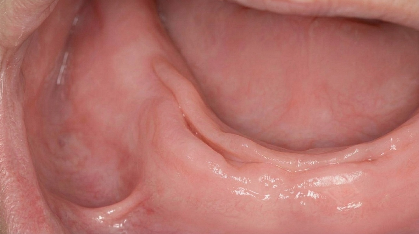
What it looks like
Most lateral periodontal cysts are completely invisible to the patient. Larger lesions, or those close to the gum surface, may produce:
A small, painless swelling at the side of a tooth, sometimes near the gum margin.
A slight bulge in the gum or alveolar ridge.
Slight separation of two adjacent teeth if the cyst has pushed the roots apart.
What it feels like
Most patients have no symptoms at all. When symptoms occur, they are mild and may include:
Mild discomfort if the cyst has grown large enough to press on adjacent structures.
A subtle change in tooth position noticed by the patient or dentist.
Soreness if the overlying gum is bumped during chewing.
Pain or swelling if the cyst becomes secondarily infected (uncommon).
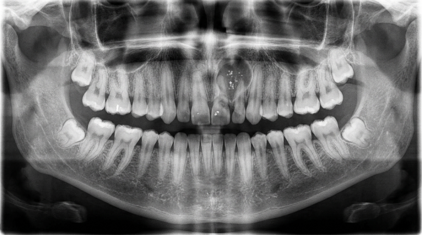
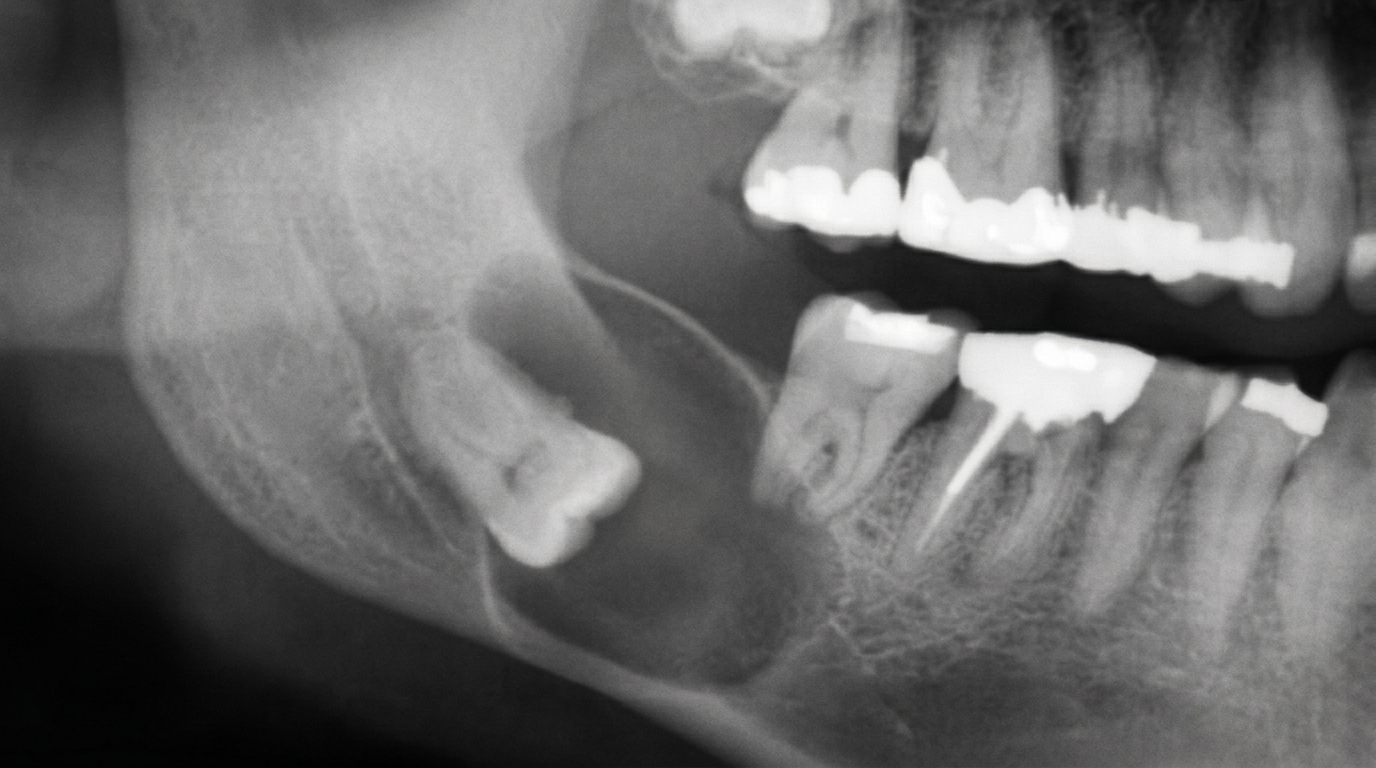
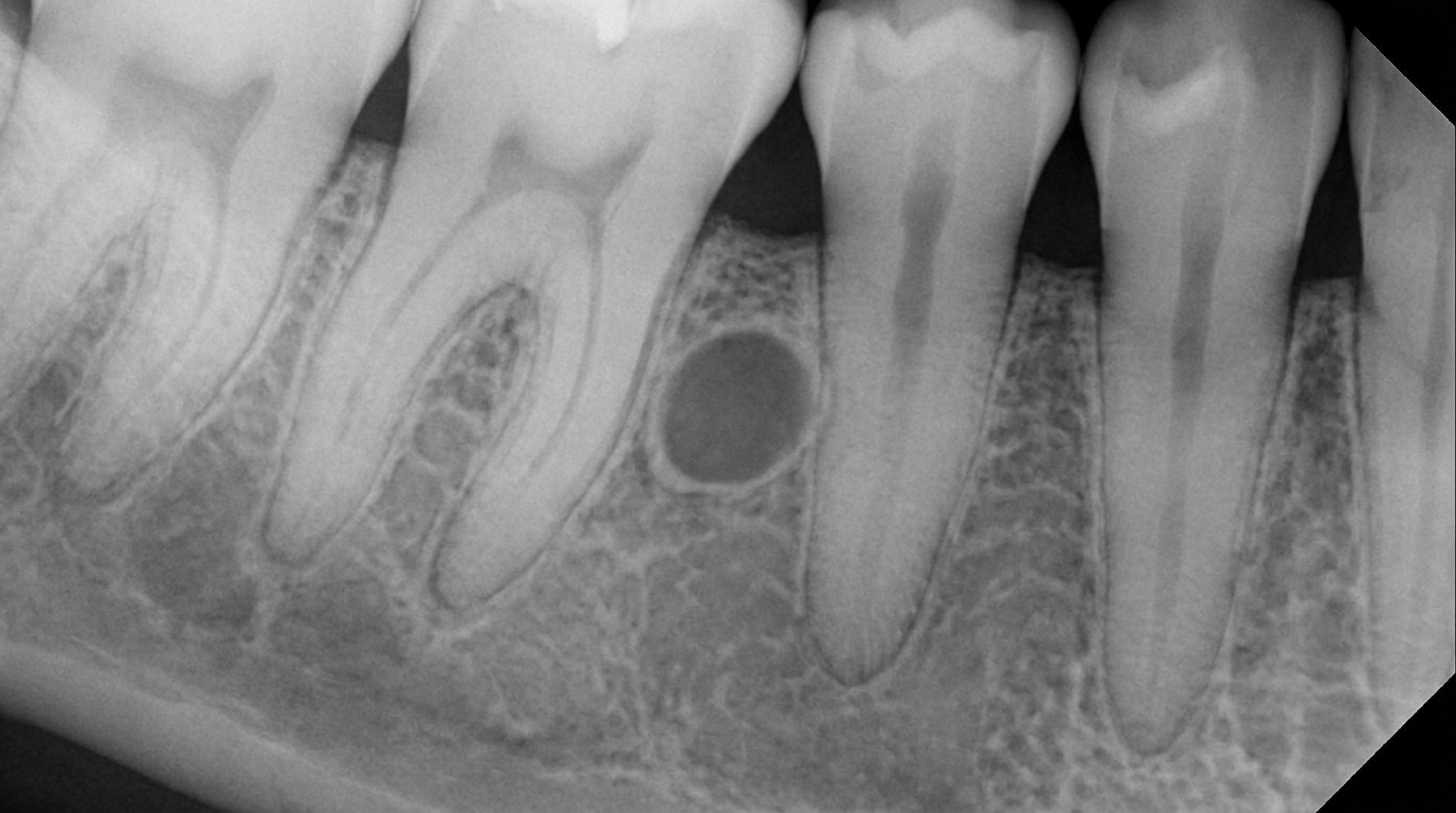
What an X-ray might show
The diagnosis is usually first made on a routine X-ray. Typical findings:
A well-circumscribed, often round or oval radiolucent (darker on X-ray, indicating soft tissue or fluid rather than bone) area beside the side of a vital tooth, with a clear bony border.
The lesion is most often less than 1 cm in greatest diameter.
No association with caries or pulp death in the adjacent tooth.
Vital pulp testing of the adjacent tooth confirms that the tooth is alive, a key clue that distinguishes this from a lateral radicular cyst.
The botryoid variant can show a multilocular (multi-chambered) appearance that resembles a small bunch of grapes on imaging.
What happens at the dentist?
A lateral periodontal cyst is most often picked up at a routine dental check-up and clean at ArtSmiles, often on a panoramic or periapical X-ray. The dentist will typically:
Examine the area for any swelling, bulging or tooth movement.
Check the vitality of the adjacent teeth with cold and electric pulp testing, a critical step, since a vital tooth strongly suggests a developmental cyst rather than an inflammatory one.
Take additional X-rays, including a small-volume cone-beam CT (CBCT) scan in selected cases, to clarify the size and exact relation of the cyst to the roots.
Discuss biopsy and removal, since the imaging features alone are not specific and a definitive diagnosis depends on histopathology.
Refer to an oral and maxillofacial surgeon for the surgical removal, particularly when the cyst sits close to the roots of teeth that should be preserved.
Is this serious?
🟢 A lateral periodontal cyst is benign. It is not cancer, does not spread, and is not contagious. The botryoid variant has a higher tendency to recur after enucleation (a minor surgical procedure where the cyst is removed cleanly from its bony cavity), but even that is not life-threatening. Most patients, once the cyst has been removed and confirmed on histopathology, can expect a very good long-term outcome and can usually keep the adjacent tooth.
If a routine X-ray has shown a small shadow between the roots of healthy-looking teeth, it is worth booking an assessment so the right imaging and biopsy can be planned.
Could it be something else?
Several other conditions can produce a similar X-ray appearance. The textbooks list these as the main differentials:
Lateral radicular cyst, looks similar but arises beside a non-vital tooth as a result of pulp death and inflammation. Vitality testing distinguishes the two.
Odontogenic keratocyst (OKC), also occurs between the roots of teeth and can look identical on a 2D radiograph. Has a higher recurrence rate; histopathology is essential.
Gingival cyst of the adult, the soft-tissue counterpart of a lateral periodontal cyst, sitting in the gum rather than the bone.
Glandular odontogenic cyst, a rare but more aggressive cyst that can show a multilocular appearance, mimicking the botryoid variant.
Calcifying odontogenic cyst (Gorlin cyst), can also occur in this region; usually shows mixed radiolucent and radiopaque areas.
Small ameloblastoma, can sometimes resemble a small cyst on imaging, especially when unilocular.
How is it treated?
Treatment is generally surgical, but conservative.
At-home measures and habits:
Maintain excellent oral hygiene so any pre-treatment review is straightforward and to reduce the risk of secondary infection.
Attend regular check-ups and X-rays so any new lesion or change can be picked up early.
Continue normal eating and brushing, there is generally no need to change daily habits while a small lateral periodontal cyst is being assessed.
Professional steps your dentist may consider:
Conservative enucleation of the cyst, with preservation of the adjacent vital tooth wherever possible. The cyst is shelled out of the bone, and the cavity heals with new bone formation.
Histopathological examination of the removed tissue, essential to confirm the diagnosis and rule out a botryoid variant or another lookalike lesion.
Slightly more extensive (but still conservative) excision for botryoid odontogenic cysts, which have a higher chance of recurrence if any small cyst is left behind.
Regular follow-up X-rays at intervals of 1, 2 and sometimes 5 years, particularly after removal of a botryoid variant, to confirm the lesion has not returned.
Endodontic care of the adjacent tooth is generally not needed, since the tooth is vital and the cyst arises independently of any pulp problem.
A patient-centred approach matters here. Many patients are anxious to hear that a "cyst beside a healthy tooth" has been spotted on a routine X-ray. Honest, unhurried discussion of what the lesion likely is, what the surgery involves, and how the tooth can usually be saved is itself part of effective care, values that sit at the heart of our clinical philosophy.
What's the long-term outlook?
The outlook is excellent. Once a lateral periodontal cyst has been enucleated and confirmed on histopathology, recurrence is uncommon. The botryoid variant has a higher recurrence rate, but with careful excision and long-term follow-up the lesion can usually be controlled. The adjacent tooth is generally healthy, requires no special treatment, and remains in place. Most patients return to a fully functional jaw within a few months of surgery and need only routine dental review thereafter.
A note on this article
This article is for educational purposes only and does not constitute a clinical diagnosis. Please consult a registered dental practitioner for assessment and treatment advice.
The cover image above is an AI-generated illustration based on the most common visible features of this condition described in clinical pathology references. It is not a photograph of a real case and should not be used to diagnose or rule out the condition in your own situation. If you are concerned about something you have noticed, please book an assessment with a registered dental practitioner.
References
Cawson, R. A., & Odell, E. W. (2017). Cawson's essentials of oral pathology and oral medicine (8th ed.). Elsevier. Chapter 7, Cysts of the Jaws: Lateral Periodontal Cysts and Botryoid Odontogenic Cysts, with key features in Box 7.14, pp. 129 to 130.
Neville, B. W., Damm, D. D., Allen, C. M., & Chi, A. C. (2023). Oral and maxillofacial pathology (5th ed.). Elsevier. Chapter 15, Odontogenic Cysts and Tumors: Lateral Periodontal Cyst (Botryoid Odontogenic Cyst), pp. 699 to 701.
Regezi, J. A., Sciubba, J. J., & Jordan, R. C. K. (2017). Oral pathology: Clinical pathologic correlations (7th ed.). Elsevier. Chapter 10, Cysts of the Jaws and Neck: Lateral periodontal cyst as differential of cyst-like radiolucencies.
Frequently asked questions
Is a lateral periodontal cyst dangerous?
No. A lateral periodontal cyst is a benign developmental cyst that grows slowly and almost never causes problems beyond its local pressure on the jaw. It is not cancer and does not spread. Surgical removal is mainly to confirm the diagnosis and prevent the cyst from enlarging further.
How is a lateral periodontal cyst found?
Most are picked up by chance on a routine dental X-ray, where they appear as a small, well-defined dark area along the side of a tooth root, usually between the lower canine and premolar. Because they are painless, patients rarely notice them on their own.
How is it treated?
Treatment is a simple surgical excision (enucleation) under local anaesthetic. The cyst is lifted out of the jaw, the wound is closed, and the tissue is sent for microscopic examination to confirm the diagnosis. Most patients recover within a couple of weeks.
Will a lateral periodontal cyst come back after removal?
Recurrence after complete enucleation is uncommon for the classic single lateral periodontal cyst. A multi-chambered variant called the botryoid odontogenic cyst recurs more often, so periodic X-ray follow-up is recommended for that variant.