Compiled from clinical pathology references. Medically reviewed by Dr Cristian Dunker, Principal Dentist, ArtSmiles Cosmetic Dentistry.
Most hereditary tooth disorders affect the enamel, the outer, visible layer. Dentin dysplasia is one of the rare conditions that affects the layer underneath: the dentin (the hard tissue that makes up the bulk of the tooth beneath the enamel). The enamel can look almost normal, but the inner structure of the tooth and its root develops abnormally, which can affect how the tooth functions and how long it survives.
This article from the team at ArtSmiles, reviewed by Dr Cristian Dunker, explains what dentin dysplasia is and how it is managed.
Quick summary
At a glance | Detail |
|---|---|
Also called | Rootless teeth (Type I, radicular); coronal dentin dysplasia (Type II) |
How urgent? | 🟡 Worth careful long-term planning, affected teeth are at risk of early loss, but the condition itself is not dangerous |
Common or rare? | Rare, about 1 in 100,000 in the general population |
Who it affects | People who inherit the gene change (autosomal dominant); both sexes equally |
Who treats it | General dentist working with paediatric dentist, prosthodontist and geneticist; long-term staged care |
Based on | Neville, Cawson, Regezi |
What is it?
Dentin dysplasia is a rare hereditary disorder of dentin formation. The tooth crowns can look normal, but the roots, pulp chambers, or both are malformed. Two main types are described:
Type I (radicular dentin dysplasia), affects the roots. Roots are very short or absent, and the pulp chamber appears as a small crescent on X-ray. Sometimes called "rootless teeth".
Type II (coronal dentin dysplasia), affects the pulp chambers (the soft nerve and blood vessel core of each tooth) and the colour of the baby teeth. Permanent teeth show a thistle-tube shape of the pulp; baby teeth are often brown or amber.
Both types are inherited in an autosomal dominant pattern (meaning each child of an affected parent has a 50% chance of inheriting the condition).
Who tends to get it?
It is uncommon, about 1 in 100,000 in the general population. It affects:
People with a family history of the condition.
Both sexes equally.
Both deciduous (baby) and permanent dentitions, though the pattern differs by type.
What causes it?
It is caused by mutations in genes that control dentin formation, including DSPP (dentin sialophosphoprotein, the gene that codes for a key dentin protein) for Type II. Some cases of Type I have been linked to mutations in VPS4B and other genes.
How does it develop?
The defect is set during tooth development. Dentin is laid down abnormally, producing the characteristic features. Crowns of teeth tend to look normal in Type I; in Type II the baby teeth are discoloured (brown or amber) and look similar to dentinogenesis imperfecta. Once teeth erupt, problems arise from short roots, abnormal pulps and increased risk of pulp infection.
What might you notice?
What it looks like
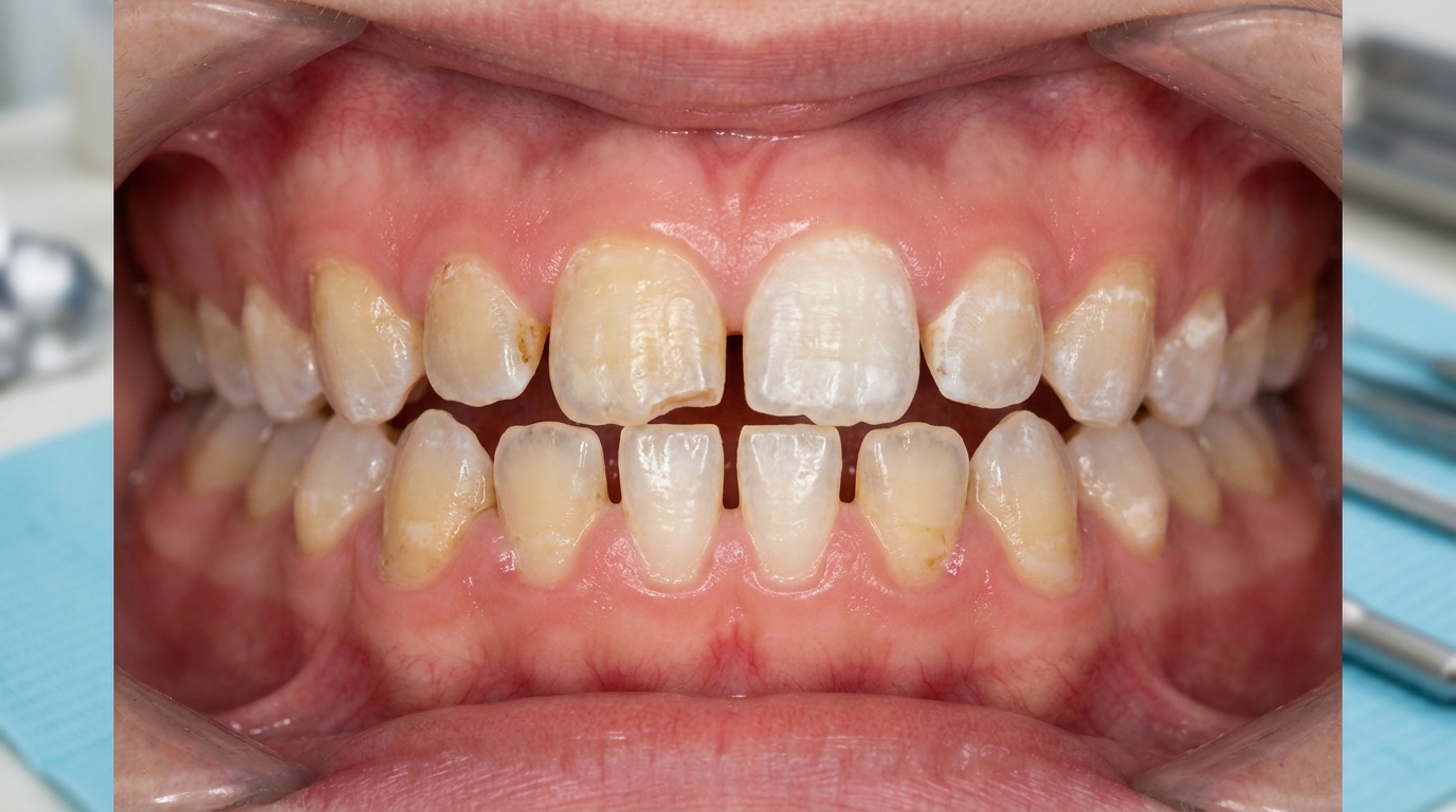
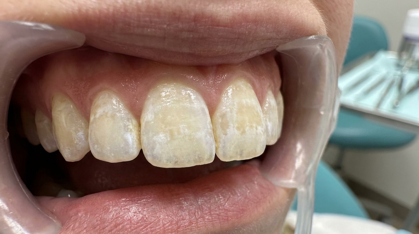
In Type I, the teeth look largely normal externally, the unusual features are inside the bone. In Type II, the deciduous teeth often appear brown or amber, sometimes with a translucent bluish tint, while the permanent teeth look largely normal.
What it feels like
In Type I:
Teeth that look normal on the outside but appear "floating" with little or no root on X-ray.
Loose teeth appearing in childhood or early adulthood without obvious gum disease.
Teeth lost early with little warning.
Cysts and abscesses at the tip of the (very short) roots, sometimes in healthy-looking teeth.
In Type II:
Brown or amber baby teeth that may look bluish under certain lighting.
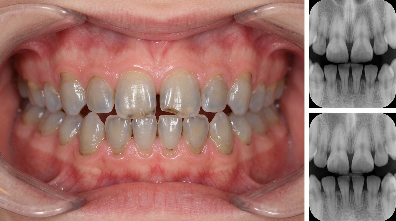
Adult teeth that look largely normal externally but show characteristic pulp shapes on X-ray.
Pulp stones and obliteration (gradual filling-in of the pulp chamber with extra dentin) of pulp chambers over time.
Less likely than Type I to result in early tooth loss.
What an X-ray might show
Dental X-rays are the key to diagnosis. In Type I, short or absent roots with small crescent-shaped pulp remnants. In Type II, thistle-tube shaped pulp chambers in adult teeth, often with pulp stones. Periapical (root-tip) radiolucencies may be present in either type.
What happens at the dentist?
When dentin dysplasia is suspected at ArtSmiles, the visit usually involves:
Detailed history including family history of similar dental issues.
Clinical examination of crowns, gum health and tooth mobility.
Comprehensive X-rays, periapical and panoramic, to see roots, pulps and any cysts.
Comparison with siblings or parents if family members can attend.
Referral to specialists as needed, paediatric dentist, prosthodontist, geneticist.
Long-term care plan with frequent reviews.
Is this serious?
🟡 In itself it is not life-threatening, but the dental consequences can be significant. Type I in particular can lead to early tooth loss in adolescence or early adulthood. Long-term planning, sometimes including dental implants supported by bone grafts, may be required.
Could it be something else?
Conditions that can mimic dentin dysplasia include:
Dentinogenesis imperfecta, also affects dentin but usually with more obvious crown discolouration and bulbous crowns.
Amelogenesis imperfecta, primarily affects enamel.
Vitamin D-dependent rickets, produces enlarged pulp chambers and similar pulp pathology.
Hypophosphatasia, affects the enamel-cementum junction and tooth attachment.
Trauma or chronic periodontal disease with secondary root loss.
A careful look at the X-rays, family history and pattern usually distinguishes them.
How is it treated?
There is no cure. Management focuses on:
Excellent oral hygiene to prevent decay and periodontal disease.
Frequent professional cleaning and review every three to four months.
Conservative restoration of any decay or wear.
Endodontic care can be challenging because pulp chambers may be abnormal.
Prosthodontic planning for teeth that are lost early, full-mouth oral rehabilitation combining implants, bridges and dentures gives durable, attractive results in adulthood.
Genetic counselling for affected families.
What's the long-term outlook?
The outlook is more guarded than for many dental conditions, but careful, planned care makes a significant difference. Some patients keep their natural teeth into adulthood; others need implant-supported solutions earlier. Early diagnosis and long-term partnership with the dental team are the key to the best outcome.
A note on this article
This article is for educational purposes only and does not constitute a clinical diagnosis. Please consult a registered dental practitioner for assessment and treatment advice.
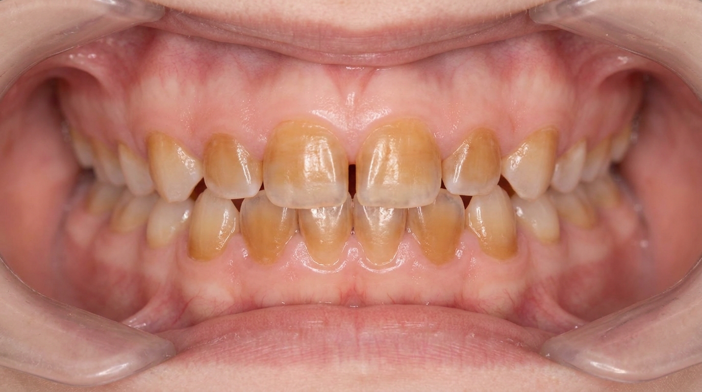
The cover image above is an AI-generated illustration based on the most common visible features of this condition described in clinical pathology references. It is not a photograph of a real case and should not be used to diagnose or rule out the condition in your own situation. If you are concerned about something you have noticed, please book an assessment with a registered dental practitioner.
References
Neville, B. W., Damm, D. D., Allen, C. M., & Chi, A. C. (2023). Oral and maxillofacial pathology (5th ed.). Elsevier. Chapter 2, Abnormalities of Teeth: Dentin Dysplasia.
Cawson, R. A., & Odell, E. W. (2017). Cawson's essentials of oral pathology and oral medicine (8th ed.). Elsevier. Chapter 2, Disorders of Development.
Regezi, J. A., Sciubba, J. J., & Jordan, R. C. K. (2017). Oral pathology: Clinical pathologic correlations (7th ed.). Elsevier. Chapter 16, Abnormalities of Teeth.
Frequently asked questions
What is dentin dysplasia?
Dentin dysplasia is a rare inherited disorder where the inner layer of the tooth (dentin) develops abnormally while the outer enamel looks fairly normal. There are two main types: Type I (radicular) where the roots are short, blunt or missing; and Type II (coronal) where the crowns of the baby teeth look amber and the adult teeth have unusual pulp shapes on x-ray.
How is dentin dysplasia diagnosed?
Diagnosis is mostly radiographic. X-rays show short, blunted or missing roots, obliterated pulp chambers (filled with extra abnormal dentin) and sometimes small periapical radiolucencies on teeth without obvious decay or trauma. Family history confirms inheritance. Genetic testing is available for confirmation.
What problems does dentin dysplasia cause?
Type I teeth tend to be mobile because of the short roots, may shed early in childhood and are very challenging to restore with implants or fixed work because of the abnormal root structure. Type II usually has fewer functional problems but the baby teeth can look amber and translucent. Periapical infection can develop without obvious cause.
How is dentin dysplasia treated?
Treatment is conservative and lifelong. Goals are to preserve teeth, manage early periapical lesions, replace lost teeth with carefully designed prostheses (often partial dentures or implants placed away from compromised areas), and provide cosmetic restoration of discoloured teeth. A multidisciplinary team and meticulous oral hygiene are essential.