Compiled from clinical pathology references. Medically reviewed by Dr Cristian Dunker, Principal Dentist, ArtSmiles Cosmetic Dentistry.
Most lumps in the mouth are painless. A small, firm lump that has appeared after a tooth was removed, after dental surgery, or after a knock, and that hurts when pressed, is unusual enough to warrant a careful look. The most common cause in this scenario is a traumatic neuroma.
This article from the team at ArtSmiles, reviewed by Dr Cristian Dunker, explains what a traumatic neuroma is and how it is treated.
Quick summary
At a glance | Detail |
|---|---|
Also called | Amputation neuroma; post-traumatic neuroma |
How urgent? | 🟡 Worth a check-up, the lump is benign but painful and is easily confirmed and removed |
Common or rare? | Uncommon overall, but a recognised cause of a painful lump after extractions or oral surgery |
Who it affects | Adults of any age; most often after lower premolar extractions, wisdom tooth removal, or denture wear |
Who treats it | General dentist; oral and maxillofacial surgeon for lesions near the mental nerve (the small sensory nerve that supplies the chin and lower lip) |
Based on | Neville, Cawson, Regezi |
What is it?
A traumatic neuroma is not a true tumour. It is a reactive proliferation of nerve fibres (a disorganised regrowth of nerve tissue) that occurs when a peripheral nerve (a nerve outside the brain and spinal cord) has been cut or crushed and the fibres on the proximal side (the part still connected to the brain or spinal cord) regrow without successfully meeting the distal end (the cut-off end further from the brain). The regrowing nerve fibres, with their supporting Schwann cells (the insulating cells around peripheral nerves) and connective tissue, form a small tangled nodule.
In the mouth, common features include:
A small (usually under 2 cm) firm nodule in soft tissue.
Tenderness on touch, a hallmark feature.
Pain that may radiate along the path of the affected nerve.
Stable size over months to years.
A close link with a previous extraction, surgery or injury.
Who tends to get it?
Traumatic neuromas can affect anyone who has had a nerve injured, but in dentistry are most often seen in:
Patients after lower premolar extractions (mental nerve area).
Patients after extraction of impacted lower wisdom teeth (inferior alveolar nerve, the main sensory nerve of the lower jaw and lip).
Patients with a history of intraoral trauma, bites, lacerations, surgery.
Patients with poorly fitting dentures that have caused chronic minor injury to a nerve.
Older adults, in whom dental and surgical procedures are more frequent.
What causes it?
Whenever a peripheral nerve is divided, by surgery, trauma or pressure, the nerve attempts to regenerate. If the cut ends are close enough and aligned, the nerve usually heals well. If the cut ends are separated, scar tissue intervenes, or the distal end is no longer present (as after an extraction), the regrowing nerve fibres form a disorganised tangle. This tangle is the traumatic neuroma.
How does it develop?
The course is gradual:
The nerve is cut or damaged.
Over weeks to months, the nerve fibres begin to regrow.
Without a clear path to the distal end, fibres curl into a small ball within the surrounding scar tissue.
The patient gradually notices a tender lump.
Once formed, the neuroma stays roughly the same size, often for years.
What might you notice?
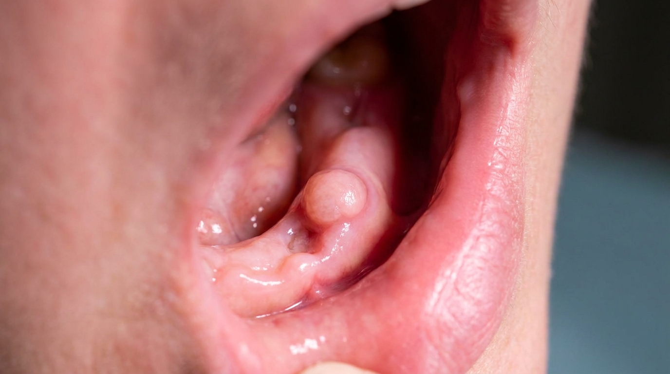
What it looks like
A small, smooth, firm, pink or flesh-coloured bump in the gum tissue, most often near the site of a previous extraction or surgery. There may be a faint scar at the gum surface from the original procedure. The lump is usually under 2 cm and stays roughly the same size over time.
What it feels like
A small firm lump at the site of a previous extraction or surgery.
Sharp pain when the lump is touched, brushed or knocked.
Pain that shoots along the path of the nerve.
Numbness or pins-and-needles in the area supplied by the nerve.
Discomfort with denture wear if the neuroma sits under a denture flange.
A history of dental work or trauma in the same area.
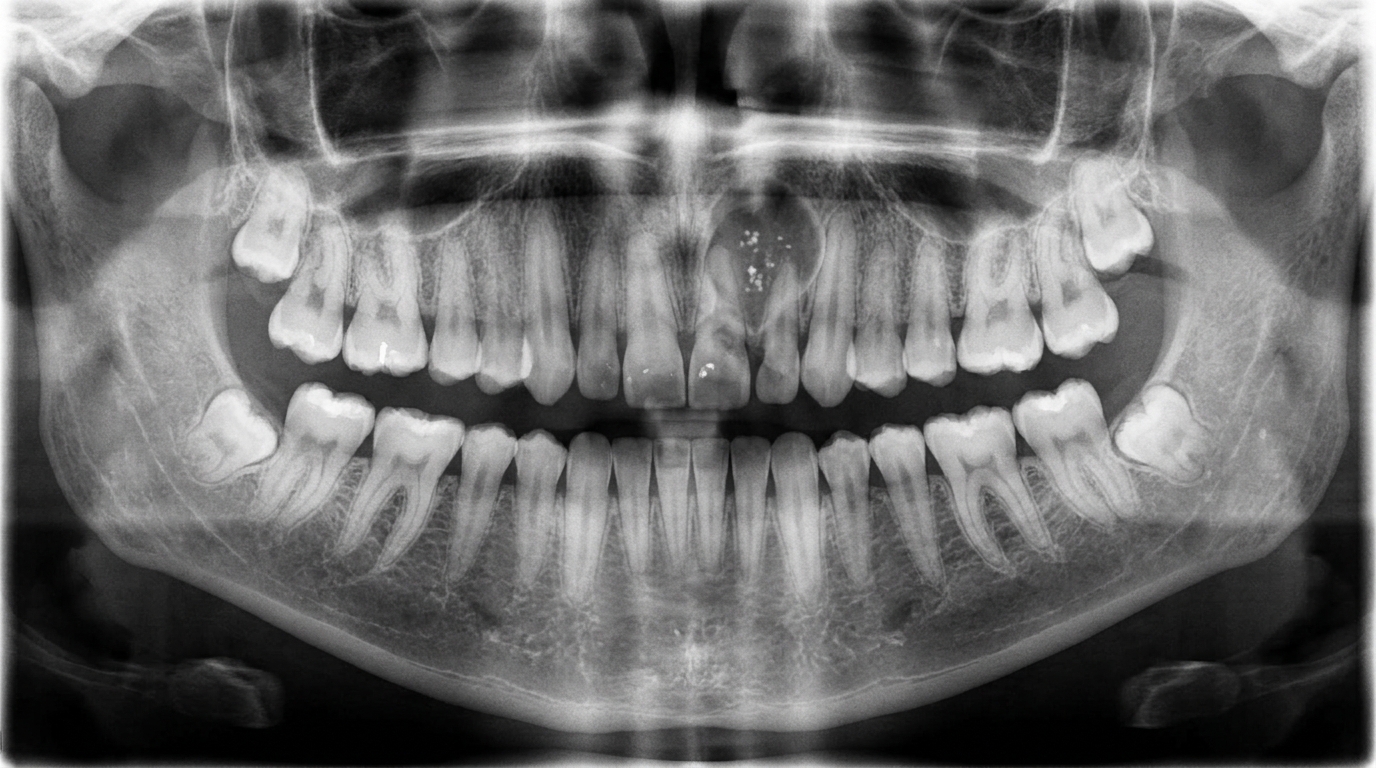
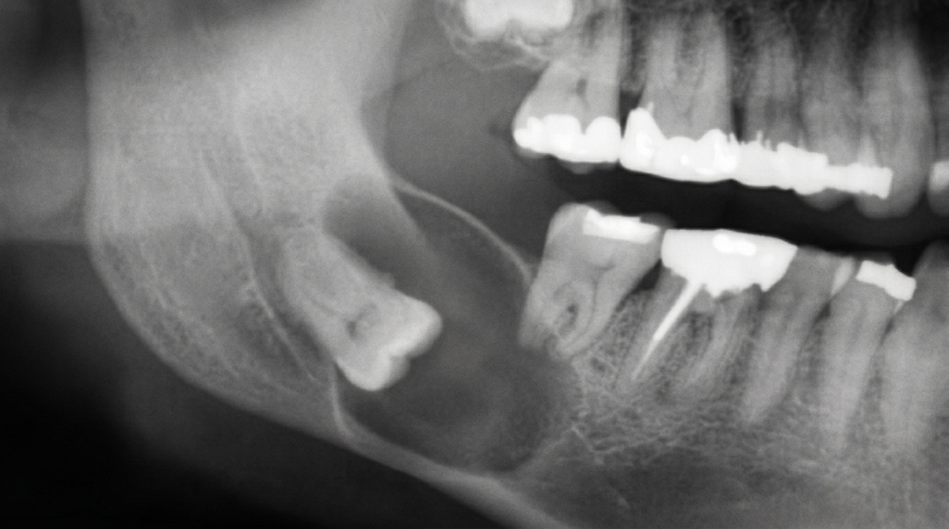
What an X-ray might show
A traumatic neuroma is a soft-tissue lesion, so dental X-rays usually show no abnormality. The role of imaging is to confirm there is no underlying bone problem (for example, a retained root tip near the site) that could explain the symptoms.
What happens at the dentist?
When a traumatic neuroma is suspected at ArtSmiles, the visit usually involves:
A history conversation about previous dental work, surgery or trauma.
A careful examination with attention to pain on touch and any numbness pattern.
A test for tenderness, gentle pressure on the lump often reproduces the symptoms.
An X-ray to exclude any underlying bone pathology.
A treatment plan, usually surgical excision with histopathological confirmation (examination of the removed tissue under the microscope).
Coordination with an oral surgeon for cases close to a major nerve such as the mental nerve.
Is this serious?
🟡 Traumatic neuromas are benign, they are not cancer and do not spread. The reasons they still deserve attention are:
Pain that affects eating, talking or sleeping.
Confirmation of diagnosis, several other lumps need to be excluded.
Improving denture or other prosthesis comfort when the lump interferes.
Could it be something else?
Other lumps that may be confused with a traumatic neuroma include:
Schwannoma and neurofibroma, true benign nerve tumours, often painless.
Granular cell tumour, usually painless, on the dorsum of the tongue.
Traumatic fibroma, firm, painless lump from chronic minor trauma.
Mucocele, soft, bluish, painless cyst.
Salivary gland tumour, usually painless and at typical sites.
Recurrent infection or abscess, tender but with redness, warmth and possibly pus.
Histopathological examination of the excised tissue gives the definitive diagnosis.
How is it treated?
The standard treatment is surgical excision under local anaesthetic. The lump is removed along with a small section of the affected nerve, allowing the cut end to retract into healthier tissue. The specimen is sent for histopathology to confirm the diagnosis.
Adjuncts in selected cases include:
Local anaesthetic injections for diagnosis and short-term pain relief before surgery.
Topical or systemic medicines (gabapentin, amitriptyline) for ongoing nerve pain in patients where surgery is not appropriate.
Adjustment of dentures to relieve pressure on the lump.
What's the long-term outlook?
The outlook is good. After excision, most patients have lasting relief, and the wound heals without significant scarring. Recurrence is uncommon. Some patients have a small area of permanent numbness in the affected nerve's territory, which is usually well tolerated.
If you have a small painful lump in your mouth, particularly at the site of a previous extraction or surgery, please book a visit. We can confirm the diagnosis and arrange the right treatment.
A note on this article
This article is for educational purposes only and does not constitute a clinical diagnosis. Please consult a registered dental practitioner for assessment and treatment advice.
The cover image above is an AI-generated illustration based on the most common visible features of this condition described in clinical pathology references. It is not a photograph of a real case and should not be used to diagnose or rule out the condition in your own situation. If you are concerned about something you have noticed, please book an assessment with a registered dental practitioner.
References
Neville, B. W., Damm, D. D., Allen, C. M., & Chi, A. C. (2023). Oral and maxillofacial pathology (5th ed.). Elsevier. Chapter 12, Soft Tissue Tumors: Traumatic Neuroma.
Cawson, R. A., & Odell, E. W. (2017). Cawson's essentials of oral pathology and oral medicine (8th ed.). Elsevier. Chapter 13, Reactive Lesions.
Regezi, J. A., Sciubba, J. J., & Jordan, R. C. K. (2017). Oral pathology: Clinical pathologic correlations (7th ed.). Elsevier. Chapter 5, Connective Tissue Lesions.