Compiled from clinical pathology references. Medically reviewed by Dr Cristian Dunker , Principal Dentist, ArtSmiles Cosmetic Dentistry.
Quick summary
Also called | Pink tooth of Mummery, internal resorption, pink spot |
How urgent? | 🟡 Worth checking soon, usually painless but progressive; early root canal treatment can often save the tooth |
Common or rare? | Relatively rare, far less common than external resorption |
Who it affects | Adults of any age, often after dental trauma, deep decay, orthodontic or periodontal treatment, or with no identifiable trigger |
Who treats it | General dentist, often in coordination with an endodontist (a dentist who specialises in root canal treatment) when the lesion is advanced |
Based on | Cawson, Neville, Regezi |
What is it?
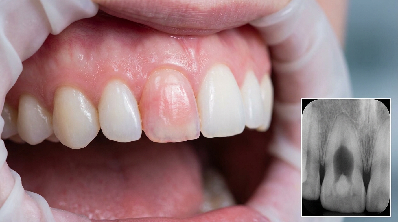
Internal tooth resorption is a condition in which the dentine (the softer layer under the enamel) of a tooth is gradually eaten away from the inside of the pulp chamber (the hollow space at the centre of the tooth that holds the nerve) or root canal. Specialised cells called osteoclasts (cells that naturally break down hard tissue, called "dentinoclasts" when working inside a tooth) become active inside the pulp and start dissolving the surrounding hard tissue. Over time, the wall of the pulp chamber becomes thinner and thinner, until the vascular pulp (the blood-vessel-rich tissue inside the tooth) can be seen through the remaining enamel as a characteristic pink shadow, the classic "pink tooth of Mummery" first described in the nineteenth century. If the process continues, the wall eventually perforates (breaks through) and the tooth becomes very difficult to save.
Who tends to get it?
Internal resorption is uncommon. Most cases the textbooks describe occur in adults, often with a history of one of the recognised triggers:
A previous knock or blow to the tooth (luxation injury, a knock that has loosened or shifted the tooth, including a sporting injury or fall), even when the tooth was not knocked out at the time.
A previous deep filling or caries that came close to the pulp, with subsequent low-grade pulpitis (inflammation of the tooth nerve).
Recent orthodontic or periodontal treatment of the affected tooth.
A history of internal bleaching (using bleaching agents inside a previously root-canal-treated tooth), a recognised, though uncommon, trigger.
No identifiable cause at all, these idiopathic cases sometimes affect more than one tooth.
There is no strong sex or ethnic predilection. Front teeth (incisors) are particularly likely to be noticed because the pink discoloration is visible.
What causes it?
Internal resorption needs vital pulp tissue (living nerve and blood-vessel tissue inside the tooth) to keep going. Once the pulp dies completely, the resorbing cells lose their support and the process stops. The textbooks list these contributing factors:
Trauma to the tooth that has damaged or partially injured the pulp.
Chronic pulpitis from deep caries, deep restorations or repeated dental procedures.
Internal bleaching of a previously root-canal-treated tooth, particularly with hydrogen peroxide-based agents that may leak through dentinal tubules (microscopic channels running through the dentine).
Orthodontic forces.
Periodontal treatment that has affected the pulp's blood supply.
Idiopathic cases, no obvious trigger identified.
A current understanding is that some form of pulp inflammation switches on the resorbing cells inside the pulp chamber. As long as the pulp keeps a partial blood supply, the resorption continues. If the inflammation completely tips the pulp into death, the process can self-arrest.
How does it develop?
The textbooks describe two main patterns of internal resorption:
Inflammatory internal resorption. The resorbed dentine is replaced by inflamed granulation tissue (fragile new tissue full of small blood vessels). This pattern can affect any part of the canal, but most commonly the cervical zone (the part of the canal closest to the gum line). The inflammation is usually driven by bacteria reaching the deeper pulp through dentinal tubules. The coronal (top) part of the pulp may have already died while the apical (deepest) part remains alive. On X-ray, the area of destruction looks like a uniform, well-defined, balloon-like enlargement of the pulp chamber or canal.
Replacement (metaplastic) internal resorption. Portions of the pulpal dentine wall are resorbed and replaced by bone or cementum-like tissue (tissue resembling the thin layer that normally covers the tooth root). On X-ray, the canal looks enlarged but partially obliterated by a hazier material that is less dense than normal dentine. The outline is less crisp than in inflammatory resorption.
When the resorption affects the crown of an upper front tooth, the vascular tissue inside the pulp eventually becomes visible through the thinning enamel as a pink area, the pink tooth of Mummery. If left long enough, the resorption can perforate through the side of the tooth into the periodontal ligament, at which point the lesion essentially becomes a combined internal-external defect and the tooth is much harder to save.
What might you notice?
What it looks like
In the early stages, internal resorption is usually invisible. The classic appearances, when they occur, are:
A pink shadow showing through the enamel of an upper or lower front tooth, the hallmark "pink tooth of Mummery". This is most easily seen against a white wall or in good daylight.
A slight pink or grey discoloration of the crown that the patient may notice in the mirror.
A darker shade to a tooth that previously matched its neighbour.
Less commonly, a visible chip if the lesion has eroded close to the surface of the enamel.
In back teeth (premolars and molars), there is rarely any visible change at all, internal resorption there is almost always picked up only on an X-ray.
What it feels like
Most people feel nothing. The textbooks consistently note that internal resorption is asymptomatic in early stages and that pain only develops if there is significant pulpal inflammation, an established infection, or a perforation. When symptoms do occur they may include:
A dull ache or low-grade discomfort, particularly when biting.
Sensitivity to cold or sweet foods if dentinal tubules are involved.
Sharper pain or swelling if the pulp has died and an infection has developed at the root tip.
What an X-ray might show
The diagnosis is usually made on a periapical or bite-wing X-ray (an X-ray showing the top half of upper and lower teeth in one image). Typical findings:
A balloon-like expansion of the pulp chamber or canal, uniform, well-defined and rounded.
A loss of the original outline of the canal at the affected level.
The radiolucency (a darker area on the X-ray) is centred on the canal itself, which is characteristically enlarged. (This contrasts with external resorption, where the canal stays its normal size and the radiolucency is superimposed on top of it.)
For complex or borderline cases, a small-volume cone-beam CT (CBCT) scan provides a 3D view that often clarifies whether resorption is internal, external, or has perforated.
What happens at the dentist?
Internal resorption is usually picked up at a routine dental check-up and clean at ArtSmiles, often on a routine X-ray rather than because of any complaint. The dentist will typically:
Examine the suspected tooth for any pink shadow, discoloration, or change in shade.
Review the dental and trauma history, including past hits to the tooth, deep fillings, recent orthodontics or bleaching.
Take periapical X-rays to map the lesion. A CBCT scan is often the most useful next step where the picture is unclear.
Test the pulp with cold and electric pulp testing (a small electric stimulus used to check whether the nerve still responds), many internally resorbing teeth still respond, though responses can be variable.
Distinguish internal from external resorption carefully, since the treatment differs.
Refer to an endodontist in complex cases, or where the resorption appears to have perforated.
Is this serious?
🟡 Internal resorption itself is not life-threatening, but it is progressive in most cases and tends to silently destroy the tooth from within. Caught early, while the pulp wall is still intact, root canal treatment usually stops the process and the tooth can be retained for many years. Caught late, after the resorption has reached the root surface or perforated through the side of the tooth, the prognosis becomes much poorer and extraction may be needed.
If you have noticed a pink shadow on a front tooth, a sudden change in tooth colour, or have been told an X-ray shows an unusual hollow inside one of your teeth, it is worth booking an assessment so the cause can be identified and the right next step recommended.
Could it be something else?
Several conditions can mimic internal resorption on X-ray or clinical examination. The textbooks list these as the main differentials:
External cervical resorption, looks similar at first glance but the resorption begins on the outside of the root, with the canal staying its normal size. CBCT imaging is often needed to tell them apart.
Deep dental caries with pulpal involvement, can produce a radiolucent area near the pulp, but caries usually starts at a chewing surface or contact point and is visible clinically.
Periapical inflammatory (at the root tip) disease, produces a radiolucency at the root tip, but it is centred outside the tooth and the pulp itself is dead and unresponsive on testing.
Calcific metamorphosis (where the pulp narrows and hardens rather than dissolving), a different post-trauma response, where the pulp narrows and calcifies rather than resorbing. The canal becomes thinner, not wider.
Dens invaginatus (a tooth that formed with an inward fold of its enamel) or other developmental defects, can produce unusual radiographic appearances inside the tooth, but they are present from the time the tooth erupts and have characteristic shapes.
How is it treated?
Once internal resorption has been confirmed, the textbooks all agree on the same general principle: stop the process by removing the vital pulp tissue. Specific options depend on the stage:
At-home measures and habits:
Avoid heavy biting forces on the affected tooth where possible until the lesion has been treated.
Maintain excellent oral hygiene to limit any contribution from periodontal inflammation.
Keep up regular check-ups so any change can be tracked over time.
Professional steps your dentist may consider:
Root canal treatment as the standard treatment. By cleaning and sealing the canal, the resorbing cells are removed and the process is halted. The textbooks particularly stress catching internal resorption before the pulp chamber is widely exposed, earlier treatment is much more predictable.
Specialist endodontic management for complex cases, for example, where the resorption defect is irregular or the canal anatomy unusual. Modern endodontic techniques use bioceramic materials (ceramic-based dental repair materials that bond to tooth structure) such as mineral trioxide aggregate (MTA) to repair perforations from the inside.
Surgical access and repair if the lesion has perforated to the outer surface of the root, particularly in front teeth where access is straightforward.
Internal bleaching may be appropriate to address residual pink discoloration after successful root canal treatment, although care is needed because aggressive bleaching agents can themselves trigger further resorption.
Extraction as a last resort when the tooth is unrestorable, with replacement options such as an implant or bridge.
Regular follow-up with X-rays to confirm the lesion has stabilised and the tooth remains healthy.
A patient-centred approach matters in a condition where the underlying cells are part of the body itself. Honest discussion of expected outcomes, particularly the difference between catching the lesion early and late, is itself part of effective care, values that sit at the heart of our clinical philosophy.
What's the long-term outlook?
The outlook depends almost entirely on stage at diagnosis. Lesions caught early, while the pulp wall is intact and the perforation has not occurred, can be successfully treated with root canal therapy and the tooth can serve for many years afterwards. Lesions caught late, after perforation, with extensive crown destruction, or with secondary infection, have a much poorer prognosis and often require extraction. In a small proportion of cases the process spontaneously arrests when the pulp dies completely, but this is unpredictable. Regular check-ups remain the single most important factor in catching internal resorption while it is still treatable.
A note on this article
This article is for educational purposes only and does not constitute a clinical diagnosis. Please consult a registered dental practitioner for assessment and treatment advice.
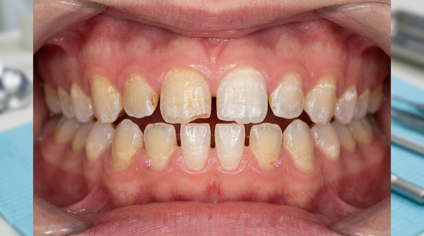
The cover image above is an AI-generated illustration based on the most common visible features of this condition described in clinical pathology references. It is not a photograph of a real case and should not be used to diagnose or rule out the condition in your own situation. If you are concerned about something you have noticed, please book an assessment with a registered dental practitioner.
References
Cawson, R. A., & Odell, E. W. (2017). Cawson's essentials of oral pathology and oral medicine (8th ed.). Elsevier. Chapter 4, Pulpitis, Apical Periodontitis, Resorption and Hypercementosis: Idiopathic internal resorption (pink spot), pp. 69 to 71.
Neville, B. W., Damm, D. D., Allen, C. M., & Chi, A. C. (2023). Oral and maxillofacial pathology (5th ed.). Elsevier. Chapter 2, Abnormalities of Teeth: Internal Resorption with inflammatory and replacement/metaplastic patterns, including pink tooth of Mummery, pp. 62 to 65.
Regezi, J. A., Sciubba, J. J., & Jordan, R. C. K. (2017). Oral pathology: Clinical pathologic correlations (7th ed.). Elsevier. Chapter 16, Abnormalities of Teeth: Internal Resorption, with pulp injury triggers and treatment options, pp. 386 to 388.