Compiled from clinical pathology references. Medically reviewed by Dr Cristian Dunker, Principal Dentist, ArtSmiles Cosmetic Dentistry.
Quick summary
At a glance | Detail |
|---|---|
Also called | Hereditary opalescent dentine; Capdepont's teeth |
How urgent? | 🟡 Worth careful long-term planning, affected teeth wear quickly and benefit from early restoration |
Common or rare? | Rare, about 1 in 8,000 in some populations of European ancestry |
Who it affects | People who inherit the relevant gene change in DSPP (and sometimes COL1A1 or COL1A2 in osteogenesis imperfecta); both deciduous and permanent teeth involved |
Who treats it | General dentist working with a paediatric dentist, prosthodontist or restorative specialist; long-term staged care |
Based on | Neville, with cross-references in Cawson and Regezi |
What is it?
Dentinogenesis imperfecta (DGI) is a group of inherited conditions in which the dentine (the hard tissue that makes up the bulk of the tooth beneath the enamel) does not form properly. Because dentine supports the overlying enamel and protects the pulp (the soft nerve and blood vessel core of the tooth) beneath it, defects in dentine produce teeth that are translucent, grey-blue or amber-brown in colour, with enamel that chips easily and underlying dentine that wears rapidly. The textbooks describe DGI as occurring either as an isolated dental disorder or as part of osteogenesis imperfecta (a wider connective tissue disease that causes brittle bones and other features). With careful staged dental care, most affected people achieve a healthy, comfortable and good-looking dentition.
Who tends to get it?
The textbooks describe a fairly recognisable profile:
People who inherit the DSPP gene mutation (autosomal dominant pattern, meaning each child of an affected parent has a 50% chance of inheriting the condition) for isolated dentinogenesis imperfecta.
People with osteogenesis imperfecta caused by COL1A1 or COL1A2 mutations, many of whom develop DGI-like teeth as part of the wider syndrome.
Both sexes affected equally.
Often a positive family history, siblings and parents may have similar features.
Both deciduous (baby) and permanent teeth affected, with the deciduous teeth typically more severely affected than the permanent dentition.
The textbooks note a historical clustering of cases in communities close to the English Channel, with an estimated prevalence of about 1 in 8,000 in US whites, although the condition occurs in all populations.
What causes it?
The textbooks describe two main genetic groups:
Isolated dentinogenesis imperfecta, caused by mutations in the DSPP (dentine sialophosphoprotein) gene. Inherited as an autosomal dominant trait, with each child of an affected parent having a 50% chance of inheriting the condition.
Dentinogenesis imperfecta as part of osteogenesis imperfecta, caused by mutations in COL1A1 or COL1A2 (the genes for type I collagen, the main organic component of dentine and bone). These patients also have brittle bones, blue sclerae (a bluish tint to the whites of the eyes), hearing loss and other features.
The DSPP gene encodes a protein essential for normal dentine mineralisation. Different mutations within different parts of the gene produce a spectrum of severity, and the textbooks now describe three grades (mild, moderate, severe) of DGI rather than the older Roman-numeral classification.
There is no environmental cause. Diet, fluoride, infection and trauma do not cause the condition.
How does it develop?
In dentinogenesis imperfecta, the layer of dentine immediately beneath the enamel forms relatively normally, but the rest of the dentine is laid down with short, misshapen tubules in an atypical granular matrix. This abnormal dentine cannot adequately support the overlying enamel, which fractures and separates from the dentine after the tooth comes through. Once exposed, the abnormal dentine wears rapidly. At the same time, the pulp chambers, which start out unusually large in many cases, gradually fill with extra dentine, eventually becoming completely obliterated on X-ray. This obliteration of the pulp creates problems for any future root canal treatment, since the canals can be hard to find and treat.
What might you notice?
What it looks like
The textbooks describe a fairly distinctive clinical picture:
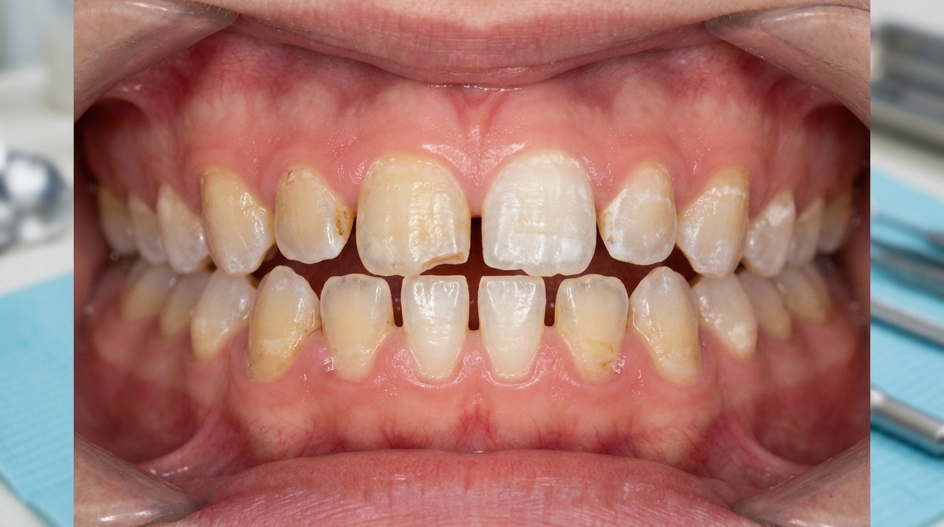
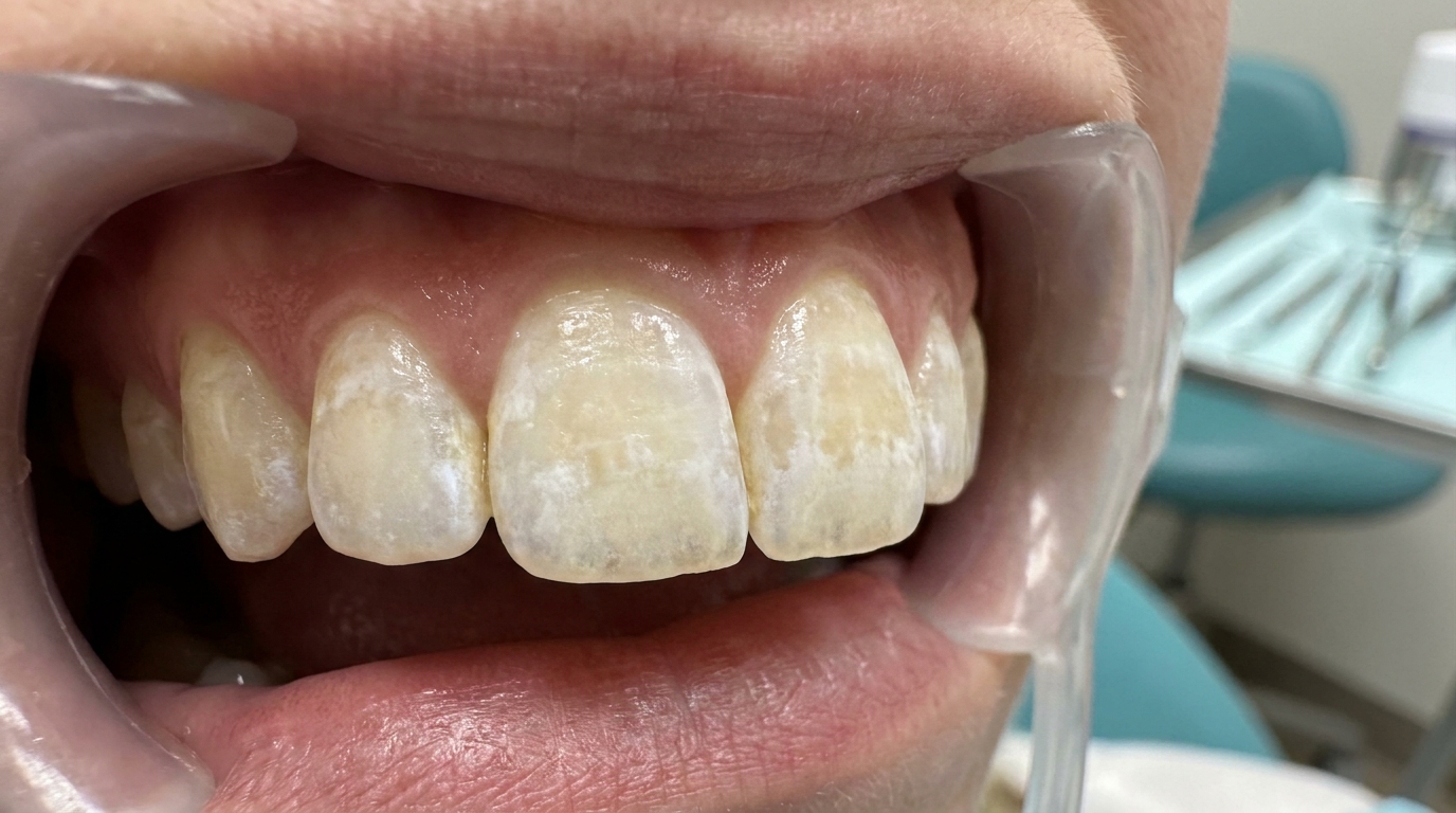
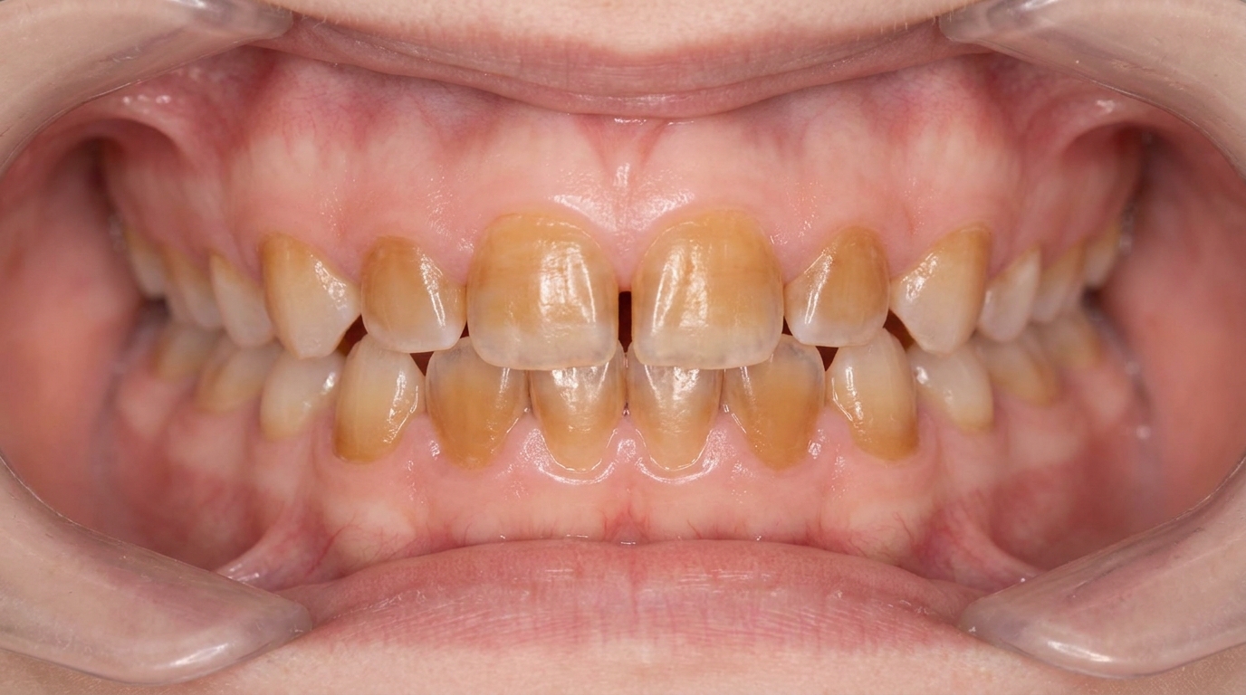
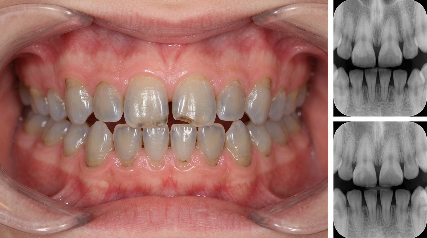
Diffuse discolouration of all teeth, ranging from yellow-brown to grey-blue to amber, with a characteristic opalescent (translucent and pearly) appearance that gives the name "hereditary opalescent dentine".
Bulbous crowns with cervical constriction (a narrowed neck where the crown meets the root) on X-ray.
Thin roots that appear short and tapered.
Rapid loss of enamel soon after the teeth come through, exposing the underlying dentine.
Severe attrition (tooth-on-tooth wear) with significant loss of crown height by adolescence in moderate to severe cases.
Both dentitions affected, with the deciduous teeth typically more severely affected.
In severe (shell teeth) form, dramatically thin dentine with very large pulps, prone to early pulp exposure.
What it feels like
Sensitivity to cold, hot and sweet foods is variable, sometimes surprisingly mild because of the obliterated pulp chambers.
Difficulty chewing when much of the crown has worn away.
Aesthetic concern, the unusual colour is often visible when smiling.
Periapical pain or swelling (pain and swelling near the root tip) can develop when the obliterated pulp's residual vessels become infected through micro-exposures.
No symptoms at all in mild cases until adulthood.
What an X-ray might show
X-rays are particularly useful in confirming the diagnosis:
Bulbous crowns with cervical constriction.
Thin, short roots.
Progressive obliteration of the pulp chambers and root canals, sometimes obvious before any symptoms develop.
Periapical inflammatory changes in some affected teeth.
Shell teeth (in severe form), paper-thin dentine with dramatically large pulp chambers, often in the deciduous dentition.
What happens at the dentist?
Dentinogenesis imperfecta is most often picked up early in childhood when the deciduous teeth come through with the characteristic translucent appearance. A dentist at ArtSmiles, typically as part of a dental check-up and clean, will commonly:
Examine all teeth carefully, photograph the appearance, and take X-rays to confirm the radiographic features.
Take a careful family history, since DGI usually shows in close relatives.
Ask about general health features of osteogenesis imperfecta, easy bone fractures, blue sclerae, hearing concerns, to determine whether the condition is isolated or part of the wider syndrome.
Refer for hearing assessment since progressive sensorineural (nerve-related) hearing loss has been associated with DGI.
Refer to a paediatric dentist or restorative specialist for staged long-term care.
Discuss genetic counselling for affected families considering further children.
Plan early protective measures, preformed metal crowns on first molars, sealants, fluoride applications.
Coordinate aesthetic and functional restoration as the patient grows.
Is this serious?
🟡 Dentinogenesis imperfecta is not life-threatening, but without dental care it can lead to severe tooth wear, sensitivity, periapical infection and early tooth loss. Combined with osteogenesis imperfecta, it is part of a more significant systemic condition that needs medical management. With timely and well-coordinated dental and medical care, most patients achieve a healthy, comfortable and good-looking dentition for life.
If you have noticed unusual translucent or grey teeth in your child from when they first came through, particularly if other family members have had similar issues or a history of brittle bones, it is worth booking an assessment so the diagnosis can be confirmed early and a long-term plan put in place.
Could it be something else?
Several conditions can produce similar tooth appearances. The textbooks list these as the main differentials:
Amelogenesis Imperfecta, a hereditary defect of enamel rather than dentine; the abnormality is in the outer covering rather than the bulk of the tooth.
Dentin dysplasia type I (rootless teeth), a different, very rare hereditary disorder of root dentine, with normal-coloured crowns but stubby, deformed roots.
Enamel Hypoplasia, a developmental enamel defect, typically not generalised across all teeth and not familial.
Tetracycline staining, yellow, grey or brown banding from medication during enamel formation.
Dental Fluorosis, bilateral lusterless white opacities; tied to fluoride exposure rather than family history.
The combination of all teeth affected, opalescent grey-blue colour, bulbous crowns with cervical constriction on X-ray, and a positive family history, sometimes with osteogenesis imperfecta, is the strongest clue toward dentinogenesis imperfecta.
How is it treated?
Treatment is long-term and staged through childhood, adolescence and into adulthood.
At-home measures and habits:
Brush twice a day with fluoride toothpaste, gently to avoid further wear.
Use a soft-bristled toothbrush and a desensitising toothpaste if needed.
Avoid heavy biting on hard foods that could fracture vulnerable teeth.
Eat a balanced diet with attention to acidic foods.
Attend regular dental check-ups, with shorter intervals in childhood and adolescence.
Professional steps your dentist may consider:
Topical fluoride applications to protect remaining enamel and dentine.
Sealants on newly erupted molars.
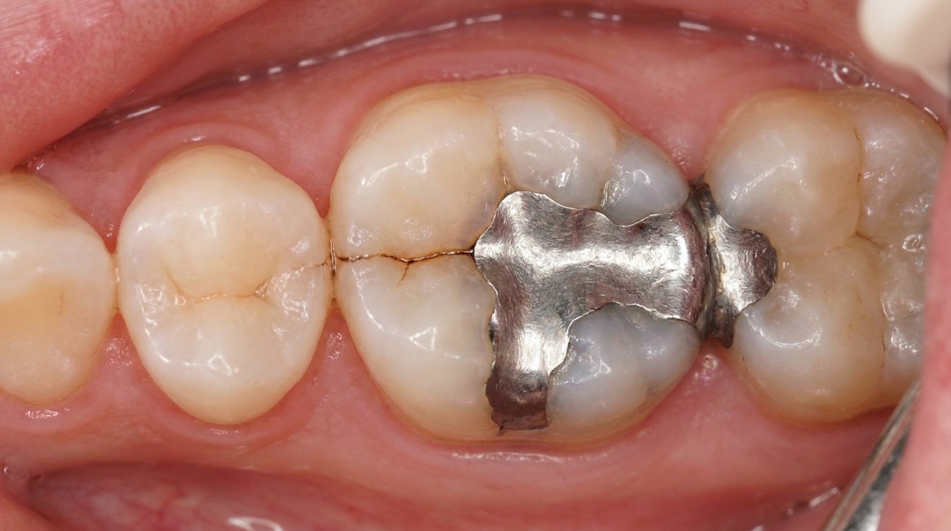
Preformed metal crowns on severely affected first permanent molars in childhood, replaced later with permanent crowns.
Composite (dental fillings) for moderate aesthetic and functional restoration.
Bonded onlays and metal castings for adolescent restoration.
Full-coverage crowns for adult restoration of severely worn teeth, although the textbooks note that cervical fracture is a risk and crowns are most successful on teeth that retain a near-normal shape.
Overlay dentures in severely affected dentitions, particularly when many crowns are not feasible, sometimes used over teeth that have been built up with fluoride-releasing glass ionomer cement.
Implant therapy in adulthood when individual teeth cannot be saved, although extra care is needed in patients with osteogenesis imperfecta because of altered bone quality.
Coordination with a paediatric medical team and geneticist in osteogenesis imperfecta cases.
Hearing assessment because of the recognised association with sensorineural hearing loss.
Full-mouth oral rehabilitation in adulthood for severely affected dentitions, combining crowns, onlays, implants and/or overlay dentures in a coordinated staged plan that restores function, comfort and appearance.
Long-term review and maintenance, affected teeth need more dental care over a lifetime.
A patient-centred approach is particularly important. Children and adolescents with visibly different teeth often feel self-conscious, and a clear, hopeful long-term plan, explaining that the appearance and function can be substantially improved with staged care, is itself part of effective care.
What's the long-term outlook?
The outlook depends on the severity of the dentinogenesis imperfecta, whether it is part of osteogenesis imperfecta, and how consistent the dental care is over a lifetime. Mild forms may need only protective care and small composite restorations. Moderate forms need staged crowns and possibly overlay dentures by adulthood. Severe (shell teeth) forms typically need early extraction of severely affected deciduous teeth and full-mouth rehabilitation in early adulthood. Across all forms, with consistent dental care, most patients can have healthy, comfortable, attractive teeth for life, and the condition's wider impact can be substantially managed. The single most important factor is engagement with regular long-term dental review.
A note on this article
This article is for educational purposes only and does not constitute a clinical diagnosis. Please consult a registered dental practitioner for assessment and treatment advice.
The cover image above is an AI-generated illustration based on the most common visible features of this condition described in clinical pathology references. It is not a photograph of a real case and should not be used to diagnose or rule out the condition in your own situation. If you are concerned about something you have noticed, please book an assessment with a registered dental practitioner.
References
Neville, B. W., Damm, D. D., Allen, C. M., & Chi, A. C. (2023). Oral and maxillofacial pathology (5th ed.). Elsevier. Chapter 2, Abnormalities of Teeth: Hereditary Disorders of Dentin and Dentin Sialophosphoprotein-Associated Dentin Defects, pp. 103 to 108.
Cawson, R. A., & Odell, E. W. (2017). Cawson's essentials of oral pathology and oral medicine (8th ed.). Elsevier. Chapter 2, Disorders of Development: cross-reference for hereditary opalescent dentine.
Regezi, J. A., Sciubba, J. J., & Jordan, R. C. K. (2017). Oral pathology: Clinical pathologic correlations (7th ed.). Elsevier. Chapter on Abnormalities of Teeth: cross-reference for dentinogenesis imperfecta.
Frequently asked questions
What is dentinogenesis imperfecta?
Dentinogenesis imperfecta (DI) is an inherited disorder of dentin development that gives teeth an opalescent blue-grey or amber colour and makes them weak and prone to wear. There are three classical types: Type I, associated with osteogenesis imperfecta (brittle bone disease); Type II, isolated DI; and Type III, the rare 'Brandywine' subtype with shell-like teeth.
What do teeth with dentinogenesis imperfecta look like?
The teeth often look translucent and grey-blue or brownish-amber, sometimes described as opalescent. Enamel can chip off because it does not bond well to the abnormal dentin, exposing softer dentin that wears down rapidly. X-rays show bulbous crowns, short narrow roots and pulp chambers that are obliterated by abnormal dentin.
How is DI different from amelogenesis imperfecta?
In amelogenesis imperfecta, the primary defect is in the enamel itself; the dentin is normal. In dentinogenesis imperfecta, the enamel structure is normal at first but it fractures off the abnormal dentin underneath. Both conditions affect all teeth and run in families, and genetic testing can confirm which gene is involved.
How is dentinogenesis imperfecta treated?
Treatment is lifelong restorative dentistry to protect the teeth from wear, restore appearance and maintain function. Children may need composite or stainless-steel crowns on the back teeth. Adolescents and adults usually need full-coverage crowns or veneers, occasionally combined with orthodontics, and sometimes implant-supported prostheses for missing teeth. Coordination with paediatricians and bone specialists is important when DI is part of osteogenesis imperfecta.