Compiled from clinical pathology references. Medically reviewed by Dr Cristian Dunker, Principal Dentist at ArtSmiles Cosmetic Dentistry.
Quick summary
Also called | Torus palatinus, torus mandibularis, buccal exostoses, palatal exostoses (palatal tubercles), bony tori |
How urgent? | 🟢 Usually harmless, these are normal bony growths, not tumours, but a one-off check-up is sensible if you've just noticed one |
Common or rare? | Very common, palatal tori affect roughly 20-35% of adults in many populations |
Who it affects | Adults, usually first noticed in the second or third decade of life; palatal tori twice as common in women, mandibular tori slightly more common in men |
Who treats it | General dentist (most cases need no treatment); occasional referral to an oral surgeon if removal is required |
Based on | Regezi, Neville, Cawson, Laskaris |
What is it?
Tori and exostoses are slow-growing lumps of normal bone that sit on the surface of the jaw. They are covered by ordinary gum tissue and are not tumours, infections, or cancers, just a normal variation in how some people's jawbone is shaped.
The three main types are named after where they appear:
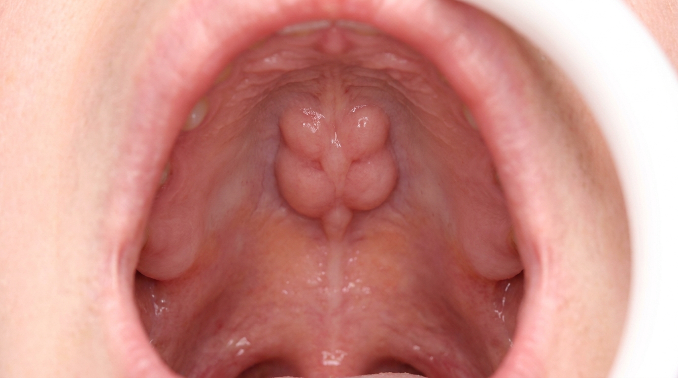
Torus palatinus, a bony bump in the middle of the roof of the mouth.
Torus mandibularis, bony bumps on the inside (tongue-side) of the lower jaw, usually behind the lower eye-teeth, and very often on both sides.
Buccal (and palatal) exostoses, rows of small bony nodules along the cheek-side of the upper or lower jaws, or along the palate-side of the upper back teeth.
Who tends to get it?
Tori and exostoses are surprisingly common. Studies cited in Neville and Regezi report palatal tori in anywhere from 4% to 60% of people surveyed, with most United States studies landing around 20-35%. Mandibular tori are reported in roughly 6-12% of adults in the United States, and buccal exostoses in nearly 1 in 5 adults in some recent surveys.
A few patterns show up consistently across the source books:
Palatal tori are about twice as common in women as in men.
Mandibular tori show a slight male preference.
Bilateral involvement of mandibular tori occurs in more than 90% of cases, meaning they almost always appear on both sides.
Higher prevalence has been documented in Asian, Native American and Inuit populations.
Age of onset is typically the second or third decade of life, with prevalence peaking in early adult life and tapering slightly later on.
Most people first notice them in their twenties or thirties, often by accident, a tongue exploring a new texture, or a dental hygienist pointing it out at a check-up.
What causes it?
The honest answer from the source textbooks is that no single cause has been pinned down. Both Neville and Regezi describe the cause as multifactorial, a combination of genetic and environmental influences.
The genetic side is well documented. Family studies, including work in Venezuelan and Japanese populations cited in Regezi, suggest a dominant inheritance pattern for palatal tori. If a parent has a torus, their children have a higher chance of developing one too.
The environmental side is largely about mechanical stress on the jawbone. Tori and exostoses are thought to form, at least in part, in response to functional or parafunctional loading of the teeth, for example:
Heavy chewing forces from a strong bite.
Bruxism (clenching or grinding the teeth, often during sleep).
The total number of teeth still present, since prevalence has been correlated with how many teeth a person has remaining.
Neville also notes a positive association between mandibular tori and conditions like migraines and temporomandibular disorders, hinting again at a parafunctional habit link. Importantly, none of these factors mean a person has done anything wrong, tori are best thought of as the bone simply responding to the load it carries.
How does it develop?
Think of jawbone as a building material that responds to how it is used. When a particular area is loaded repeatedly, from a strong bite, grinding, or just decades of chewing, the bone may lay down extra layers in that spot to reinforce itself. Over years, those extra layers gradually build into a smooth, hard mound covered by a thin layer of gum.
The build-up is dense, lamellar (layered) cortical bone, sometimes with a small inner pocket of spongy bone and a little fatty marrow. Because the growth is so gradual, most people never notice it happening. The supporting evidence in Regezi and Neville, that prevalence peaks in early adult life and then often tapers in older age, especially after teeth are lost, fits with the idea that tori are dynamic structures that grow and remodel with the forces placed on the jaw.
What might you notice?
What it looks like
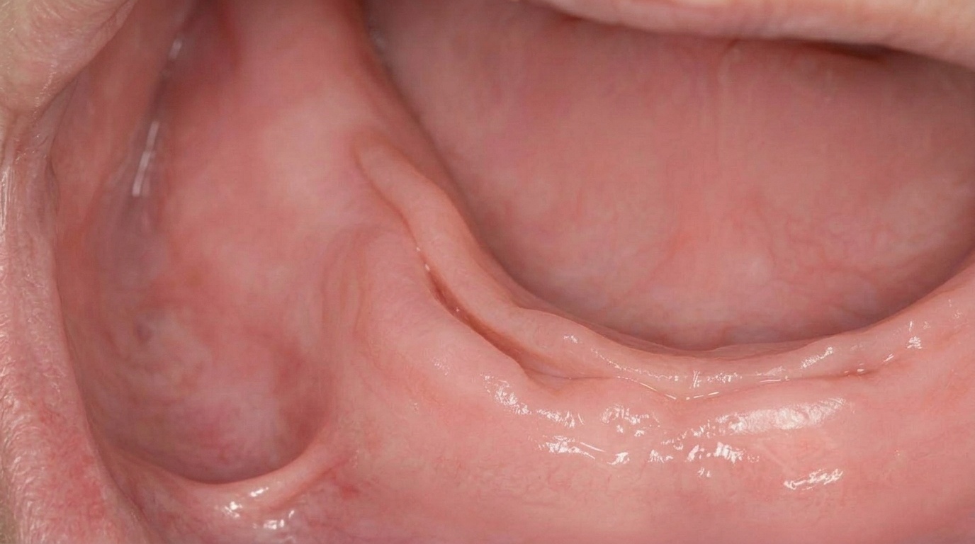
A torus or exostosis appears as a smooth, hard, rounded lump under normal-looking pink gum. The covering mucosa is thin, particularly over the top of the lump, and so it may look slightly pale or shiny compared with the surrounding tissue.
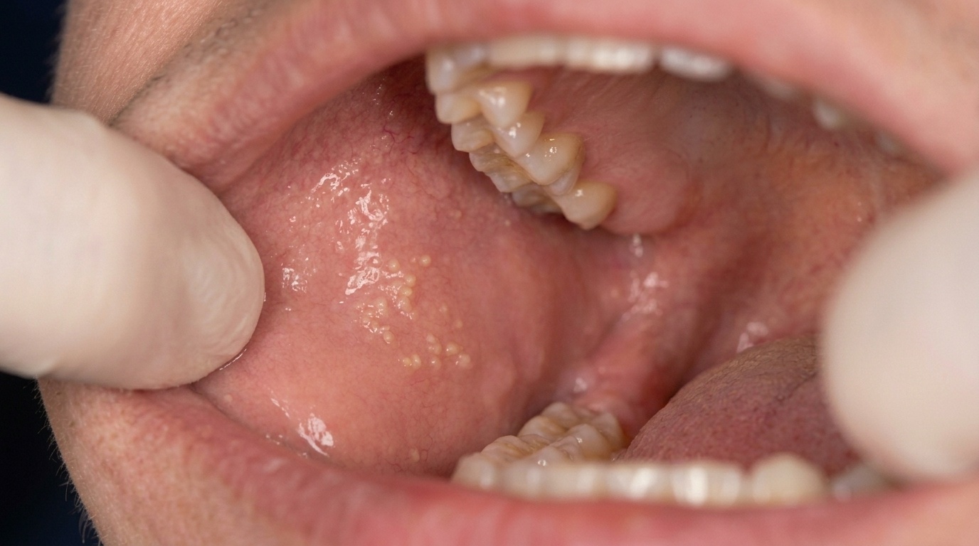
Torus palatinus sits dead-centre on the roof of the mouth. It can be flat and broad, ridge-like along the midline, knobbly with several smaller bumps, or a single multi-lobed mound. Most are under 2 cm across, but some can fill the entire palatal vault.
Torus mandibularis appears as one or more bony bumps on the tongue-side of the lower jaw, usually opposite the premolars and above the gum line. In rare "kissing torus" cases, the two sides grow large enough to almost meet in the middle.
Buccal exostoses look like a row of small, hard nodules along the cheek-side of the upper or lower back teeth.
Palatal exostoses (palatal tubercles) form on the inside of the upper back teeth, often on both sides.
What it feels like
Most tori and exostoses are completely asymptomatic, patients are often unaware of them until a dentist mentions them or until the tongue explores the area. Specific things people sometimes report:
A hard, immovable lump that feels different from gum.
An intermittent ulcer or sore patch on the surface, usually after biting on something hard or sharp like a chip or a piece of toast. Because the gum covering is thin and the underlying bone has limited blood supply, these ulcers can be slow to heal.
Discomfort with dentures, particularly if the torus interferes with the fit of an upper or lower denture base.
Anxiety, many people are alarmed when they first feel the lump and worry that it might be a tumour. Reassurance is often the most important part of the conversation.
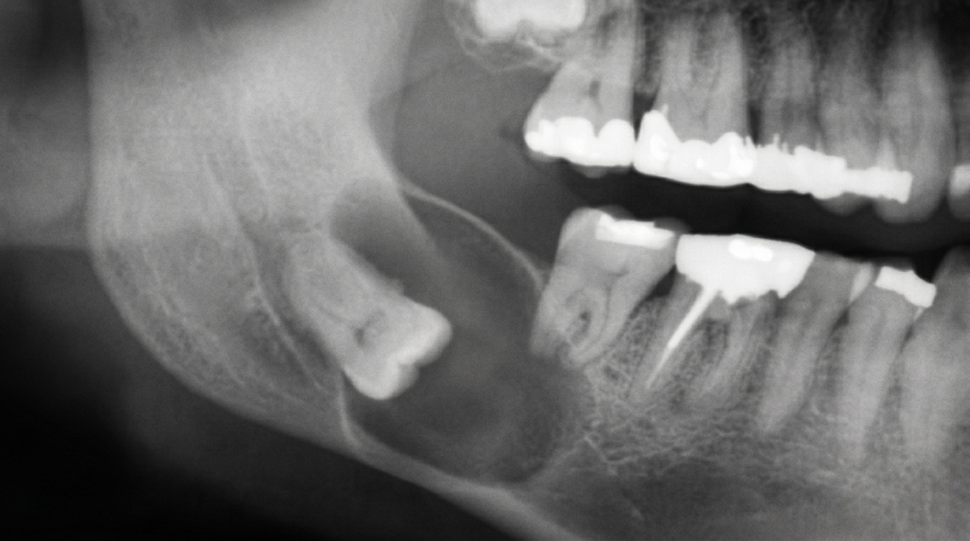
What an X-ray might show
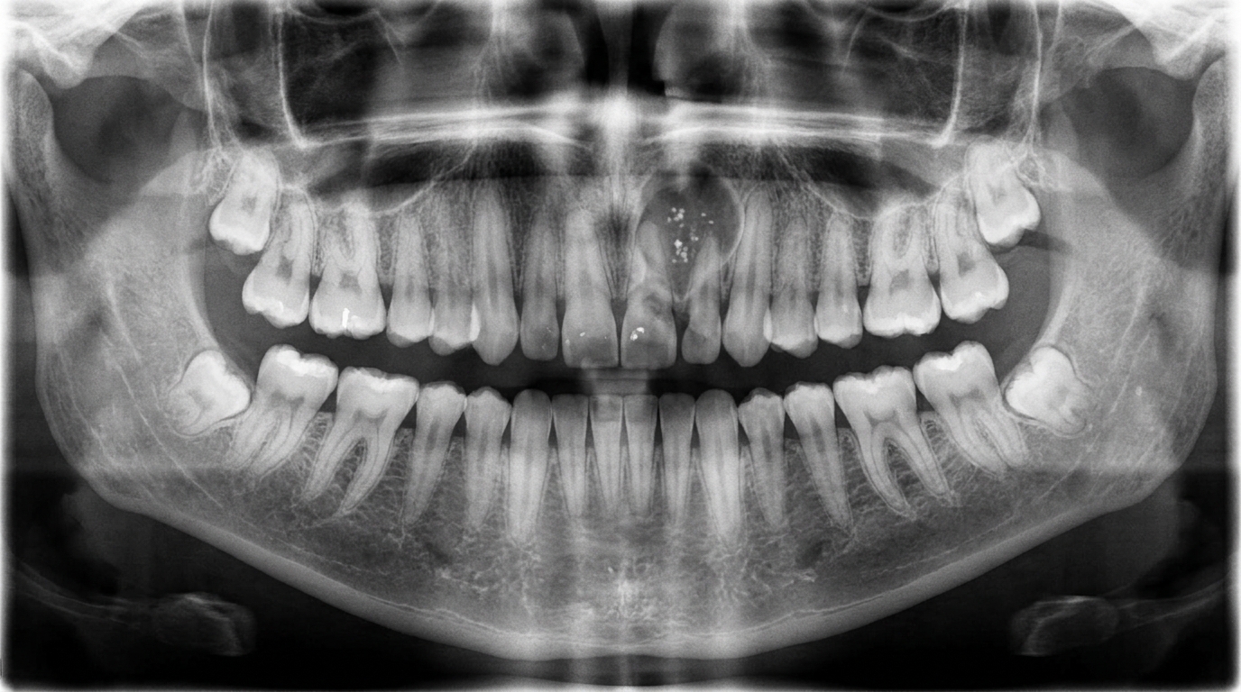
Most tori are diagnosed clinically and don't need imaging. When they are large enough, they may show up on dental radiographs as a diffuse, well-defined radiopacity (a whiter area). A mandibular torus can appear as a radiopaque shadow superimposed over the roots of the lower teeth on a periapical film, and is easily seen on a lower occlusal radiograph.
What happens at the dentist?
Tori and exostoses are usually diagnosed at a routine dental examination at ArtSmiles. The pathway is generally straightforward:
Visual and tactile examination. Your dentist will look at and gently feel the lump. The classic features, a smooth, bony-hard, mucosa-covered swelling in a typical location, are usually enough to make the diagnosis.
Reassurance and documentation. A clinical photograph or a note in your records helps track whether the lump changes over time.
Imaging if needed. Dental radiographs are not required to diagnose a torus, but they may be taken if the size, location, or appearance is unusual.
Biopsy in rare cases. Most tori are distinctive enough that a biopsy is not necessary. If anything about the lump is unusual, for example, rapid growth, an unusual location, multiple osteoma-like lesions, or other systemic findings, your dentist may recommend referral and a small tissue sample to rule out other bony conditions.
Specialist referral to an oral surgeon may be considered if removal is required for prosthetic, hygiene or symptomatic reasons.
Is this serious?
🟢 In almost all cases, no. Tori and exostoses are non-neoplastic, they are not cancer and have no potential to become cancer. The source textbooks consistently describe them as having little clinical significance.
There are, however, a few situations where they do matter:
Recurrent trauma. Because the gum covering is thin, the surface can be ulcerated by hard food. Healing can be slow and occasionally painful, and very rarely the area can become infected (osteomyelitis).
Denture problems. A large torus can prevent a denture from seating properly or being comfortable.
Medication-related concerns. Neville notes that palatal tori are prone to medication-related osteonecrosis of the jaw in patients taking certain bone-modifying medications (such as bisphosphonates), where pressure or minor trauma over the torus can trigger exposed bone.
Could it be something else?
Most tori are obvious to a dentist, but there are a few conditions that can look or feel similar. The differentials below are drawn directly from the source textbooks:
Osteoma, A true benign bone tumour, also slow-growing and often hard to distinguish from a very large exostosis. Osteomas tend to grow more progressively and may occur in atypical locations; multiple osteomas of the jaws can be a sign of Gardner syndrome and warrant further investigation.
Osteochondroma, Another benign bony overgrowth, but typically arising from the coronoid or condylar process near the jaw joint rather than the alveolar bone. It can interfere with jaw movement, which a torus does not.
Fibrous dysplasia, A developmental bone condition that causes expansion of part of the jaw. It usually involves a wider area than a discrete torus and produces a characteristic "ground-glass" pattern on radiographs.
Ossifying fibroma, A benign fibro-osseous tumour that is well-circumscribed on radiographs and tends to expand the jaw rather than sit as a surface bump.
Osteoma associated with Gardner syndrome, Multiple jaw osteomas, particularly at unusual sites, can be an early marker of this inherited condition, which carries a high risk of colorectal cancer; investigation beyond the mouth is recommended.
Reactive subpontine exostosis (subpontic osseous hyperplasia), A bony nodule that grows up beneath the false tooth (pontic) of a fixed bridge. It is a related entity, distinguished by its specific location under a bridge.
Exostosis under a graft, A localised bony growth can develop beneath a free gingival graft or skin graft, presumably stimulated by the graft itself. Distinguished by the patient's surgical history.
Coronoid hyperplasia, Enlargement of the coronoid processes of the mandible that limits mouth opening. Distinguished by reduced jaw movement and its characteristic appearance on imaging.
Fibrous developmental malformation, A rare fibrous overgrowth at the upper tuberosity, described in Laskaris. It feels firm rather than bony-hard and is composed of fibrous tissue, not bone.
Soft tissue swellings (fibroma, lipoma, abscess), These feel soft or rubbery and are mobile against the underlying bone; a torus is bony-hard and immobile.
Oral cancer, A common patient worry. A tumour generally appears as an ulcer or growth that changes rapidly, may bleed, often feels firm but not bony-hard, and rarely sits exactly in the classic torus locations.
How is it treated?
The consistent message across all four source textbooks is that most tori and exostoses need no treatment at all. The right approach is usually monitoring and reassurance.
At home, you can help by:
Being gentle with very hot or hard, sharp foods (chips, crusty bread, hard nuts) that may scrape the thin gum over the lump.
Keeping the area clean as part of your normal brushing routine, it can be slightly trickier to brush around a mandibular torus, so a soft-bristled toothbrush helps.
Letting your dentist know if you grind or clench your teeth, as managing bruxism with a night-time splint may reduce ongoing functional load.
Mentioning any new bone-modifying medications (such as bisphosphonates), which can change how the body responds to bony lumps.
Professional treatment may include:
Routine monitoring at your regular check-ups, the most common approach by far.
Surgical removal, considered when a torus or exostosis:
Is repeatedly traumatised or chronically ulcerated.
Interferes with the construction or fit of a denture.
Gets in the way of a periodontal procedure or proper flap adaptation during gum surgery.
Is associated with reactive subpontine exostosis under a bridge that is causing oral hygiene problems.
When surgery is needed, it is typically a same-day minor procedure performed under local anaesthetic, with healing over a few weeks. Recurrence after removal is rare, although tori associated with continued tooth function may regrow if the underlying stress remains.
What's the long-term outlook?
The outlook for tori and exostoses is excellent. They are benign, non-cancerous bony variations that pose no risk to general health. Most people live their entire lives with them and never need any treatment.
Things worth knowing about the long term:
Tori grow slowly over years, usually peaking in early adult life and sometimes shrinking slightly after teeth are lost or chewing forces decrease.
The covering gum will occasionally be ulcerated by trauma, but these sores generally heal with conservative care.
If surgical removal is performed for a clear reason, the result is usually long-lasting, with only rare recurrence.
They do not transform into cancer.
They are simply part of how some people's jawbone is built, and once you know what they are, they tend to stop being a worry.
A note on this article
This article is for educational purposes only and does not constitute a clinical diagnosis. Please consult a registered dental practitioner for assessment and treatment advice.
The cover image above is an AI-generated illustration based on the most common visible features of this condition described in clinical pathology references. It is not a photograph of a real case and should not be used to diagnose or rule out the condition in your own situation. If you are concerned about something you have noticed, please book an assessment with a registered dental practitioner.
References
Regezi, J. A., Sciubba, J. J., & Jordan, R. C. K. (2017). Oral pathology: Clinical pathologic correlations (7th ed.). Elsevier. Chapter 12, Benign Nonodontogenic Tumors, Tori and Exostoses section, pp. 309-310.
Neville, B. W., Damm, D. D., Allen, C. M., & Chi, A. C. (2023). Oral and maxillofacial pathology (5th ed.). Elsevier. Chapter 1, Developmental Defects of the Oral and Maxillofacial Region, Exostoses / Torus Palatinus / Torus Mandibularis sections, pp. 19-23.
Cawson, R. A., & Odell, E. W. (2017). Cawson's essentials of oral pathology and oral medicine (8th ed.). Elsevier. Chapter 9, Non-odontogenic Tumours of the Jaws, Osteoma and Other Bony Overgrowths, p. 156.
Laskaris, G. Pocket atlas of oral diseases. Chapter 2, Developmental Anomalies, Torus Palatinus / Torus Mandibularis / Multiple Exostoses, pp. 8-10.
Frequently asked questions
Are tori and exostoses dangerous?
No. They are benign, slow-growing overgrowths of normal bone with no cancer risk. Most people are completely unaware of them and only notice them when their tongue or finger brushes over a firm lump on the palate or jaw.
Do tori need to be removed?
Most of the time, no. Tori only need surgical removal when they interfere with denture fit, get repeatedly ulcerated by food or appliances, or affect speech and oral hygiene. Otherwise they can be left alone and simply monitored.
Why do I have a hard lump in the roof of my mouth?
A bony lump on the midline of the hard palate is most often a torus palatinus, a benign bony growth that affects up to 20-25% of people in some populations. It tends to develop slowly through adult life. If you have noticed a hard, painless lump there, a dental check confirms what it is.
Will a torus come back after it is removed?
Recurrence after complete surgical removal is uncommon. The tendency to form tori or exostoses is partly genetic, so very occasionally a small new lump appears in the same area years later, but it does not usually need further treatment.