Compiled from clinical pathology references. Medically reviewed by Dr Cristian Dunker, Principal Dentist, ArtSmiles Cosmetic Dentistry.
Most jaw infections start in a tooth and end with the tooth, once the tooth is treated, the infection settles. Occasionally, however, a deep infection travels into the jawbone itself and takes hold. This is osteomyelitis, an inflammation of bone caused by infection. While modern dentistry has made it much less common than it once was, it remains an important condition to recognise, especially in patients with conditions that affect healing.
This article from the team at ArtSmiles, reviewed by Dr Cristian Dunker, explains what osteomyelitis of the jaw is, why it happens, and how it is treated.
What is it?
Osteomyelitis is the medical term for an inflammatory process of bone, typically caused by infection. In the jaw, the lining of the bone (periosteum), the soft inner spaces (marrow), and the surrounding bone all become involved.
There are two broad clinical patterns:
Acute osteomyelitis, a recent infection with severe pain, fever, swelling and pus.
Chronic osteomyelitis, a long-standing infection that has become a low-grade smoulder, sometimes punctuated by acute episodes.
Special variants include chronic sclerosing osteomyelitis, diffuse sclerosing osteomyelitis and chronic osteomyelitis with proliferative periostitis (Garré osteomyelitis), each with characteristic imaging features.
Who tends to get it?
Osteomyelitis of the jaw is uncommon in healthy adults today, thanks to widespread use of antibiotics and modern dentistry. It is more often seen in:
Adults with poorly controlled diabetes, who heal less well.
Smokers, in whom blood supply to bone is reduced.
Patients with a history of radiotherapy to the head and neck, where bone is permanently more vulnerable to infection.
Patients on long-term bone-strengthening medicines such as bisphosphonates and denosumab, used for osteoporosis or some cancers, a related but distinct condition called medication-related osteonecrosis of the jaw (MRONJ) can mimic osteomyelitis.
Patients on long-term steroids or immunosuppressive therapy.
Patients with sickle cell disease or other haematological conditions.
The lower jaw is overwhelmingly more affected than the upper jaw because of differences in blood supply and bone density.
What causes it?
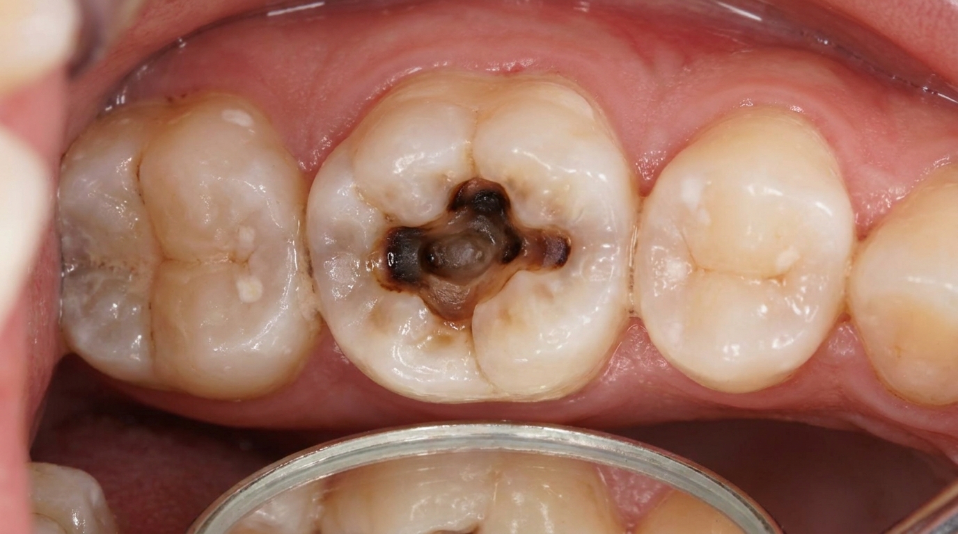
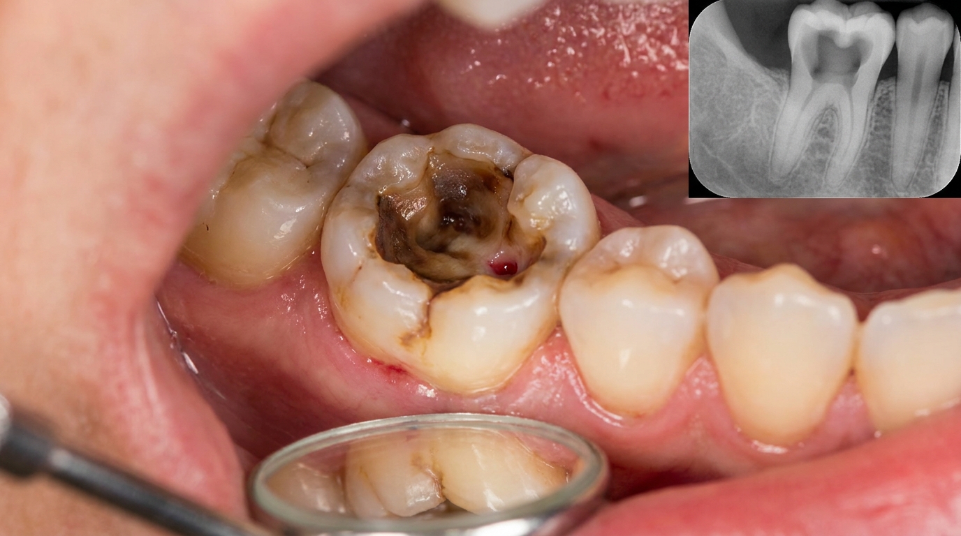
Osteomyelitis of the jaw is most often caused by:
Spread from a tooth infection, a deep cavity reaching the nerve, an abscess that has not drained, a chronic apical infection.
Spread from a periodontal abscess.
Trauma, such as a jaw fracture allowing bacteria into the bone.
Postoperative infection after extraction or jaw surgery.
Direct inoculation during dental procedures in patients with reduced healing.
Spread from sinus infections in the upper jaw (uncommon).
Bloodstream spread from infection elsewhere in the body (rare in adults).
The bacteria most often involved are mixed populations of mouth bacteria, streptococci, anaerobes and others, rather than the Staphylococcus aureus that typically causes osteomyelitis in long bones.
How does it develop?
The course begins with infection breaching the bone surface and gaining access to the soft inner marrow:
Bacteria reach the marrow through a tooth, a fracture or a surgical wound.
Inflammation increases pressure inside the bone, which is rigid and cannot expand.
Small blood vessels in the marrow are compressed, reducing the blood supply.
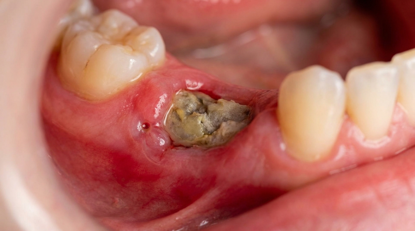
Areas of bone die because they have lost their blood supply, forming pieces of dead bone called sequestra.
Pus drains through the bone and overlying soft tissue, sometimes appearing as a lump or sinus on the gum or skin.
If untreated, the area expands, neighbouring teeth become loose, and the affected jaw segment may weaken.
With treatment, infection clears, sequestra are removed, and the bone gradually heals.
What might you notice?
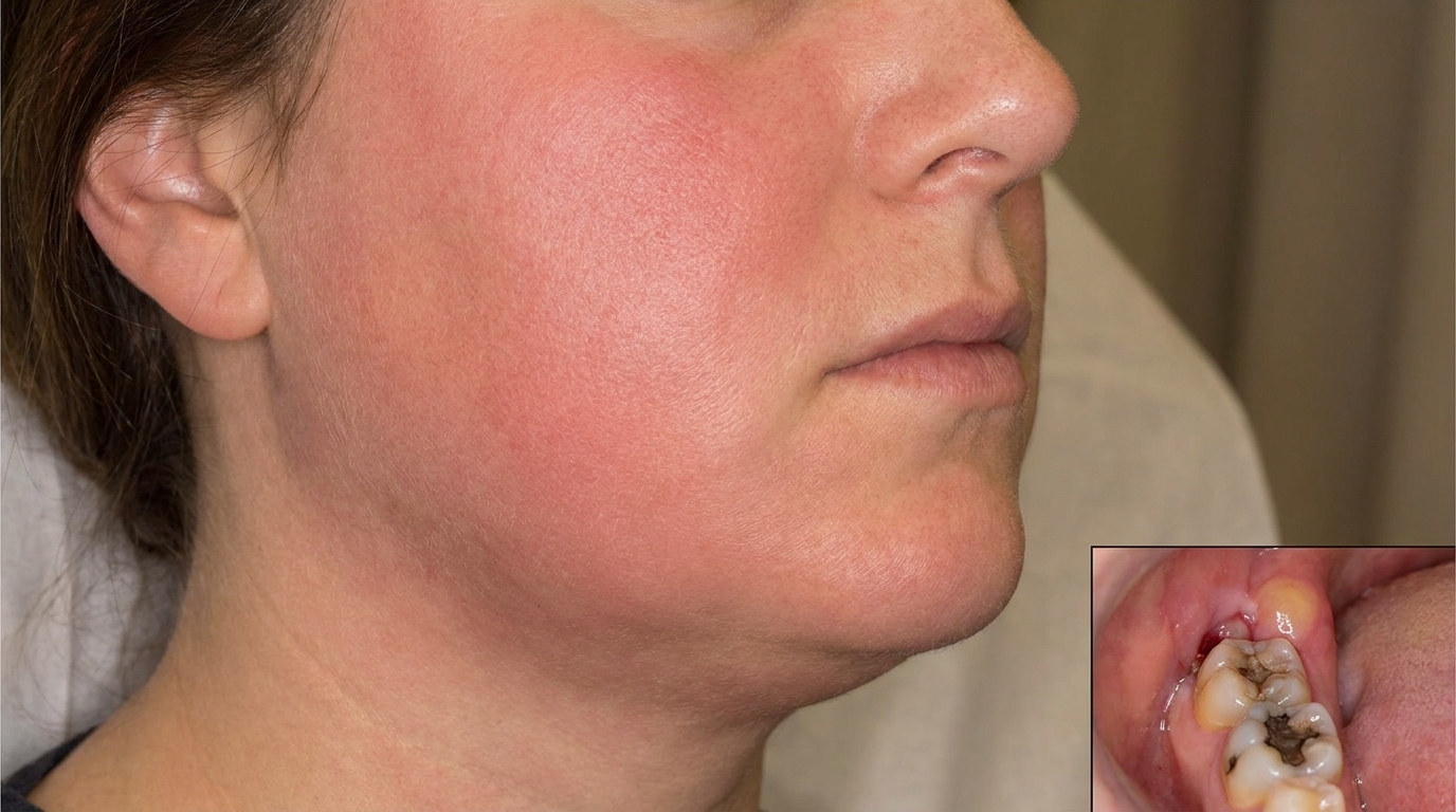
In acute osteomyelitis, common symptoms include:
Severe, deep, throbbing jaw pain.
Swelling of the cheek or under the jaw.
Fever, malaise and feeling unwell.
Tender lymph nodes under the jaw.
Loose teeth in the affected area.
Numbness or tingling of the lower lip (a particularly important sign indicating pressure on the inferior alveolar nerve), the main nerve running through the lower jaw.
Pus drainage from the gum or through the skin.
Difficulty opening the mouth (trismus).
Chronic osteomyelitis often presents more quietly, a persistent dull ache, a small sinus on the gum that intermittently discharges pus, occasional flare-ups of swelling, and changes seen on imaging.
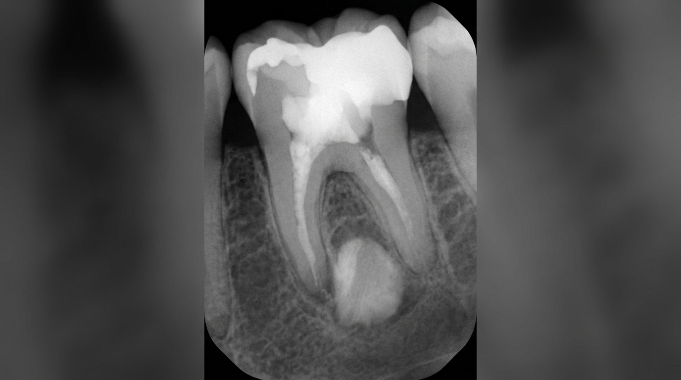
What an X-ray might show
In the early days, an X-ray of an acute osteomyelitis may look surprisingly normal, because changes in bone density take time to appear. After about two weeks, a panoramic X-ray or cone-beam CT can show a moth-eaten loss of bone density, areas of denser sclerotic bone in chronic cases, and small pieces of dead bone (sequestra) sitting separately from the rest of the jaw. A widened periodontal ligament space around an offending tooth is a common early clue.
What happens at the dentist?
When osteomyelitis is suspected at ArtSmiles, the visit usually involves:
A careful history and examination. Pain pattern, swelling, lip numbness, fever, prior dental procedures and medical history are documented.
A check for underlying tooth infection or a wound source.
Imaging, usually a panoramic X-ray (a single X-ray that shows both jaws end-to-end) initially, followed by a cone-beam CT (CBCT) to see the extent of bone involvement.
Referral to an oral and maxillofacial surgeon (specialist in face, jaw and mouth surgery) for further assessment. Most cases need surgical involvement.
Sampling of pus or bone for laboratory analysis so the right antibiotics can be chosen.
Coordination with your medical doctor to manage diabetes, immune status or relevant medicines.
A discussion of the plan, expected duration of treatment and follow-up.
Is this serious?
Osteomyelitis of the jaw is a serious condition that needs prompt attention because:
Bone destruction can be extensive if untreated.
Pathological fracture (a break in bone that has been weakened by disease rather than impact) of the jaw is possible in advanced cases.
Loss of teeth in the affected area is common.
Spread to neighbouring spaces (face, neck) can occur.
Rarely, septicaemia (bloodstream infection) can develop in immunocompromised patients.
With timely care, however, the great majority of patients respond well to combined antibiotic and surgical treatment.
Could it be something else?
Several conditions can mimic osteomyelitis on examination or imaging:
Medication-related osteonecrosis of the jaw (MRONJ) in patients on bisphosphonates, denosumab or some anti-cancer drugs.
Osteoradionecrosis (ORN) in patients who have had radiotherapy to the head and neck.
Chronic dental abscess without true bone infection.
Odontogenic cysts and tumours, especially when they become infected.
Ewing sarcoma and osteosarcoma, uncommon but important.
Metastatic cancer to the jaw in patients with known cancer elsewhere.
Fibrous dysplasia with secondary infection.
Imaging, biopsy and microbiology together separate these.
How is it treated?
Treatment combines several elements:
Antibiotics, usually started empirically (based on the most likely bacteria before lab results return) with a broad-spectrum agent and adjusted once microbiology results are available. Treatment may continue for several weeks.
Drainage of any pus collection, either through a tooth socket or via a small surgical incision.
Removal of the source. A non-restorable infected tooth is extracted; an old infected root tip is removed.
Surgical removal of dead bone (sequestrectomy). Loose pieces of dead bone are removed under anaesthesia.
Saucerisation of the affected bone segment in chronic cases, saucer-shaping the wound to allow access for ongoing care.
Hyperbaric oxygen therapy, considered in selected cases, particularly osteoradionecrosis.
Medical optimisation, control of diabetes, smoking cessation, nutrition support.
Long-term follow-up with imaging at intervals to confirm bone healing.
The whole journey, from acute presentation to full bone healing, can take weeks to months. Patience and adherence to follow-up appointments matter.
What's the long-term outlook?
The outlook is generally good for most patients with timely treatment. Pain settles within days to weeks of starting antibiotics and removing the source. Bone gradually fills in over months, although a small scar in the bone may remain visible on later X-rays. Patients with significant risk factors (radiation, bone-modifying drugs, poorly controlled diabetes) need closer follow-up and a more cautious approach to future dental procedures in that area.
If you have persistent jaw pain, lip numbness or a non-healing area in your mouth, particularly after a recent extraction or in a high-risk medical context, please book an early appointment. Early recognition is the most important factor in keeping treatment straightforward.
A note on this article
This article is for educational purposes only and does not constitute a clinical diagnosis. Please consult a registered dental practitioner for assessment and treatment advice.
The cover image above is an AI-generated illustration based on the most common visible features of this condition described in clinical pathology references. It is not a photograph of a real case and should not be used to diagnose or rule out the condition in your own situation. If you are concerned about something you have noticed, please book an assessment with a registered dental practitioner.
References
Neville, B. W., Damm, D. D., Allen, C. M., & Chi, A. C. (2016). Oral and maxillofacial pathology (4th ed., Ch. 3: Pulpal and Periapical Disease; Ch. 14: Bone Pathology, Osteomyelitis). Elsevier.
Cawson, R. A., & Odell, E. W. (2017). Cawson's essentials of oral pathology and oral medicine (8th ed., Ch. 6: Periapical Inflammatory Disease; Ch. 12: Bone Disease). Elsevier.
Regezi, J. A., Sciubba, J. J., & Jordan, R. C. K. (2017). Oral pathology: clinical pathologic correlations (7th ed., Ch. 9: Bone Lesions). Elsevier.
Frequently asked questions
What causes osteomyelitis of the jaw?
Most cases follow a long-standing dental infection (such as an untreated abscess or after a tooth extraction) that has spread into the jawbone. Other triggers include jaw fractures, radiation therapy to the head and neck, and certain bone-related medications. People with diabetes, immune suppression or heavy alcohol use are at higher risk.
How is osteomyelitis of the jaw different from a simple dental abscess?
A dental abscess is a localised pus collection that drains with treatment of the source tooth. Osteomyelitis is a deeper infection that has actually invaded and damaged the bone itself. It tends to cause persistent pain, swelling, draining sinuses, sometimes loose teeth, and characteristic 'moth-eaten' changes on jaw X-rays. It needs longer and more involved treatment.
How is osteomyelitis of the jaw treated?
Treatment combines a long course of antibiotics (often 4-6 weeks or more) with surgical removal of the dead and infected bone (sequestrectomy), control of any source teeth, and improving overall health. Severe or chronic cases may need hospital admission, hyperbaric oxygen therapy or jaw reconstruction.
What is the long-term outlook?
With prompt, complete treatment most cases settle, although follow-up over months is normal. Chronic forms can recur, and people on certain bone medications or with previous head-and-neck radiation may face slower healing. Regular dental care and early treatment of infected teeth is the best prevention.